1. 2/17/2010
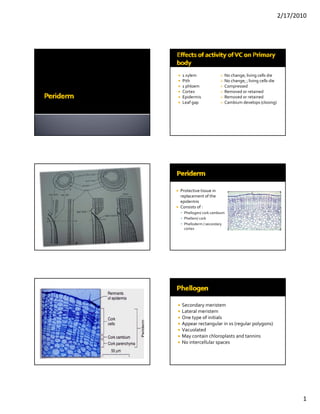
1 xylem No change; living cells die
Pith No change; ; living cells die
1 phloem Compressed
Cortex Removed or retained
Epidermis Removed or retained
Leaf gap Cambium develops (closing)
Protective tissue in
replacement of the
epidermis
Consists of :
Phellogen/ cork cambium
Phellem/ cork
Phelloderm / secondary
cortex
Secondary meristem
Lateral meristem
One type of initials
Appear rectangular in xs (regular polygons)
l l l
Vacuolated
May contain chloroplasts and tannins
No intercellular spaces
1
2. 2/17/2010
Legend:
1 lenticel
3 cork Polygonal in TS and XS
4 cambium
5 collenchyma Compact radial rows
7 phloem
8 secondary xylem
y y
Devoid of intercellular spaces
9 primary xylem Dead cells
2 epidermis
6 sclerenchyma Lined with a layer of suberin
10 pith
SUBERIN‐ impermeable to water and gases;
withstands actions of acids
Phelloids= non suberized
Living cells; non suberized
Arranged in radial rows ORIGIN
Similar to cortical parenchyma Epidermal
cells Subepidermal Parenchyma and Collenchyma
Parenchyma of phloem or pericycle, phloem rays
Lose their Volume of Undergo Starch grains
Cells become
central cytoplasm periclinal and tannins
meristematic
vacuole increases division are lost
Number of phellem layers > phelloderm
layers
Epidermis – Nerium
Immediately below the epidermis
2nd or 3rd cortical layers – Aristolochia
Near the phloem or in phloem parenchyma
h hl hl h
Roots – pericycle (dicot; gymnos); outer
layers of cortex (monocot)
*first phellogen
2
4. 2/17/2010
Depends on the
manner of growth of
periderm, structure of
phellem, etc
phellem etc
Determined by the
type of rhytidome
A. Scaly bark
B. Ring bark
Suberized cortical cells (when epidermis is
sloughed off)
e.g. Gramineae
Storied cork –derived from the outer cortex
e.g palms
‐ repeated division of cortical parenchyma
cells and subsequent suberization of the
products of division
‐ without formation of an initial layer, or
phellogen.
Formed in the pericycle of the root or
underground stem of certain families
Formed by a special phellogen
‐‐[centrifugal prod.]‐‐Layers of thin‐walled
f l d f h ll d
non‐suberized cells alternating with a layer of
endodermal‐like cells
Endodermal‐like cells become cork
Cork cells are living and may serve as storage
4
5. 2/17/2010
Restricted areas of relatively loosely arranged
cells [suberized or non‐suberized]
May develop
a. under a stoma
b. group of stoma
c. between stomata [if sparsely distributed]
b f l d b d
d. under some stoma [if numerous]
May appear in longitudinal rows or horizontal
rows
• Division progresses in the cortex
• Periderm formation or shortly before – inwards
dependent on the persistence of epidermis
• Orientation of divisions becomes
• Cells under stoma or group of stomata more periclinal
begin to divide in different directions
• Lenticel phellogen is formed
• Chlorophyll disappears
COMPLEMENTARY CELLS
5
6. 2/17/2010
• Inc. in number of complementary
cells
May be suberized or non‐suberized
More or less spherical
• Rupture of epidermis Thin‐walled
*Lenticel phellogen—may produced
• Complementary cells are pushed phelloderm below
out and rise above the organ
* CLOSING LAYERS– compact tissue, alternate
with complementary cells
Closing layer Complementary cells
Origin‐‐ Epidermis
6