

Recommandé
Contenu connexe
Tendances
Tendances (20)
Similaire à 01 Gametogenesis
Similaire à 01 Gametogenesis (20)
Plus de Jaya Kumar
Plus de Jaya Kumar (20)
Dernier
Dernier (20)
01 Gametogenesis
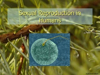
- 1. Sexual Reproduction in Humans ALBIO9700/2006JK
- 2. Female urinogenital system ALBIO9700/2006JK
- 3. Female reproductive system • Ovaries: – site of production of the female gametes (ova) and female sex hormones (oestrogen and progesterone) • Oviducts (Fallopian tubes): – collect secondary oocytes released by ovaries – sweeps egg towards uterus – site for fertilisation – end in funnels fringed with feathery processes called fimbriae • Uterus (womb): – expands during pregnancy – consist of myometrium and endometrium (lining which is shed every month unless pregnant) ALBIO9700/2006JK
- 4. • Cervix: – narrow junction between uterus and vagina where ring of muscle can close off uterus • Vagina: – site where semen is deposited from the penis during sexual intercourse – the birth canal during childbirth – can enlarge to allow entry of erect penis or exit of baby due to elastic tissue and folded epithelium lining – epithelium secretes acid which deter growth of harmful microorganisms ALBIO9700/2006JK
- 5. • Vulva: – labia majora and labia minora – protect the openings of the vagina and urethra • Clitoris: – female equivalent of the penis – contains many nerve endings and swells with blood, becoming erect when sexually stimulated – source of sexual arousal during sexual intercourse ALBIO9700/2006JK
- 6. Male urinogenital system ALBIO9700/2006JK
- 7. Male reproductive system • Testis: – site of production of the male gametes (spermatozoa/sperm) • Seminiferous tubules: – cells lining the walls produce the sperm • Epididymis: – sperm are stored while completing their maturation – fluid is absorbed to make sperm more concentrated – where sperm become mobile ALBIO9700/2006JK
- 8. • Scrotal sac/scrotum: – sac of skin containing the 2 testes which hangs from the main body cavity and helps keep the sperm about 3ºC cooler than normal body temperature • Vas deferens/vas deferentia: – tube which carries sperm out of the testis to the urethra – sperm storage ALBIO9700/2006JK
- 9. • Prostate gland, Cowper’s glands and seminal vesicles: – secrete fluid for carrying the sperm and in which sperm can swim (semen) – alkaline fluids (Cowper’s and seminal vesicles) to neutralise the acidity of remaining urine in urethra – secretes mucus (prostate) – secretes fructose (vesicles) • Penis: – contains urethra which carries sperm to outside world – contains special spongy tissue which can fill with blood when the male is sexually stimulated, causing it to become erect and rigid – ejaculates semen into vagina during sexual intercourse ALBIO9700/2006JK
- 10. Gametogenesis • Formation of gametes in the gonads (testes in the male and ovaries in the female) • Involves meiosis in nuclei of diploid ‘mother cells’ to form haploid gametes • Formation of sperm – spermatogenesis • Formation of eggs – oogenesis • Mother cells known as oocytes and spermatocytes ALBIO9700/2006JK
- 11. Spermatogenesis ALBIO9700/2006JK
- 12. • Process starts between the ages of 11 and 15 and will continue for life • Gametes produced in vast numbers in a continuous production line of several thousand per second (> 100 million/day) • In testes (walls of seminiferous tubules) • From outer layer of tube (germinal epithelium) towards centre of tube where mature sperm break away from wall and float down towards epididymis for storage • Sertoli cells: – Secrete fluid found in lumen of tubes – Sperm development take place in close association – Assist in complex modelling of sperm, particularly spermatids (phagocytosis of spermatid cytoplasm) • Interstitial cells (cells of Leydig): – Secrete the male sex hormone (testosterone) ALBIO9700/2006JK
- 13. Photomicrograph of t.s. of mammalian testis Layer of spermatogonia attached to epithelium and dividing by mitosis Primary spermatocytes dividing by meiosis and moving towards lumen Haploid spermatids maturing into spermatozoa Mature spermatozoa released into lumen ALBIO9700/2006JK
- 14. Seminiferous tubules and interstitial cells ALBIO9700/2006JK
- 15. Sperm ALBIO9700/2006JK
- 16. • Acrosome: contains hydrolytic enzymes needed to digest a path to the egg at fertilisation • Nucleus: carries haploid set of chromosomes • Axial filament: responsible for the wave- like beating of the tail which propels through fluid surroundings ; microtubules have ‘9+2’ arrangement • Centriole: form microtubules • Middle piece and tail: propulsion • Mitochondria: energy for beating of tail ALBIO9700/2006JK
- 17. Oogenesis ALBIO9700/2006JK
- 18. • Process starts before birth • Outer layer of ovary (germinal epithelium) produces primary oocytes and follicle cells which multiply and cluster around the oocytes, forming structures know as primary follicles (~2 million already present at birth) • By birth, each primary oocyte has started to divide by meiosis but stops once chromosomes pair up in prophase I • Once sexually mature, one primary follicle per month is stimulated to complete development into a Graafian follicle (surrounded by layer called theca which secretes the hormone oestrogen) • Follicle cells multiply and several fluid-filled spaces develop which fuse to form one space • Primary oocyte completes meiosis I and divides unequally into 2 • Smaller daughter cell (polar body) disintegrates • Larger daughter cell (secondary oocyte) – released at ovulation with some of the surrounding follicle cells • After ovulation, empty follicle develops into a corpus luteum (yellow body) which disintegrates unless fertilisation takes place • Corpus luteum secretes hormone progesterone ALBIO9700/2006JK
- 19. Sequence from a primary follicle to a secondary follicle ALBIO9700/2006JK
- 20. Photomicrograph of mature follicle Ovary tissue Follicle cells secreting oestrogen Fluid-filled cavity Haploid secondary oocyte held at metaphase II of meiosis, ready to be released at ovulation ALBIO9700/2006JK
- 21. Secondary oocyte and egg • Egg is largest cell in human body • Contains no conspicuous yolk • Obtains nutrients from surrounding cells • Lysosomes function at fertilisation ALBIO9700/2006JK
- 22. ALBIO9700/2006JK