
Recommandé
Contenu connexe
Similaire à ultrasound JP.pptx
Similaire à ultrasound JP.pptx (20)
Plus de jayapandiyan Paraman
Plus de jayapandiyan Paraman (17)
Dernier
Dernier (20)
ultrasound JP.pptx
- 1. ULTRASOUND
- 2. Frequency: ▫ Audible sound – 20 to 20000Hz ▫ Ultrasound – Greater then 20000Hz ▫ Infrasound – Less than 20Hz ▫ Medical ultrasound – 2.5 - 40 MHz In physics, the term "ultrasound" applies to all acoustic energy (longitudinal, mechanical wave) with a frequency above the audible range of human hearing. The audible range of sound is 20 hertz-20 kilohertz. Ultrasound is frequency greater than 20 kilohertz.
- 3. WHY ULTRASOUND? Ultrasound (US) is the most widely used imaging technology worldwide due to • availability • Speed • low cost, • patient-friendliness (no radiation)
- 4. Ultrasonography is widely utilized in medicine, primarily in Gastroenterology Cardiology Gynecology and obstetrics Urology •Bursitis (Bursitis is the inflammation of a bursa, typically one in a shoulder joint) •Tendonitis (A condition in which the tissue connecting muscle to bone becomes inflamed) •Muscle Strain and tears •Osteoarthritis •Ligament and tendon injuries Where do we use USG
- 5. Ultrasound can help relax tight muscles that are sore and warms muscles and soft tissues, which increases circulation that helps to heal. sound waves penetrate the skin's surface causing soft tissues to vibrate, creating heat. The heat induces vasodilation: drawing blood into the target tissues. Increased blood flow delivers needed oxygen and nutrients, and removes cell wastes. The heat helps relieve pain and inflammation, reduce muscle spasms, and accelerate healing. Depending on the treatment area, the range of motion may be increased.
- 6. Ultrasound is also used to: Guide procedures such as needle biopsies, in which needles are used to extract sample cells from an abnormal area for laboratory testing. Diagnose a variety of heart conditions and assess damage after a heart attack or diagnose valvular heart disease. Image the breasts and guide biopsy of breast cancer
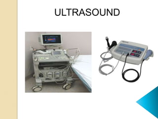
- 8. ULTRASOUND MACHINE A basic ultrasound machine has the following parts: 1. Transducer probe - probe that sends and receives the sound waves 2. Central processing unit (CPU) - computer that does all of the calculations and contains the electrical power supplies for itself and the transducer probe 3. Transducer pulse controls - changes the amplitude, frequency and duration of the pulses emitted from the transducer probe 4. Display - displays the image from the ultrasound data processed by the CPU 5. Keyboard/cursor - inputs data and takes measurements from the display 6. Disk storage device (hard, floppy, CD) - stores the acquired images 7. Printer - prints the image from the displayed data
- 9. HOW IS THE PROCEDURE PERFORMED? For most ultrasound exams, the patient is positioned lying face-up on an examination table that can be tilted or moved. A clear water-based gel is applied to the area of the body being studied to help the transducer make secure contact with the body and eliminate air pockets between the transducer and the skin that can block the sound waves from passing into your body. The sonographer (ultrasound technologist) or radiologist then presses the transducer firmly against the skin in various locations, sweeping over the area of interest or angling the sound beam from a farther location to better see an area of concern.
- 10. ATTENUATION OF ULTRASOUND IN THE TISSUES: • The loss of energy from the ultrasound beam in the tissues is called attenuation and depends on both absorption and scattering • Absorption accounts for some 60 – 80% of the energy loss. The scattered energy may also be absorbed other than in the region to which the ultrasound beam is applied. • Scattering is caused by reflections and refractions, which occur at interfaces throughout the tissues. This is particularly apparent where there is a large difference in acoustic impedance.
- 11. ABSORPTION OF SONIC WAVES • Kinetic energy is converted to heat energy as it passes through the material. • The energy will decrease exponentially with distance from the source because a fixed proportion of it is absorbed at each unit distance so that the remaining amount will become a smaller and smaller percentage of the initial energy • The conversion of sonic energy to heat is due to increased molecular motion
- 12. Kinetic energy is converted to heat energy as it passes through the material. The conversion of sonic energy to heat is due to increased molecular motion Absorption of sonic energy is greatest in tissues with largest amounts of structural protein and lowest water content. Blood – least protein content and least absorption Bone - greatest protein content and greatest absorption
- 14. • Effect of Frequency: Increasing the frequency of Ultrasound causes a decrease in its depth of penetration and concentration of the Ultrasound energy in the superficial tissues. 1MHz 3MHz Muscle 9.0mm 3.0mm Fat 50.0mm 16.5mm Tendon 6.2mm 2.0mm
- 15. Thermal effects: • The advantage of using ultrasound to achieve heating is due to the preferential heating of collagen tissue and to the effective penetration of this energy to deeply placed structures. • Heating fibrous tissue structures such as joint capsules, ligaments, tendons, and scar tissue may cause a temporary increase in their extensibility, and hence a decrease in joint stiffness. • Mild heating can also have the effect of reducing pain and muscle spasm and promoting healing processes.
- 16. Therapeutic Uses:- Ultrasound promotes healing of varicose ulcers and pressure sores (decubital ulcer). • Varicose Ulcer: Ulcer in the leg • associated with varicose veins • is known as varicose ulcer. • Pressure Sore: A bed sore appearing on dependent sites usually on lumbosacral region, most commonly in bed- ridden elderly persons is known as pressure sore.
- 17. Pain relief: • Ultrasound is used in herpes zoster, low backache, prolapsed intervertebral disc (PIV) and many other condition • (Herpes zoster, is a viral disease characterized by a painful skin rash with blisters in a localized area) Acute tissue injury:- • Ultrasound is used in soft tissue and sport injuries, in occupational injuries and post-natal injuries. It is used for perineal post-natal pain, for painful shoulders and for both neurogenic & chronic pain.
- 18. Scar Tissue:- • Ultrasound improves the quality of scar tissue and excessive fibrous tissue. It is used in conditions like Dupuytren’s contracture and plantar fasciitis. (Dupuytren's contracture is a condition in which one or more fingers become permanently bent in a flexed position) (Plantar fasciitis is An inflammation of a thick band of tissue that connects the heel bone to the toes)
- 19. Bone injury:- • Ultrasound therapy in the first 2 weeks after bone injury can increase bones union. Ultrasound has also been used in the early diagnosis of stress fractures (A stress fracture is a small crack in a bone). Chronic Indurated Oedema: • The mechanical effect of ultrasound has an effect on chronic oedema and helps in its treatment. (Oedema - a condition characterized by an excess of watery fluid collecting in the cavities or tissues of the body)
- 20. Contraindications • Tumors – it might encourage neoplastic growth(abnormal growth) and provoke metastases or over precancerous tissue should be avoided • Pregnant Uterus – avoid applying ultrasound over a pregnant uterus, the probable risk to the rapidly dividing and differentiating cells of the embryo and fetus • Spread of Infection - Bacterial or viral infection could be spread by ultrasound, presumably by facilitating microorganism movement across membranes and through the tissues. • Tuberculosis - Due to the possible risk of reactivating encapsulated lesions tuberculous regions should not be treated. (a region in an organ or tissue which has suffered damage through injury or disease, such as a wound, ulcer, abscess, or tumour)
- 21. Dangers of Ultrasound: • There are very less evidences of dangers of ultrasound but it may occur in some conditions only. – Burns could occur if the heat generated exceeded the physiological ability to dissipate it. – Tissue destruction would result from transient cavitation. – Blood cell stasis and endothelial damage (a type of coronary artery disease) may occur if there is standing wave formation. (cell stasis -A stoppage or slowdown in the flow of blood)
- 22. Strengths of ultrasound imaging: It images muscle and soft tissue very well and is particularly useful for indicating the exact position, and the interfaces between solid and fluid-filled spaces. It renders "live" images, where the operator can dynamically select the most useful section for diagnosing and documenting changes, often enabling rapid diagnoses. It shows the structure as well as some aspects of the function of organs. It has no known long-term side effects and rarely causes any discomfort to the patient. Equipment is widely available and comparatively flexible; examinations can be performed at the bedside.
- 23. Weaknesses of ultrasound imaging: Ultrasound cannot penetrate bone and performs poorly when there is air between the scanner and the organ of interest. For example, overlying gas in the gastrointestinal tract often makes ultrasound scanning of the pancreas difficult. Even in the absence of bone or air, the depth penetration of ultrasound is limited, making it difficult to image structures that are far removed from the body surface, especially in obese patients. The method is operator-dependent. A high level of skill and experience is needed to acquire good- quality images and make accurate diagnoses.
- 24. Ultrasonography is generally considered a "safe" imaging modality. Diagnostic ultrasound studies of the fetus are generally considered to be safe during pregnancy. World Health Organizations technical report supports that ultrasound is harmless.
- 25. THANK YOU