
Recommandé
Contenu connexe
Tendances
Tendances (20)
Similaire à Embroylogy
Similaire à Embroylogy (20)
Dernier
Dernier (20)
Embroylogy
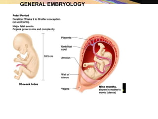
- 2. EMBRYOLOGY: • Embryology – study of the origin and development of single individual • Prenatal period • Embryonic period – first 8 weeks • Fetal period – remaining 30 weeks
- 3. • Embryogenesis starts with fertilization • Fertilization: process of fusion of male gamete(germ cell) sperm with female gamete(germ cell) to form zygote
- 4. Fertilization: • Two sex cells fuse to form a new cell containing genetic material derived from both parents. • Restores the diploid number of chromosomes. • Determines the sex of the organism • Initiates cleavage. • Occurs in the widest part of the uterine tube (the ampulla).
- 5. Fertilization: • Millions of sperm cells are deposited in the female reproductive tract during intercourse. • Only a few hundred have a chance at fertilization. • Only the first sperm to enter the secondary oocyte is able to fertilize it. • The remaining sperm are prevented from penetrating the oocyte.
- 6. The Stages of Embryogenesis • Cleavage. The zygote divides by mitosis to form a multicellular structure called blastocyst. • Gastrulation. The blastocyst cells form three primary germ layers, which are the basic cellular structures from which all body tissues develop. • Organogenesis. The three primary germlayers arrange themselves in ways that give rise to all the organs within the body.
- 7. The Embryonic Period • Week 1 – from zygote to blastocyst • Conception – in lateral third of uterine tube • Zygote (fertilized oocyte) moves toward the uterus • Blastomeres – daughter cells formed from zygote • Morula – at about 12–16 cell (blastomeres)stage • Blastocyst –further division, fluid-filled structure – about 60 cells
- 8. Fertilization and the Events of the First 6 Days of Development Figure 3.3
- 9. Blastocyst: Formation of blastocyst: by 5th day, blastomere reach to 107, small space appear between them -- • The blastocyst is a sphere of about 150 cells, with an outer layer (the trophoblast), a fluid-filled cavity (the blastocoel), and a cluster of cells on the interior (the inner cell mass).
- 11. Implantation: • By the end of the first week after fertilization, the blastocyst enters the lumen of the uterus. • Implantation is the process by which the blastocyst burrows into and embeds within the endometrium.
- 14. Week 2 – The Two-Layered Embryo • Bilaminar embryonic disc – inner cell mass divided into two sheets • Epiblast and the hypoblast • At the site of implantation,the outer cell mass of blastocyst start proliferating and give rise to trophboblast • Placenta: is made up of finger like process known as villi and inetvillious space. An organ that joins mother and off spring.
- 15. Placenta: • Functions in exchange of nutrients, waste products, and respiratory gases between the maternal and fetal bloodstreams. • Transmission of maternal antibodies to the developing embryo or fetus. • Production of hormones to maintain and build the uterine lining.
- 16. Week 2 – The Two-Layered Embryo • Amniotic sac – formed by an extension of epiblast • Outer membrane – forms the amnion • Inner membrane – forms the amniotic sac cavity • Filled with amniotic fluid
- 17. Week 2 – The Two-Layered Embryo • Yolk sac – formed by an extension of hypoblast • Digestive tube forms from yolk sac • Tissues around yolk sac • Gives rise to earliest blood cells and blood vessels
- 18. Implantation of the Blastocyst Figure 3.4
- 19. Week 3 – The Three-Layered Embryo • Primitive streak –the embryonic disc become elongated.The primitve streak is a midline proliferative region of epiblast cells which may break free from the epithelium and migrate beneath the epiblast. • Ingression: it’s a process by which cell become part of the streak and then migrate away from it • Gastrulation –formation of primitive streak is the beginning of gastrulation, a process of where epiblast give rise to a trilaminar structure with a define cranio-caudal axis and formation of embryonic shape
- 20. The Primitive Streak Figure 3.5e-h
- 21. Gastrutlation: • Occurs during the third week of development immediately after implantation. • One of the most critical periods in the development of the embryo. • Cells of the epiblast migrate and form the three primary germ layers which are the cells from which all body tissues develop. • The three primary germ layers are called ectoderm, mesoderm, and endoderm