The nervous system is composed of neurons that transmit signals throughout the body via electrical and chemical signals. The central nervous system includes the brain and spinal cord, while the peripheral nervous system includes all other neurons. Sensory neurons collect information and transmit it to the central nervous system, while motor neurons transmit signals from the central nervous system to tissues. The brain controls homeostasis and interprets sensory information. It is composed of regions like the cerebrum, brainstem, and cerebellum. The peripheral nervous system includes sensory receptors, motor neurons, and the autonomic nervous system. Neurons transmit signals via action potentials generated by ion flow across membranes. Neurotransmitters transmit signals between neurons. Sensory systems allow the detection of stimuli
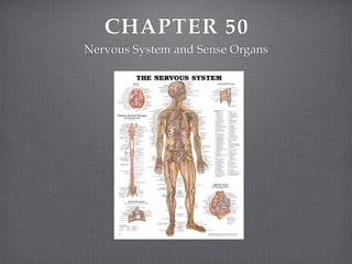
3. CENTRAL NERVOUS
SYSTEM
Mental and physical activity and many aspects of
homeostasis are controlled by the nervous system, a
complex network of cells that communicate with one
another. Within this communication network, a
carefully organized division of labor exists so that
each component of the nervous system operates
effectively.
4. The nervous system is
composed of neurons,
specialized cells that
transmit information
throughout the body. The
axon, a threadlike
projection of the cell
body, enables the neuron
to transmit signals
rapidly over relatively
long distances.
5. ORGANIZATION
The nervous system includes two major divisions: the
central nervous system and the peripheral nervous
system. The brain and spinal cord make up the central
nervous system, and all the other neurons that are not
included in the brain and spinal cord make up the
peripheral nervous system. Some peripheral neurons
collect information from the body and transmit it toward
the central nervous system. These neurons are called
afferent neurons. Other peripheral neurons transmit
information away from the central nervous system.
These are called efferent neurons.
6. BRAIN
The human brain is responsible for overseeing the daily
operations of the human body and for interpreting the vast
amount of information it receives.
The adult human brain weighs about 2% of the total body weight.
The brain contains about 100 BILLION neurons.
These neurons make up the most complex and highly organized
structure on earth.
The brain is responsible for not only controlling emotions, but
maintaining homeostasis.
7. CEREBRUM
The largest portion of the human brain consists of the cerebrum.
The cerebrum, which is easily identified by its highly folded
outer layer, is composed of two cerebral hemispheres.
The cerebral hemispheres are connected by the corpus callosum,
a heavy band of the axons and many neurons.
The corpus callosum lies deep in the central groove that
separates the right hemisphere from the left hemisphere.
Other deep grooves separate each hemisphere into four lobes:
frontal, parietal, temporal, and occipital lobes.
9. The folded outer layer of the cerebral hemisphere is called the cerebral cortex.
In humans, the cerebral cortex is a sheet of neurons about 2 to 4mm thick.
(contains mearly 10%, or 10 billion, of the brain’s total neurons)
The surface area of the cerebral cortex is too large to lie smoothly over the
lower structure of the brain.
The cerebral cortex is very important in sensory processing and in motor
responses. (some functions are not localized in a symmetrical way; right
handed and left handed individuals)
Below the wrinkled surface of the cerebral cortex lies white matter, which is
composed of the axons of the cortical neurons. These axons link specific
regions of the cortex with each other and with neural centers.
There is a great deal of crossover of axons in the spinal cord and brain.
10. UPPER BRAIN STEM-
DEINCEPHALON
Below the cerebrum, the brain stem links the cerebrum with the spinal cord.
The upper part of the brain stem, the diencephalon contains important
relay centers for information relay centers for information entering and
exiting the brain.
The upper relay center, the thalamus, directs most incoming sensory signals
to the proper region of the cerebral cortex.
The hypothalamus, located just below the thalamus, is linked to several
other parts of the brain.
The hypothalamus is very important in maintaining homeostasis, and it
both directly and indirectly controls much of the body’s hormone
production.
11.
12. Parts of the diencephalon and the cerebrum are
included in an important group of connected brain
centers called the limbic system.
The limbic system includes the thalamus, the
hypothalamus, some deeper parts of the cerebral
cortex, and centers in the temporal lobes.
the limbic system plays an important role in emotion,
memory, and motivation.
13. LOWER BRAIN STEM
Below the diencephalon,
the brain stem narrows.
This region of the brain
stem has three main
divisions: the midbrain,
the pons, and the
medulla oblongata.
14. The pons serves as a relay center between the neurons of the cerebral
hemispheres and those of the cerebellum.
The medulla oblongata contains neurons that serve as both a relay center
and a control center. also within the medulla oblongata are centers that
control various homeostasis activities, including heart rate and respiration
rate.
Lying throughout the brainstem is a diffuse network of neurons called the
reticular formation. The reticular formation helps to control respiration and
circulation and serves as a filtering system for incoming sensory signals.
The reticular formation separates signals that demand attention form those
that are unimportant. Some of this function of the reticular formation is
modified by learning.
15. CEREBELLUM
Just below the occipital lobes of the cerebral hemispheres lies the
cerebellum, a region of the brain that plays a vital role in the
coordination of muscle action, particularly the timing of muscle
contractions.
The cerebellum receives sensory impulses from muscles, tendons,
joints, eyes, ears, as well as input from other brain centers.
The cerebellum coordinates rapid and ongoing movements. It
acts together with brain stem motor centers and with the section
of the cerebral cortex that is responsible for motor responses.
Nerve impulses are sent down through the spinal cord and
stimulate or inhibit the contraction of skeletal muscles.
16. PROTECTION
The delicate neurons of the brian and spinal chord are surrounded by three
protective layers called the meninges.
The outer layer, the dura mater, consists of connective tissue, blood vessels,
and neurons.
The middle layer, the arachnoid layer, is elastic and weblike.
The thin inner layer, the pia mater, adheres to the brain and the spinal cord,
and contains many blood vessels and neurons.
A clear liquid called cerebrospinal fluid provides a cushion that protects the
brain an spinal cord from injury. Cerebrospinal fluid separates the middle and
inner meninges and fills four interconnected ventricles, or cavities, in the
brain. Within the ventricles, cerebrospinal fluid acts as a transport medium
for substances that are important to the brain function.
17.
18. SPINAL CORD
The spinal cord is a column of the nerve tissue that starts in the medulla oblongata and runs
down through the vertebral column.
The spinal cord is composed of an outer sheath of white matter. As it does with the brain, the
white matter surrounds a rigid inner core of gray matter, which is composed of the cell
bodies of neurons.
31 pairs of spinal nerves, part of the peripheral nervous system, originate in the spinal cord
and branch out to both sides of the body. When bundled together, these axons form a nerve.
Each spinal nerve consists of a dorsal root and a ventral root. The dorsal roots contain
neurons that carry signals to the central nervous system from various kinds of sensory
receptor neurons. A sensory receptor is a neuron that is specialized to detect a stimulus, such
as pressure or heat.
The ventral roots contain the axons of motor neurons, which are neurons that contact and
carry information to muscles and glands.
Within the spinal cord and elsewhere in the body are interneurons, which are neurons that
connect other neurons to each other.
21. PERIPHERAL NERVOUS
SYSTEM
The sensory division of the peripheral nervous
system is composed of sensory receptors and the
interneurons that connect them to the central nervous
system.
Sensory receptors acquire information from the
external and internal environments of the body.
Spinal and cranial nerves enable the flow of sensory
information to the central nervous system.
22. MOTOR DIVISION
The motor division of the peripheral nervous system
allows the body to react to the sensory information.
The motor division is composed of two independent
systems- the somatic nervous system and the
autonomic nervous system.
23. SOMATIC NERVOUS
SYSTEM
The somatic nervous system of the motor division consists of motor
neurons that control the movement of skeletal muscles.
The somatic system is said to be voluntary, but can also operate
automatically, as it does when you maintain your balance.
One important function of the peripheral nervous system is the relay of
signals in reflexes, which are involuntary and often self-protective
movements.
The tap you get by doctors on your knee is a true spinal reflex; that is, it
involves only neurons in the body and spinal cord, and completely
bypasses the brain. Many of the body’s involuntary actions, however,
originate in a second subdivision of the motor division of the peripheral
nervous system- the autonomic nervous system.
24. AUTONOMIC NERVOUS
SYSTEM
The autonomic system of the motor division consists of
nerves that control the body’s internal conditions by affecting
smooth muscles, both in blood vessels and in organs.
The main function of the autonomic nervous system is the
control of respiration, heartbeat, and other functions involved
in homeostasis.
Stimulation and inhibition of body systems are the
responsibility of two different subdivisions of the autonomic
system- the sympathetic division and the parasympathetic
division.
25. A major function of the sympathetic division is the
shunting of blood from one part of the body to another.
The sympathetic division of the autonomic nervous system
is activated by conditions of physical or emotional stress.
Fight-or-flight
The parasympathetic division of the autonomic nervous
system controls the internal environment during routine
conditions. After a threat of danger has passed, nerves
from the parasympathetic division signal organs to revert
to normal levels of activity.
27. TRANSMISSION OF
NERVE IMPULSES
The ability of the nervous system to monitor and respond
to the environment, both external and internal, depends on
the transmission of signals within a neuron and from one
neuron to another.
The transmission of a signal along the axon of a neuron is
known as an action potential.
The transmission of action potentials depends on
electrochemical energy- electrical energy that is generated
as charged chemical substances move into and out of
neurons.
28. NEURON STRUCTURE
Extending out from the cell body in different directions are membrane-
covered extensions called dendrites. Dendrites receive action potentials from
other neurons.
A neuron may have a single axon or branching axons that contact several
other neurons. The end of an axon is called the axon terminal. It may lie near
a muscle, a gland, dendrite, or cell body of another neuron.
The axons of most neurons are covered with a lipid layer known as the
myelin sheath. It both insulates the axon much like the rubber coating of an
electrical cord and seeds up transmission of action potentials through the
axon. In the peripheral nervous system, myelin is produced by cells called
Schwann cells, which surround the axon.
Gaps in the myelin sheath along the length of the axon are known as the
nodes of Ravier.
29. Neurons do not touch each other,
instead a small gap called the
synaptic cleft is present between the
end of the axon of one neuron and
the dendrite or cell body of another
neuron.
In most neurons, electrical activity
in the neuron causes the release of
chemicals, called neurotransmitters,
into the synaptic cleft; thus
signaling activity of the nervous
system is composed of electrical
activity within neurons and
chemical flow between neurons.
30. NEURON FUNCTION
Neuron function is dependent on electrical activity.
Neurons have an electrical charge different form the
extracellular fluid that surrounds them. A difference in the
electrical charge between two locations is called a
potential.
In neurons, potentials are produced by a complex
interplay of several different ions. The movement of these
ions is affected by their ability to pass through the cell
membrane, their concentrations inside and outside of the
cell, and their charge.
31. RESTING POTENTIAL
A neuron is at rest when it is not receiving or transmitting a
signal.
In a neuron at rest, the concentration of large, negatively-
charged proteins and potassium, K+, ions is greater inside the
cell than outside.
In contrast, the concentration of sodium, Na+, ions is greater
outside the cell than inside.
This imbalance of Na+ and K+ ions is, in part, due to the
action of the sodium-potassium pump. (it actively moves Na
+ out of cells while moving K+ in)
32. The cell membrane is permeable to some ions, but not to others.
Na+ ions do not readily diffuse through the membrane, and the
accumulate outside the cell. The - charged proteins remain
inside because they are too large to exit. K+ ions, however, pass
freely through the membrane and tend to diffuse out of the cell,
down their concentration gradient.
This exit of + charged K+ ions, coupled with the retention of -
charged protein ions, eventually causes the interior of the
neuron to become - charged with respect to the exterior.
This change difference is called the resting potential of the
membrane.
33. ACTION POTENTIAL
When a dendrite of the cell body of a neuron is stimulated, a sudden
change occurs in the permeability of its cell membrane.
At the point where it is stimulated, the cell membrane becomes more
permeable to Na+ ions.
The rush of Na+ ions into the cell opens voltage gated channels in the
membrane that allow even more Na+ ions to diffuse rapidly from the
outside of the membrane to the inside of the neuron.
As a result of the inward diffusion of Na+ ions, the interior of the
neuron’s cell body becomes more + charged than the outer surface. The
interior, once - charged, is now + charged.
This reversal of polarity across the membrane begins an action potential.
34. This action potential starts at the point where the cell body of the neuron joins the
axon.
As the first small segment of the axon becomes + charged, the rise in voltage opens
channels in the adjacent segment of the axon membrane.
As before, Na+ ions enter, driving the voltage in a + direction and opening channels in
the next segment of the axon. (axon potentials travel in one direction only- away from
the cell body, where they begin, and toward the axon terminal)
Shortly after they open, the gated channels for Na+ ions close. Then voltage gated
channels for K+ ions open. The result is an abrupt outward flow of K+ ions. The outer
space becomes + charged, and the inner surface regains its - charge. This signals the
end of the action potential.
The neuron cannot generate another action potential until the resting membrane
potential is restored. This period, during which the neuron can not fire is called the
refractory period.
35. After the action potential, the concentration of Na+ inside
the cell is abnormally high, and the concentration of K+
inside the cell is abnormally low.
The sodium-potassium pump reestablishes the original
concentrations of Na+ ions and K+ ions on both sides of
the membranes.
But, this is not without a price. This requires a continuous
supply of ATP to keep the s-p pump operating. Neurons
consume a great deal of energy in the body.
36. NEUROTRANSMITTER
FUNCTION
When an action potential reaches the axon terminal,
vesicles that are stored in the axon terminal and that
contain neurotransmitters fuse with the presynaptic
membrane.
The fusion releases neurotransmitter molecules into
the synaptic cleft.
These molecules diffuse across the short distance of
the synaptic cleft and bind to receptor molecules
embedded in the postsynaptic membrane.
37. The interaction of neurotransmitter molecules and receptor
molecules changes the permeability of the postsynaptic membrane
by affecting chemically-gated ion channels.
The opening of Na+ channels in the postsynaptic membrane cause
the neuron to become more + in charge.
If this + change of the membrane potential is great enough, a new
action potential is generated in the receiving neuron, effectively
continuing the electrical signal.
If a sufficient number of Na+ channels in the postsynaptic
membrane fail to open, the membrane potential of the receiving
neuron will not rise appreciably or will become more negative.
39. SENSORY SYSTEMS
Human experience is affected by both internal and external stimuli.
Humans are able to distinguish among many different types of stimuli by
means of highly developed system of sense organs.
Sensory systems represent an integration of the functions of the
peripheral and central nervous systems.
The sensory division of the peripheral nervous system gathers
information about the body’s internal conditions and the external
environment.
Sensory systems translate light, sound, temperature, and other aspects of
the environment to electrical signals and transmit these signals, in the
form of action potentials, to the central nervous system, where they are
interpreted.
40. RECEPTORS AND SENSE
ORGANS
A sensory receptor is a neuron that is specialized to detect a stimulus.
The different types of receptors are:
Mechanoreceptors respond to movement, pressure, and tension
Photoreceptors respond to variations in light
Chemoreceptors respond to chemicals
Thermoreceptors respond to changes in temperature
Pain receptors respond to tissue damage
Sensory receptors are found in high concentrations in the sense organs: the eyes, ears,
nose mouth, and skin.
When a particular sense organ receives appropriate stimulation, it sensory receptors
convert the stimulus into electrical signals that are sent to specific regions of the brain.
41. HEARING AND BALANCE
The ear is specialized for two functions: detecting sound and maintaining
balance.
Sound is transmitted as vibrations of air in humans.
Sound is directed by the fleshy structure of the external ear into the ear
itself, where the vibrations are translated into action potentials.
The auditory canal connects the external ear with the tympanic
membrane (the eardrum).
Air pressure in the chamber beyond the tympanic membrane, the middle
ear, can be regulated and balanced by the amount of air allowed into the
ear through the Eustachian tube (an opening in the throat that enables
you to equalize pressure on both sides of your tympanic membrane)
42.
43. Vibrations of the tympanic membrane are transmitted to three small bones of
the middle ear: the hammer, the anvil, and the stirrup.
The stirrup transfers the vibrations to a membrane called the oval window,
which separates the middle ear from the inner ear.
The inner ear contains the cochlea, a coiled tube consisting of three fluid-filled
chambers that are separated by membranes. The middle chamber contains the
organ of Corti, which is the organ of hearing. It rests on the bottom
membrane in the cochlea and contains sensory receptors known as hair cells.
Vibrations of fluid in the cochlea cause the hear cells to move and produce
action potentials which travel along the auditory nerve to the auditory region
of the brain stem, then to the thalamus, and finally to the auditory cortex,
where they are interpreted as sound.
44. Different sounds stimulate different parts of the
cochlea.
Hair cells are very delicate, repeated or sustained
loud noise destroys them, and once they are
destroyed, they can not be replaced.
45. Balance is maintained with the help of machanoreceptors in
three semicircular canals of the inner ear.
These canals are filled with fluid, and their interriors are lined
with hair cells that have tiny particles of calcium carbonate of
top of them.
When the head moves, the hair cells are bent by the action of
gravity of the calcium carbonate particles.
The brain decodes the degree and direction of bend in the
hair cells, interpreting the motion and the orientation of the
head in space.
46. VISION
The eyes are specialized organs that function by receiving light and
transmitting signals to visual processing areas of the brain.
The structures of the eye act together to focus light on the retina, the
light sensitive inner layer of the eye.
Light first passes through the cornea, then through the pupil which
becomes larger when light is dim (controlled by muscles in the
pigmented iris)
After light passes through the pupil, it travels to the lens that is
attached to muscles that adjust the lens’s shape to bend the rays of
the incoming light. This bending focuses the complete image
formed by the light onto the retina.
47.
48. Lying deep within the retina are rods and cones
(photoreceptors) that translate light energy into electrical
signals that can be interpreted by the brain.
Cones initiate the production of sharp images and
respond to different colors.
The optic nerve carries visual information in the form of
action potentials form the retina to the thalamus.
Visual information is processed in the cortex of the
occipital lobe.
49. TASTE AND SMELL
The chemoreceptors for taste are clustered in the taste buds.
Most of the taste buds are imbedded between bumps called
papillae on the tongue. (more found on roof of mouth and
throat)
Chemicals from food dissolved in saliva enter a taste bud
through a small opening and stimulate a signal in the
neurons that line the inner surface of the taste buds.
Taste signals travel through a relay in the brain stem, then to
the thalamus, and finally to the cortex where they are
interpreted.
50. OTHER SENSES
Mechanorereceptors located throughout the skin
make it possible to sense touch, pressure, and
tension.
The receptors for touch are concentrated in the face,
tongue, and fingertips.
Body hair also plays an important role in the ability
to sense touch. Large numbers of merchanoreceptors
are found in the skin at the base of two hair folicles.