2. 1296 Riley et al.: Unifying dose specification between clinical BNCT centers 1296
TABLE I. Results of comparative measurements performed by the dosimetry groups from MIT and CAB in the
hyperthermal neutron beam facility at the RA-6 reactor. Measurements of the thermal neutron flux, photon, and
fast neutron dose rates were performed on central beam axis in air and in a 40 40 20 cm3 water phantom
positioned against the 15 cm diameter circular field aperture. The CAB dosimetry group specifies thermal
neutron flux using an absorption cross section in Au that is averaged over the energy spectrum of the beam and
varies between 82.4 barns in air and 86.0 barns at 5 cm depth in phantom. These data were adjusted using a
constant value of 98.8 barns for the absorption cross section to give the 2200 m s−1 neutron flux for direct
comparison with the MIT method of reporting. The results are scaled to a reactor operating power of 500 kW.
Thermal neutron
Depth 2200 ms−1 flux 108 Photon dose rate Fast neutron dose rate
cm cm−2 s−1 cGy min−1 cGy min−1
MIT CAB MIT CAB MIT CAB
In-air 2.4 0.1 2.3 0.2 3.8 0.2 3.6 0.1 2.2 0.4 1.4 0.4
1.2 7.5 0.3 7.9 0.5 7.9 0.4 7.3 0.2 1.1 0.4 1.4 0.4
2.0 6.6 0.3 6.7 0.4 7.4 0.3 6.9 0.1 0.78 0.39 1.1 0.3
3.0 5.2 0.2 5.1 0.3 6.4 0.3 6.1 0.1 0.53 0.36 0.84 0.25
4.0 3.6 0.2 3.8 0.2 5.5 0.2 5.3 0.1 0.44 0.35 0.62 0.20
5.0 2.6 0.1 2.7 0.2 4.6 0.2 4.5 0.1 0.38 0.34 0.46 0.14
capture reactions in tissue i.e., 14N n , p 14C that may also Although identical ionization chambers are employed, the
have a RBE different from those for fast neutrons or photons. two groups convert ionization response to absorbed dose dif-
The kerma resulting from the 14N n , p 14C reaction accounts ferently. The CAB group empirically determines the thermal
for approximately 98% of the thermal neutron dose to nor- neutron sensitivities of each chamber5,7 and subtracts a
mal tissue when no boron is present. This dose component is scaled response from each measurement based upon the mea-
also needed to correct the response of A-150 walled ioniza- sured thermal neutron fluence to then determine the photon
tion chambers in determining the fast and, hence, total neu- and fast neutron doses from the residual ionization currents
tron dose in tissues such as skin and brain that have mark- as initially proposed by Rogus et al.8 The MIT group, how-
edly different nitrogen concentrations. ever, now includes the thermal neutron response as part of
A series of measurements was performed to determine the the neutron sensitivity of each chamber and determines the
thermal neutron flux as well as photon and fast neutron dose from neutrons of all energies.9
kerma rates free in-air and the thermal neutron flux together To complete the intercomparison, CNEA computed a
with the photon and fast neutron absorbed dose rates in- treatment plan using the Monte Carlo based NCTPlan ordi-
phantom. Measurements of the separate thermal neutron, fast narily used for dose calculations in their clinical trials.10,11
neutron all neutrons with energies above the cadmium cut Calculations were performed pertinent to the measurement
off of approximately 0.5 eV and photon dose components conditions on central axis in the large water phantom for a
were performed on the central axis of the beam in a large, direct comparison.
water filled rectangular box 40 W 40 H 20 D cm3
that was aligned with the larger face against the 15 cm di-
ameter beam aperture. Identical graphite and A-150 walled III. RESULTS
ionization chambers IC-18s manufactured by Far West The results of the comparative measurements performed
Technology, Goleta, CA were used by both groups as well both in-air and in-phantom for the three principal dose com-
as bare and Cd covered Au activation foils to determine the ponents are given in Table I. Results are scaled to the maxi-
thermal neutron flux which is used to separately account for mum beam monitor voltage that therefore represent beam
the boron and thermal neutron dose. Activated foils were intensities realizable during therapy and are given with the
counted using a HPGe detector at Bariloche that was cross respective experimental uncertainties 1 determined by
checked with subsequent measurements at MIT. Certified each group. Agreement between the two groups is generally
mixtures of methane based tissue equivalent 64.4% CH4, satisfactory although the estimated uncertainties for the fast
32.4% CO2, and 3.2% N2 and research grade CO2 flush neutron component are large. The data measured by CAB in
gases were used with the A-150 and graphite walled cham- this phantom are used to benchmark the treatment planning
bers, respectively. Irradiations were performed with the software NCTPlan utilized by the CNEA for dose
RA-6 operating at nominal full power of 500 kW and were calculations.5 Examining the calibrated output from NCT-
normalized to a response of 320 mV from a compensated Plan enabled a quantitative comparison between the dose
ionization chamber beam monitor used for delivering beam prescribed by CNEA to that measured and so prescribed by
fluence during therapy. The ionization current from the beam MIT for the four individual dose components including that
monitor was collected across a RC circuit with a long time from thermal neutron capture in boron. A fourth-order poly-
constant so that the change in voltage across a charging ca- nomial was fit using the least-squares method to the MIT
pacitor was proportional to the integrated ionization charge. dose measurements and the CNEA NCTPlan calculation was
Medical Physics, Vol. 35, No. 4, April 2008
3. 1297 Riley et al.: Unifying dose specification between clinical BNCT centers 1297
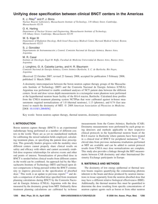
FIG. 1. Thermal neutron circle and photon triangle absorbed dose rates
measured by the MIT dosimetry group as a function of depth on the central
axis of a 15 cm diameter circular field in a large 40 40 20 cm3 water FIG. 2. Fast neutron absorbed dose rates as a function of depth on the central
phantom at the hyperthermal neutron beam of the RA-6 reactor Bariloche. axis of a 15 cm diameter circular field in a large 40 40 20 cm3 water
Experimental uncertainties are illustrated when larger than the symbols. Cal- phantom at the hyperthermal neutron beam of the RA-6 reactor Bariloche.
culated curves from the BNCT treatment planning system NCTPlan used by Data measured by the visiting MIT dosimetry group are shown with experi-
CNEA are also shown scaled to match the MIT measurements. The scaling mental uncertainties that increase with depth in-phantom ranging from 32%
factors normalize the CNEA calculations to the MIT measurements and to 100%. The curve was calculated by CNEA with the treatment planning
enable a quantitative comparison between the CNEA and MIT prescription system NCTPlan routinely used for treatment prescriptions during BNCT
doses. and is shown normalized to the MIT measurements. The scaling factor re-
quired to normalize the CNEA calculations to the MIT measured data is
0.74.
scaled to this polynomial expression by a single factor to
yield the smallest sum in the squared residuals between the turn would be reported by MIT. This procedure improves
measured MIT results and the CNEA TPS calculation. The precision in the specification of absorbed dose between the
thermal neutron dose was determined from the measured two centers. The magnitude of these results for the individual
thermal flux. To enable this comparison for the thermal neu- dose components are similar to those from the earlier com-
tron dose the measured neutron flux was multiplied by the parisons performed in Europe.4
appropriate neutron kerma coefficient for soft muscle con- To examine the combined effect of the scaling factors on
taining 3.5% nitrogen.12 Figures 1 and 2 show the depth-dose the total biologically weighted dose, the scaling factors were
curves calculated by CNEA with NCTPlan, scaled to the applied to the prescription of a previously treated patient
MIT measurements. Scale factors of 1.0, 1.13, and 0.74 were from the CNEA melanoma trial. This dose conversion or
determined for the thermal neutron, photon and fast neutron adjustment reduced the total weighted dose to the patient by
dose components with uncertainties due principally to the less than 2%. However, to properly convert CNEA prescrip-
experimental uncertainties in the MIT measurements which tions to those from other clinical protocols will first require
are 4.0% thermal neutrons , 4.4% photons , and depending independent validation of the reported scaling factors and
upon depth 30%–100% fast neutrons . Multiplying each of then application of the biological weighting factors appropri-
the dose components calculated by the CNEA TPS with ate for each dose component as specified in those protocols.
these scale factors converts the CNEA specified dose to a Reanalyses will necessitate determining the dose parameters
MIT measured dose that is reproducible to within 2%–3% of clinical relevance such as dose volume histograms, maxi-
and facilitates a more precise and direct comparison of the mum delivered dose, maximum tolerable dose, etc. for the
administered doses between the two centers.4 particular dose response relationship or adverse events being
investigated. These will include data for normal tissue and
tumor as well as the dose to the various organs at risk.
IV. CONCLUSIONS The reported scale factors should enable dose prescrip-
The physical dosimetry pertinent to recent irradiations tions administered at RA-6 to be expressed in terms of dose
during the current BNCT clinical trials in Argentina com- specified at MIT and in turn to those from the previous clini-
pares well with that of MIT. Good agreement was found for cal programs at the Brookhaven Medical Research Reactor
the in-phantom thermal neutron flux dose , fast neutron, and as well as those in Europe.
photon depth dose profiles obtained under reference irradia-
tion conditions that are needed to determine the total patient
dose. To normalize the individual dose components to those ACKNOWLEDGMENTS
measured by MIT requires that the CNEA data be multiplied The authors thank the staff of the RA-6 reactor Bariloche
by scale factors of 1.0 thermal neutrons , 1.13 photons , for their cooperation and assistance throughout the course of
and 0.74 fast neutrons . These adjustments effectively reca- this study. This work was supported in part by the Depart-
librate the treatment planning system used by CNEA, con- ment of Energy Grant Nos. DE-FG02-87ER-6060 and DE-
verting the output to doses that are measured and which in AC02-06CH11357.
Medical Physics, Vol. 35, No. 4, April 2008
4. 1298 Riley et al.: Unifying dose specification between clinical BNCT centers 1298
a
Author to whom correspondence should be addressed. Present address: edited by Y. Nakagawa, T. Kobayashi, and H. Fukuda International So-
Nuclear Reactor Laboratory, 138 Albany Street, Cambridge, MA 02139. ciety for Neutron Capture Therapy, Osaka, Japan, 2006 , pp. 14–17.
7
Telephone: 617 258-5938; Fax: 617 253-7300. Electronic mail: J. Longhino, O. Calzetta Larrieu, and H. Blaumann, “Experimental deter-
flavor@mit.edu mination of the thermal neutron sensitivity of a TE ionisation chamber,”
1
P. J. Binns, K. J. Riley, O. K. Harling, J. R. Albritton, and W. S. Kiger III, in Proceedings of the Tenth International Congress on Neutron Capture
“Normalization of prescribed dose in BNCT,” Radiat. Prot. Dosim. in Therapy, edited by W. Sauerwein, R. Moss, and A. Wittig Monduzzi
press . Editore, International Proceedings Division, Bologna, 2002 , pp. 489–
2
K. J. Riley, P. J. Binns, D. D. Greenberg, and O. K. Harling, “A physical 493.
8
dosimetry intercomparison for BNCT,” Med. Phys. 29, 898–904 2002 . R. D. Rogus, O. K. Harling, and J. C. Yanch, “Mixed field dosimetry of
3
P. J. Binns, K. J. Riley, O. K. Harling, I. Auterinen, M. Marek, and W. S. epithermal neutron beams for boron neutron capture therapy at the
Kiger III, “Progress with the NCT international dosimetry exchange,” MITR-II research reactor,” Med. Phys. 21, 1611–1625 1994 .
9
Appl. Radiat. Isot. 61, 865–868 2004 . K. J. Riley, P. J. Binns, and O. K. Harling, “Performance characteristics
4
P. J. Binns, K. J. Riley, O. K. Harling, W. S. Kiger III, P. M. Munck af of the MIT fission converter based epithermal neutron beam,” Phys. Med.
Rosenschöld, V. Giusti, J. Capala, K. Sköld, I. Auterinen, T. Serén, P. Biol. 48, 943–958 2003 .
10
Kotiluoto, J. Uusi-Simola, M. Marek, L. Viererbl, and F. Spurny, “An S. J. González, G. A. Santa Cruz, W. S. Kiger III, J. T. Goorley, M. R.
international dosimetry exchange for boron neutron capture therapy. Part Palmer, P. M. Busse, and R. G. Zamenhof, “NCTPlan, the New PC ver-
I: Absorbed dose measurements,” Med. Phys. 32, 3729–3736 2005 . sion of MacNCTPlan: Improvements and verification of a BNCT treat-
5
H. R. Blaumann, S. J. González, J. M. Longhino, G. A. Santa Cruz, O. A. ment planning system,” in Proceedings of the Tenth International Con-
Calzetta Larrieu, M. R. Bonomi, and B. M. C. Roth, “Boron neutron gress on Neutron Capture Therapy, edited by W. Sauerwein, R. Moss, and
capture therapy of skin melanomas at the RA-6 reactor: A procedural A. Wittig Monduzzi Editore, Bologna, 2002 , pp. 557–561.
11
approach to beam set up and performance evaluation for upcoming clini- M. R. Casal, S. J. González, H. R. Blaumann, J. Longhino, O. A. Calzetta
cal trials,” Med. Phys. 31, 70–81 2004 . Larrieu, and C. A. Wemple, “Comparison of the performance of two NCT
6
B. M. Roth, M. R. Bonomi, S. J. González, R. J. Rebagliati, P. Menéndez, treatment planning systems using the therapeutic beam of the RA-6 reac-
G. A. Santa Cruz, M. R. Casal, H. R. Blaumann, O. A. Calzetta Larrieu, tor,” Appl. Radiat. Isot. 61, 805–810 2004 .
12
D. Feld, D. Batistoni, J. Longhino, S. Castiglia, and S. J. Liberman, ICRU, “Nuclear data for neutron and proton radiotherapy and for radia-
“BNCT clinical trials of skin melanoma patients in Argentina,” in Pro- tion protection,” Report No. 63, International Commission on Radiation
ceedings of the 12th International Congress on Neutron Capture Therapy, Units and Measurements, Bethesda, MD, 2000.
Medical Physics, Vol. 35, No. 4, April 2008