
Recommandé
Recommandé
Contenu connexe
Similaire à Multiple Submandibular Duct Calculi: A Case Report
Similaire à Multiple Submandibular Duct Calculi: A Case Report (18)
Plus de komalicarol
Plus de komalicarol (20)
Dernier
Dernier (20)
Multiple Submandibular Duct Calculi: A Case Report
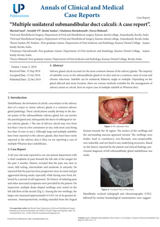
- 1. “Multiple unilateral submandibular duct calculi: A case report”. Shermil Sayd1* , Sreejith VP2 , Resmi Sankar3 , Chaitanya Harindranath4 , Navya Mukund5 , 1 Oral and Maxillofacial Surgery, Department of Oral and Maxillofacial surgery, Kannur dental college, Anjarakandy, Kerala, India. 2 Oral and Maxillofacial Surgery, Department of Oral and Maxillofacial Surgery, Kannur dental college, Anjarakandy, Kerala, India. 3 Resmi Sankar, PG Student , Post-graduate trainee, Department of Oral medicine and Radiology, Kannur Dental College, Anjara- kandy, Kerala, India. 4 Chaitanya Harindranath, Post-graduate trainee, Department of Oral medicine and Radiology, Kannur Dental College, Anjara- kandy, Kerala, India. 5 Navya Mukund, Post-graduate trainee, Department of Oral medicine and Radiology, Kannur Dental College, Kerala, India. Volume 1 Issue 3- 2018 Received Date: 15 Sep 2018 Accepted Date: 15 Oct 2018 Published Date: 22 Oct 2018 1. Abstract Salivary gland calculi account for the most common disease of the salivary glands. The majority of sialoliths occur in the submandibular gland or its duct and are a common cause of acute and chronic infections. Sialolith can be unilateral, bilateral, single or multiple. Depending on the gland affected and stone location, there are various methods available for the management of salivary stones or calculi. here we report case of multiple sialolith in Wharton duct. Annals of Clinical and Medical Case Reports Citation: Sayd S, Multiple Submandibular Duct Calculi: A Case Report. Annals of Clinical and Medical Case Reports. 2018; 1(3): 1-3. United Prime Publications: http://unitedprimepub.com *Corresponding Author (s): Shermil Sayd, Department of Oral and Maxillofacial Surgery, Kunhitharuvai memorial charitable trust (KMCT) Dental College and Hospitals, India, Tel: +919446230425; Fax: +91495 2294726; Email:shermil12@gmail.com Case Report 2. Introduction Sialolithiasis, the formation of calcific concretions in the salivary duct of a major or minor salivary gland, is a common salivary gland pathology. These calcifications usually develop in the duc- tal system of the submandibular salivary gland, but can involve the parotid gland and, infrequently the ducts of sublingual or mi- nor salivary glands. 1 The size of salivary calculi may vary from less than 1 mm to a few centimeters in size, with most cases being less than 10 mm in size.2 Although large and multiple sialoliths have been reported in the salivary glands, they have been rarely reported in the salivary duct.2 Here we are reporting a case of multiple Wharton duct sialolithiasis. 3. Case Report A 40-year-old male reported to our out-patient department with a chief complaint of pain beneath the left side of the tongue for the past 3 months. History revealed that the pain was slow in onset, dull aching, intermediate and moderate in intensity. He reported that the pain has been progressive since its onset and got aggravated during meals, especially while having sour food, fol- lowed by a period of self regression. No history of radiating pain and other associated symptoms were provided by the patient. On inspection, multiple dome-shaped swellings were noted on the left side floor of the mouth (Fig 1). Among the two swellings, the larger one measured approximately 0.5×0.5 cm at its greatest di- mension. Anteroposteriorly, swelling extended from the lingual Figure 1: Pre-operative view. frenum towards the 36 region. The surface of the swellings and the surrounding mucosa appeared normal. The swellings were tender, hard in consistency, non-fluctuant, non-compressible, non-reducible, and not fixed to any underlying structures. Based on the history reported by the patient and clinical findings, pro- visional diagnosis of left submandibular gland sialolithiasis was made. Figure 2: Occlusal View of the lesion. Mandibular occlusal radiograph and ultrasonography (USG) followed by routine hematological examinations were suggest-
- 2. Copyright ©2018 Sayd S et al. This is an open access article distributed under the terms of the Creative Commons Attribution License, which permits unrestricted use, distribution, and build upon your work non-commercially. 2 Volume 1 Issue 3 -2018 Case Report 4. Discussion Submandibular salivary glands are bilaterally placed on the floor of the mouth, drains saliva up into the floor of the mouth through Wharton’s duct. Eighty percent of all salivary duct calculi form in Figure 3: USG view of the lesion. Figure 4: Surgical removal of the sialolith. Figure 5: Removed sialolith. the Wharton’s duct. While 70-80% of cases reports with solitary stones, about 5-10 % of patients reports with multiple stones.2 Salivary calculi are usually unilateral in occurrence, round to ob- long in shape, have an irregular or smooth surface, vary in size, and are usually yellow.3 They are found more often in adults, al- though they also occur in children. The classic symptoms of sali- vary calculi are manifested as pain and swelling of the involved gland during eating.4 There are currently two theories about the pathogenesis of salivary calculi. The first theory states that intra- cellular micro calculi develop within autophagosomes in normal salivary tissue and are naturally voided through the duct system. However, if these became impacted during their discharge, they will act as a nidus for stone formation.5 The submandibular gland is most susceptible because its saliva is more alkaline, has a higher concentration of calcium and phosphate, and has a higher mucus content than other major salivary glands. Also, the sub- mandibular duct is longer with a kink over the posterior border of the mylohyoid muscle and has an anti-gravity flow.6 Investigative modalities that can be used include plain radiograph, sialography, computed tomography and ultrasonog- raphy (USG). In the present case, we performed a mandibular occlusal radiography and USG. In the occlusal radiograph, two radiopaque structures were noted on the left submandibular re- gion. Calculi that are less radiodense cannot be accurately appre- ciated in the occlusal radiograph- hence we performed an USG, which represents an excellent first-level diagnostic technique be- cause it can reveal mineralized stones with a diameter of at least 1.5 mm with an accuracy of 99 %.7 The sialolith should be removed with a minimally in- vasive procedure, usually through an intraoral sialolithectomy, to avoid morbidity associated with sialadenectomy.2 Palpabil- ity of the calculi, regardless of its location or size, is considered to the most crucial factor in the successful intraoral removal of the stone.2 In our case the sialolith was palpable, so intraoral re- moval was successfully performed. Recently, minimally invasive techniques including lithotripsy, basket retrieval, and sialendos- copy have been developed with proven efficacy. However, these techniques are known to have some limitations on large or in- fected calculi. 8 Intraoral removal can be performed successfully regardless of the location, size, presence of infection, or recur- rence of calculi if the calculi themselves are palpable.9 5. Conclusion Sialoliths are common salivary gland pathology, which common- ly occurred in the submandibular gland, usually presented as solitary calculi, but rarely as multiple. Regardless of the number, intraoral removal of sialolith can be performed. The palpability of calculi is the most critical factor influencing the successful in- traoral removal of submandibular calculi. ed. Mandibular occlusal radiographs revealed two radiopaque structures of sizes approximately 0.7×0.5 cm and 1×0.6 cm in the left submandibular region corresponding to the lingual as- pect of 36 & 37 regions (Fig 2). USG revealed two heterogenous hyperechoic structure in the left submandibular duct measuring approximately 6.5 mm and 8 mm with dilatation of the proximal duct (Fig 3). Routine hematological reports were noncontribu- tory. Considering the investigative reports we came to the diag- nosis of submandibular sialolithiasis. After obtaining consent from the patient, sialolithectomy was performed under local anesthesia. Removal was done after plac- ing a tie suture behind the posterior-most sialolith, followed by an incision over the most prominent swelling. Both the sialoliths were removed through the same incision reducing the surgical trauma (Fig 4 & Fig 5). Once the sialoliths were removed, milk- ing of the gland was done by bi-digital palpation to ensure pa- tency of the duct. This was followed by the placement of single suture for the approximation of the oral mucosa. The patient was followed up for 2 months by both clinical and radiographic examinations, and mandibular occlusal radiograph revealed no evidence of calculi.
- 3. References 1. Louis Mandel, Salivary Gland Disorders, Dent Clin N Am 55 (2011) 121–140. 2. Krishnappa BD. Multiple submandibular duct (Wharton’s duct) cal- culi of unusual size and shape. Indian J Otolaryngol Head Neck Surg. 2008 Sep; 60(3):287-8. 3. Harold D. B, Submandibular Salivary Stones, Current Management Modalities J Oral Maxillofac Surg 62:369-378, 2004. 4. Nahlieli O, Eliav E, Hasson O, et al: Pediatric sialolithiasis. Oral Surg Oral Med Oral Pathol Oral Radiol Endod 6:709, 2000. 5. Epivatianos A, Harrison JD, Dimtiou T (1987) Ultrastructural and histochemical observations on micro calculi in chronic submandibular sialadenitis. J Oral Pathol 16:514–517. 6. Sherman JA, McGurk (2000) Lack of correlation between water hard- ness and salivary calculi in England. Br J Oral Maxillofac Surg 38:50–53 7. Yoshimura Y, Inoue Y, Odagawa T. Sonographic examination of sialolithiasis.J Oral Maxillofac Surg 1989; 47: 907-12. 8. Marmary Y. A novel and non-invasive method for the removal of sali- vary gland stones.Int J Oral Maxillofac Surg 1986;15:585–7. 9. June Sik Park, Jin Ho Sohn, and Jeong Kyu Kim, Factors influencing intraoral removal of submandibular calculi,Otolaryngology–Head and Neck Surgery, Vol 135, No 5, November 2006. Volume 1 Issue 3 -2018 Case Report 3