1. Oncogene (2003) 22, 6517–6523
& 2003 Nature Publishing Group All rights reserved 0950-9232/03 $25.00
www.nature.com/onc
Viruses and cancer: lessons from the human polyomavirus, JCV
Krzysztof Reiss1 and Kamel Khalili*,1
1
Center for Neurovirology and Cancer Biology, Temple University, Philadelphia, PA 19122, USA
The possible role of eucaryotic viruses in the development vated and causes PML when the immune system is
of cancer has been the subject of intense investigation impaired.
during the past 50 years. Thus far, a strong link between Prior to the AIDS epidemic, PML was considered an
some RNA and DNA viruses and various cancers in extremely rare disorder associated with immunocom-
humans has been established and the transforming activity promising diseases such as lymphomas, or was seen in
of several of the viruses in cell culture and their renal transplant and chemotherapy patients as a
oncogenecity in experimental animals has been well complication of immunosuppressive therapies. How-
documented. Perhaps, one of the most common themes ever, recent reports indicate that 70% of all HIV-1-
among the oncogenic viruses rests in the ability of one or infected patients will exhibit neurological disorders and
more of the viral proteins to deregulate pathways involved between 5 and 8% of all HIV-1-infected patients will
in the control of cell proliferation. For example, inactiva- develop PML (Berger and Concha, 1995). PML is
tion of tumor suppressors through their association with characterized by demyelination due to the cytolytic
viral transforming proteins, and/or impairment of signal destruction of oligodendrocytes, the subset of glial cells
transduction pathways upon viral infection and expression in brain that are responsible for myelin production.
of viral proteins are among the key biological events that Other hallmarks of PML include giant bizarre
can either trigger and/or contribute to the process of astrocytes, hyperchromatic oligodendrocyte nuclei, and
cancer. In recent years, more attention has been paid to multiple foci of demyelination (Major et al., 1992).
human polyomaviruses, particularly JC virus (JCV), The viral genome is comprised of a closed, circular,
which infects greater than 80% of the human population, double-stranded DNA that can be divided into early
due to the ability of this virus to induce a fatal and late coding sequences, and the viral regulatory
demyelinating disease in the brain, its presence in various region. The regulatory region of JCV encompasses the
tumors of central nervous system (CNS) and non-CNS viral origin of DNA replication and a bidirectional
origin, and the oncogenic potential of this virus in several promoter composed of two 98 base pair repeats. The
laboratory animal models. Here, we will focus our viral early sequence that is transcribed before DNA
attention on JCV and describe several pathways employed replication is responsible for the production of T-
by the virus to contribute to and/or accelerate cancer antigen, whereas the viral late genes that are transcribed
development. after DNA replication, produce capsid proteins
Oncogene (2003) 22, 6517–6523. doi:10.1038/sj.onc.1206959 VP1, VP2, and VP3 and the accessory Agnoprotein
(Frisque and White, 1992). The lytic cycle of JCV
Keywords: tumorigenesis; T-antigen; oncogene; JC explains some of the pathological features of PML,
virus; PML hallmarks of which are the destruction of myelin sheaths
and oligodendrocytes, the myelin-producing cells;
and the appearance of astrocytes that exhibit trans-
Introduction formed cell morphology. Evidently, lytic infection
with JCV results in cytolytic destruction of oligoden-
The human neurotropic JC virus (JCV) is the etiologic drocytes, while its abortive replication in astrocytes
agent of progressive multifocal leukoencephalopathy causes morphological changes leaving astrocytes
(PML), a fatal demyelinating disease of the central that resemble transformed cells. In cell culture systems,
nervous system (CNS) (Berger et al., 1998). JCV is a JCV exhibits a narrow tissue specificity in that the
member of the polyomaviruses whose other members virus can replicate most efficiently in primary human
include BK virus and the well-known simian virus 40 fetal glial cells. Studies have also reported weak
(SV40). JCV coexists within the human population, as replication of the virus in B cells (Monaco et al.,
greater than 80% of adults worldwide exhibit JCV- 1996). Owing to the species specificity of the DNA
specific antibodies (Major et al., 1992). Subclinical polymerase, JCV can only replicate in primates, and
infection with the virus occurs in early childhood and humans are thought to represent the natural viral host
the virus remains in a latent stage throughout life, (Frisque and White, 1992).
although on rare occasions the virus becomes reacti- In addition to its primary role in the development of
PML, JCV has been shown to be associated with several
*Correspondence: K Khalili; E-mail: kamel.khalili@temple.edu human tumors (Del Valle et al., 2001a). While the
2. Viruses and cancer
K Reiss and K Khalili
6518
etiologic role for JCV in the development of cancer in cells by JCV. Expression of the JCV early protein from
humans remains to be established, there have been the Mad-4 strain of JCV in transgenic animals contain-
several experimental animal models that verify the ing only the early DNA sequences of JCV results in
oncogenic potential of JCV in vivo. Furthermore, the the development of two distinct phenotypes. One line
ability of the JCV early protein to transform human exhibited dysmyelination of the central nervous system
fetal cells has led to the development of several cell lines due to reduced production of myelin proteins, while the
that possess tumorigenic activity in vivo. In this review, other set developed adrenal neuroblastoma (Small et al.,
we have summarized the most recent observations on 1986a, b; Franks et al., 1996). Expression of the JCV
JCV-transformed tumor cells in which the viral protein, early protein from the archetype strain of JCV in FVB/
T-antigen deregulates several cellular pathways that N mice leads to the development of primitive neuroec-
control proliferation. These observations can provide an todermal tumors resembling medulloblastoma (Krynska
alternative working platform for studying oncogenic et al., 1999b). Of note, no evidence for abnormal
pathways utilized by various viruses to induce cancer in myelination of the brain was observed in transgenic
humans. mice harboring tumors. In a different series of studies,
transgenic animals were created using the C57BL/6
JCV and cancer: in vitro and in vivo observations mouse strain expressing the JCV Mad-4 early genome.
After 6–8 months, the mice exhibited pituitary adeno-
Similar to the well-studied SV40, JCV possesses the mas (Gordon et al., 2000). Curiously, some of the
ability to infect and transform primary cultures, animals that had not developed pituitary adenomas
including primary human fetal glial cells and human instead developed malignant peripheral nerve sheath
vascular endothelial cells, albeit to a lesser degree than tumors (MPNST) (Shollar and Gordon, unpublished
SV40 (Fareed et al., 1978; Walker and Padgett, 1978). observations). Histological examination of the various
Almost all JCV-infected transformed cells express the JCV-induced tumors in transgenic mice revealed that
viral early protein and they exhibit altered morphology, while all cells contain the transgene, as expected, only a
enhanced growth rate in low serum medium, the ability subset of cells express T-antigen. Interestingly, results
to form foci in culture, and in some but not all cases, from studies on cell cultures derived from PNETs
induce tumors in Nude mice (Gallia et al., 1998a; Del showed that after several passages, T-antigen-positive
Valle et al., 2001a). Also, transformation of primary cells lose their expression of T-antigen, yet retain their
hamster brain cells by various strains of JCV, including transforming phenotype. While the mechanism involved
Mad-1 and Mad-4, have generated rapidly growing cells in the extinction of T-antigen remains unknown, its has
with several characteristics of a transformed phenotype, been suggested that a ‘hit-and-run’ mechanism may be
including growth in low serum, enhanced production of involved in JCV-induced cancer cells. Results from
plasminogen activator, and anchorage-independent in vitro studies have indicated that T-antigen-positive
growth (Frisque et al., 1980; Kang and Folk, 1992). In and T-antigen-negative medulloblastoma cells derived
addition to the cells of neural origin that seem to be from transgenic mice exhibit similar growth patterns in
readily transformed by the virus, a low incidence of monolayer cultures (Wang et al., 2001). Under ancho-
JCV-mediated transformation in baby hamster kidney rage-independent growth conditions, however, the
cells and in rat fibroblasts has been reported (Bollag T-antigen-positive cells manage to survive in the absence
et al., 1989; Haggerty et al., 1989). In laboratory animals of serum and proliferate upon treatment of cells with
JCV infection often results in the development of insulin-like growth factor 1 (IGF-1). Under similar
tumors. Newborn Syrian hamsters develop a broad conditions, T-antigen-negative cells demonstrate an
range of tumors, including medulloblastomas, primitive attenuated response when stimulated with IGF-1 (Wang
neuroectodermal tumors, astrocytomas, glioblastoma et al., 2001). Similarly, T-antigen-positive cells form
multiforme, and peripheral neuroblastomas after brain tumors when injected subcutaneously in Nude mice,
inoculation with JCV (Walker et al., 1973; Varakis, while the T-antigen-negative cells show limited, if any,
1978; Zu Rhein and Varakis, 1979). While no evidence growth (Krynska et al., 2000).
for productive infection of the tumor cells by JCV has
been observed, expression of the viral early, but not late, Molecular basis of JCV T-antigen-mediated
promoter in the tumor cells leads to the accumulation of
transformation
large amounts of T-antigen (Raj et al., 1995). Similarly,
injection of JCV into the brains of newborn rats induces While the precise mechanism responsible for cellular
undifferentiated neuroectodermal tumors in the cere- transformation by JCV is not fully understood, it is
brum of 75% of the animals (Ohsumi et al., 1986). In believed that the early proteins of the virus, particularly
owl and squirrel monkeys, intracerebral, subcutaneous, T-antigen, by associating with several cellular proteins,
or intravenous inoculation of JCV causes the develop- plays a critical role in this event (Sullivan et al., 2000).
ment of astrocytomas, glioblastomas, and neuroblasto- Like SV40 T-antigen, JCV T-antigen has a modular
mas by 16–24 months of age (London et al., 1983). In structure with multifunctional activities including
accord with the observations in the JCV hamster model, ATPase, helicase, DNA binding, and a-polymerase; all
expression of the JCV early genome was observed in the of which are essential for the process of DNA
absence of the viral late proteins and viral DNA replication (Pipas, 1992; Sullivan et al., 2000). Further-
replication ruling out productive infection of the tumor more, results from protein–protein interaction studies
Oncogene
3. Viruses and cancer
K Reiss and K Khalili
6519
tumor cells (Darbinian et al., 2001). Thus, one may
p53, pRb Tumor Suppressor Pathway
envision a role for T-antigen in the inactivation of Pura
by association in cells in which Pura contributes to the
control of cell proliferation. The cellular chaperone
hsp70 and the insulin receptor substrate 1 (IRS-1), a key
component of the type 1 insulin-like growth factor
p53 JCV-T receptor (IGF-1R), have also been shown to associate
with T-antigen (Baserga, 1999; Sullivan et al., 2000;
p53 JCV-T Lassak et al., 2002).
The association of T-antigen and IRS-1 is of
p21WAF1
X
particular interest as recent studies have suggested that
cyclin-cdk *cyclin-cdk the IGF-1 signaling pathway contributes to prolifera-
tion and survival of medulloblastoma cell lines (Patti
P et al., 2000; Wang et al., 2001; Del Valle et al., 2002a, b).
cdk Rb:E2F Rb + E2F An abundance of IGF-1R, overexpression of IRS-1, and
cyclin
phosphorylation of IGF-1R and IRS-1 have all been
E2F
detected in JCV T-antigen murine cell lines, suggesting
that IGF-1R and T-antigen may cooperate in cellular
transformation (Wang et al., 2001). The use of an
JCV-T Rb JCV-T G1 S
antisense strategy against IGF-1R mRNA and domi-
nant-negative mutants for IGF-1R in in vivo and in vitro
assays established the importance of IGF-1R in growth
Tumor of JCV T-antigen tumor cells (Wang et al., 2001; Reiss
2002). A Of interest, IRS-1 has been found in the
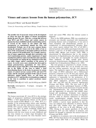
Figure 1 The p53, pRb tumor suppressor pathways. The nucleus of T-antigen-positive cell lines and human
association of T-antigen with p53 leads to inactivation of p53
and downregulation of p21WAF-1 that eventually affects the status
tumor samples, suggesting that T-antigen expression
of pRb phosphorylation by cyclin cdk and the release of E2F. may lead to nuclear localization of IRS-1 (Del Valle
Further, E2F is liberated from pRb:E2F by the interaction of et al., 2002a; Lassak et al., 2002, Tu et al., 2002).
T-antigen with pRb Mapping studies have determined that the N-terminal
portion of IRS-1 interacts with JCV T-antigen and that
this binding is independent from IRS-1 tyrosine
revealed that JCV T-antigen has the capacity to interact phosphorylation and can be strongly inhibited by IRS-
with several critical tumor suppressor proteins including 1 serine phosphorylation. Importantly, competition for
p53 and members of the pRb family (Bollag et al., 1989; IRS-1–T-antigen binding by a dominant-negative mu-
Haggerty et al., 1989; Del Valle et al., 2001a). It is tant of IRS-1 inhibited anchorage-independent prolif-
believed that the association of T-antigen with p53 and eration of JCV T-antigen-transformed medulloblastoma
the pRb family can lead to the inactivation of these cells (Lassak et al., 2002). All of these observations have
tumor suppressors in cells expressing T-antigen and led us to propose a model in which the interaction of
thus, promote uncontrolled proliferation. According to T-antigen and IRS-1 may lead to the uncoupling of
one model, the association of JCV T-antigen with p53 IRS-1 from the IGF-1R and the translocation of IRS-1
abrogates the ability of p53 to augment transcription of to the nucleus (Figure 2). Further studies are needed to
p21/WAF-1, an inhibitor of cyclin kinases, including determine the nuclear function of IRS-1 in the presence
cyclins A and E, and their associated kinases. Under and absence of JCV T-antigen.
normal conditions, a decrease in kinase activity of G1/S In light of recent studies demonstrating the involve-
cyclins:cdks maintains pRb in a hypophosphorylated ment of Wnt signaling in various cancers, including
and active state that in turn sequesters the S phase- medulloblastomas (Morin, 1999; Eberhart et al., 2000)
specific transcription factor, E2F. Not mutually exclu- and the association of JCV with these tumors, it was
sive, T-antigen’s association with pRb can liberate E2F hypothesized that JCV exerts its oncogenic activity, at
from the pRb:E2F complex and permit E2F to exert its least in part, through deregulation of the Wnt signaling
affect on cell proliferation by promoting unscheduled pathway. In normal cells, b-catenin, a key modulator of
transcription of S phase genes (Figure 1). Support for the Wnt pathway, is modified on its N-terminal serine
pRb regulation of T-antigen-induced tumors comes and threonine by phosphorylation and then ubiquiti-
from studies illustrating that overexpression of pRb2/ nated and rapidly degraded by the proteosome pathway
p130 can overcome JCV–T-antigen-mediated tumori- (for review see Polakis 2000; Gao et al., 2002).
genecity in experimental animals (Howard et al., 1998). Degradation of b-catenin occurs by a multiprotein
JCV T-antigen has also been associated with other cytoplasmic complex containing GSK3b, Axin 1, and
cellular proteins such as YB-1 and Pura (Gallia et al., APC. When cells are stimulated by Wnt proteins, the
1998b; Safak et al., 1999). The association of T-antigen function of the GSK3b/Axin1/APC complex is inhibited
with Pura is relevant to the transforming ability of by a lesser known pathway leading to the accumulation
T-antigen, as earlier results have demonstrated that of unphosphorylated and stable b-catenin in the
Pura overexpression suppresses the growth of several cytoplasm.
Oncogene
4. Viruses and cancer
K Reiss and K Khalili
6520
Wnt Signaling Pathway
IGF-1 Signaling Pathway
JCV-T
β-catenin
(GSK3β. Axin1, APC)
wt
β-catenin β-catenin P
JCV-T wt
JCV-T
mut
β-catenin
wt
LEF
IRS-1 β-catenin β-catenin
IRS-1 JCV-T mut
LEF
degradation
MAPK PI3K Nucleus
PDKs Activation of
c-myc, cycln D1
Others
Akt/PKB cell cycle
Nucleus
Apoptosis Tumor
Cell Others? Figure 3 The Wnt signaling pathway. The Wnt signaling pathway
Cycle? DNA and its well-studied cytoplasmic protein, b-catenin, is regulated by
Repair? Tumor a series of proteins with kinase activity such as GSK3b, Axin, and
APC. A mutation in b-catenin immunizes this protein against
phosphorylation in which the stable proteins associate with LEF-1
Figure 2 The IGF-1 signaling pathway. The IGF-1 signaling
and translocate to the nucleus where they stimulate c-myc, cyclin
pathway via its key component, IRS-1, alters phosphorylation and
D1, and several other cell proliferation genes. JCV T-antigen, by
activity of MAPK and PI3 K that eventually led to tumor
associating with wild-type b-catenin, increases the stability of b-
development. The interaction of T-antigen with IRS-1 translocates
catenin and accelerates its nuclear import
IRS-1 to the nucleus where it can affect cell cycle parameters as well
as other critical events such as DNA repair
small t-antigens, one may ascribe similar functions as
Cytoplasmic b-catenin translocates into the nucleus those established for SV40 small t-antigen for the JCV
where it forms a heterocomplex with DNA binding protein. For example, the cysteines and the central
transcription factors such as TCF/LEF, which have the proline of SV40 small t-antigen appear to be important
potential to stimulate transcription of genes related to for its interaction with and the inhibition of cellular
cell cycle regulation such as c-myc and cyclin D1 (He phosphatase PP2A. As a consequence of PP2A inhibi-
et al., 1998; Shtutman et al., 1999). In several cancer tion by small t-antigen, several cellular kinases are
cells, including PNETs, it was shown that mutations at hyperphosphorylated and their activity is elevated. This
the site of phosphorylation of b-catenin results in was first described for MAPK and its kinase, ERK, then
stabilization of b-catenin, its nuclear import, and extended to JNK and a key ion transporter, the Na/H
enhanced expression of cell cycle regulators (Figure 3). antiporter (Sontag et al., 1993). Activation of the
Examination of the Wnt signaling pathway in JCV T- MAPK family kinases by small t-antigen also leads to
antigen-positive cells compared to T-antigen-negative increased transcription from AP-1-driven promoters,
cells revealed enhanced levels of b-catenin, LEF-1, and consistent with activation of the Elk-1 family of
their downstream target, c-myc, suggesting a role for T- transcription factors (Wheat et al., 1994). Also, JCV
antigen in deregulating the Wnt pathway (Gan et al., small t-antigen may influence protein phosphorylation
2001). Further studies have revealed the ability of T- cascades involved in CREB-related transcriptional
antigen to associate physically with b-catenin and activities (Wheat et al., 1994). In general, the effects of
stabilize wild-type b-catenin. This event was concurrent small t-antigen on intracellular kinases promote cell
with increased levels of b-catenin in the nucleus and growth presumably by accelerating cell entry into the
enhancement of c-myc gene expression (Figure 3). S phase by a complex mechanism that involves proteo-
Through alternative splicing, the early region of JCV some degradation of p27 (Sheaff et al., 1997). Of note,
can also encode several smaller isoforms of large efficient degradation of p27 is required for the accumu-
T-antigen including small t-antigen and T0 135, T0 136, lation of cyclin A and S phase progression (Zerfass-
and T0 165 (Bollag et al., 2000; Prins and Frisque, 2001). Thome et al., 1997). Thus, by favoring the elimination of
Initial studies with the T0 proteins have suggested that p27, small t-antigen may allow cyclin A/cdk2 to
these proteins may contribute to replication of viral orchestrate cell cycle progression.
DNA and suspected to possess some oncogenic poten- In addition to multiple viral early proteins, the JCV
tial (Prins and Frisque, 2001). Little is known about the late region encodes a small protein, named Agnoprotein,
function of JCV small t-antigen, and its potential role in whose open reading frame resides in the leader of the
cellular transformation. However, based on sequence late transcripts. In SV40, Agnoprotein plays a role in the
homology between specific regions of SV40 and JCV lytic cycle of the virus by facilitating nuclear import of
Oncogene
5. Viruses and cancer
K Reiss and K Khalili
6521
the capsid protein (Carswell and Alwine, 1986; Resnick in 28 (32.9%) of 85 tested samples. In a separate series
and Shenk, 1986; Khalili et al., 1988). Similarly, of studies, the presence of JCV in medulloblastomas has
Agnoprotein plays a critical role in productive replica- been established. Parallel reports demonstrate immuno-
tion of JCV, as mutations in the Agnoprotein hamper histochemical detection of JCV T-antigen in 17.3 and
replication of JCV in primary astrocytes (Safak et al., 45% of samples obtained from two separate groups of
2001, unpublished observations). With respect to its medulloblastoma patients (Krynska et al., 1999a; Del
effect on host cells, results from in vitro studies have Valle et al., 2002c). In addition to T-antigen, analysis of
demonstrated that the expression of Agnoprotein in cells medulloblastoma samples has revealed the presence of
leads to upregulation of cyclin A and p21/WAF-1. Agno DNA sequences in 11 (69%) out of 16 samples,
Interestingly, results from protein binding studies have and immunohistochemical analysis showed the presence
revealed the ability of Agnoprotein to interact, although of Agnoprotein in cytoplasm of 11 (55%) out of 20
weakly, with p53, and that the region between residues samples. Importantly, the JCV early gene product, large
1–36 of Agnoprotein that contain a helix–loop–helix T-antigen, was detected in nine (45%) out of 20
motif is essential for this interaction. Thus, it is evident medulloblastoma cases examined.
that in addition to well-studied large T-antigen, other In addition to tumors of the nervous system, more
JCV proteins including small t-antigen, Agnoprotein, recent studies have detected JCV DNA sequences and
and perhaps T0 may play a role in the oncogenic expression of the viral proteins in colorectal carcinoma,
potential of this human polyomavirus. rather than tumors arising from within the nervous
system (Ricciardiello et al., 2001; Enam et al., 2002).
Association of JCV with human cancer Greater than 50% of the tumor samples were found to
express the viral proteins T-antigen and Agnoprotein,
As mentioned earlier, prior to the discovery of JCV, while a number of T-antigen-positive samples also
several reports indicated an association between PML contained b-catenin protein localized to the nucleus
and brain tumors. The first observations were made by (Enam et al., 2002). While VP-1 DNA sequences can be
Richardson in which the post-mortem examination of a amplified from significant numbers of these tumors, VP1
58-year-old man with chronic lymphocytic leukemia and expression by immunohistochemistry has not been
PML revealed the presence of an oligodendroglioma observed in any tumor type, suggesting that the tumors
(Richardson, 1961). In other studies of PML cases cells are not productively infected with JCV, but rather
associated JCV with multiple astrocytomas (Sima et al., transformed by the virus.
1983) and numerous foci of anaplastic astrocytes Interestingly, recent evidence has shown the presence
(Castaigne et al., 1974) were reported. In these cases, of JCV in untreated urban sewage, suggesting transmis-
viral particles were observed in both oligodendrocytes sion of the virus via the fecal–oral route (Bofil-Mas et al.,
and astrocytes, but not in the neoplastic astrocytes. 2001). In addition, JCV DNA has also been found in the
Recently, Shintaku et al. (2000) reported a case of normal human gastrointestinal tract, which further
dysplastic ganglion-like cells in association with PML. supports the possibility that JCV, and other polyoma-
Detailed immunohistochemical studies revealed that viruses, may colonize the gut (Ricciardiello et al., 2000).
the neurons were infected with JCV and expressed These data, taken together with several reports of SV40
JCV T-antigen in the absence of capsid protein, VP1. detection in human mesothelioma, osteosarcoma, and
In addition to cases of simultaneous PML and B-cell lymphoma, suggest that JCV may be observed
cerebral neoplasm, JCV has been found in human brain within other tumor types throughout the body. The
tumors in the absence of any PML lesions. In our recent observations from such clinical samples along with the
work (Del Valle et al., 2001b), 85 clinical specimens data from cell culture and experimental animals add
from the United Kingdom, Greece, and the United further evidence of the involvement of JCV in the
States have been examined for their possible association process of cellular transformation, and strongly suggest
with JCV. These multiple samples represented various a possibility that this human neurotropic polyomavirus
human brain tumors, including oligodendroglioma, may play a role in the development of human brain
astrocytoma, pilocytic astrocytoma, oligoastrocytoma, tumors.
anaplastic astrocytoma, anaplastic oligodendroglioma,
glioblastoma multiforme, gliomatosis cerebri, gliosarco-
ma, ependymoma, and subependymoma. Gene amplifi-
cation using primers that recognize the JCV DNA Acknowledgements
We express our appreciation to past and present members of
sequences followed by Southern blot hybridization have
the Center for Neurovirology and Cancer Biology for their
demonstrated the presence of the viral early sequences in contribution, and to Cynthia Schriver for editorial assistance.
49 (69%) of 71 samples. More importantly, results of This work was made possible by grants awarded by NIH to
immunohistochemical analysis have demonstrated the KR and KK.
expression of JCV T-antigen in the nuclei of tumor cells
References
Baserga R. (1999). Exp. Cell Res., 253, 1–6. Berger JR, Levy RM, Flomenhoft D and Dobbs M. (1998).
Berger JR and Concha M. (1995). J. Neurovirol., 1, 5–18. Ann. Neural., 44, 341–349.
Oncogene
6. Viruses and cancer
K Reiss and K Khalili
6522
Bofill-Mas S, Formiga-Cruz M, Clemente-Casares P, Calafell Krynska B, Del Valle L, Croul S, Gordon J, Katsetos K,
F and Girones R. (2001). J. Virol., 75, 10290–10299. Carbone M, Giordano A and Khalili K. (1999a). Proc. Natl.
Bollag B, Chuke W-F and Frisque RJ. (1989). J. Virol., 63, Acad. Sci. USA, 96, 11519–11524.
863–872. Krynska B, Otte J, Franks R, Khalili K and Croul S. (1999b).
Bollag B, Prins C, Snyder EL and Frisque RJ. (2000). Oncogene, 18, 39–46.
Virology, 274, 165–178. Krynska B, Del Valle L, Gordon J, Otte J, Croul S and Khalili
Carswell S and Alwine JC. (1986). J. Virol., 60, 1055–1061. K. (2000). Virology, 274, 65–74.
Castaigne P, Rondot P, Escourolle R, Ribadeau Dumas J-L, Lassak A, Del Valle L, Peruzzi F, Wang JY, Croul S,
Cathala F and Hauw J-J. (1974). Rev. Neurol. Paris, 130, Khalili K and Reiss K. (2002). J. Biol. Chem., 277,
379–392. 17231–17238.
Darbinian N, Gallia GL, King J, Del Valle L, Johnson EM London WT, Houff SA, McKeever PE, Wallen WC, Sever JL,
and Khalili K. (2001). J. Cell. Physiol., 189, 334–340. Padgett BL and Walker DL. (1983). Prog. Clin. Biol. Res.,
Del Valle L, Gordon J, Ferrante P and Khalili K. (2001a). 105, 227–237.
Human Polyomaviruses: Molecular and Clinical Perspectives. Major EO, Amemiya K, Tornatore CS, Houff SA and Berger
Stoner GL, Khalili K. (eds). John Wiley & Sons, Inc.: New JR. (1992). Clin. Microbiol. Rev., 5, 49–73.
York, pp. 409–430. Monaco MC, Atwood WJ, Gravell M, Tornatore CS and
Del Valle L, Gordon J, Assimakopoulou M, Enam S, Geddes Major EO. (1996). J. Virol., 70, 7004–7012.
JF, Varakis J, Katsetos CD, Croul S and Khalili K. (2001b). Morin PJ. (1999). BioEssays, 21, 1021–1030.
Cancer Res., 61, 4287–4293. Ohsumi S, Motoi M and Ogawa K. (1986). Acta Pathol. Jpn.,
Del Valle L, Enam S, Lassak A, Wang J-Y, Croul S, Khalili K 36, 815–825.
and Reiss K. (2002a). Clin. Cancer Res., 8, 1822–1830. Patti R, Reddy CD, Geoerger B, Grotzer MA, Raghunath M,
Del Valle L, Wang JY, Lassak A, Peruzzi F, Croul S, Khalili K Sutton LN and Phillips PC. (2000). Int. J. Oncol., 16,
and Reiss K. (2002b). J. Neurovirol., 8 (Suppl. 2), 577–584.
138–147. Pipas JM. (1992). J. Virol., 66, 3979–3985.
Del Valle L, Gordon J, Enam S, Delbue S, Croul S, Abraham Polakis P. (2000). Genes Dev., 14, 1837–1851.
S, Radhakrishnan S, Assimakoupoulou M, Katsetos CD Prins C and Frisque RJ. (2001). J. Neurovirol., 7, 250–264.
and Khalili K. (2002c). J. Natl. Cancer Inst., 94, 267–273. Raj G, Gordon J, Logan TJ, Hall D, Chang C-F, Sala A, De
Eberhart CG, Tihan T and Burger PC. (2000). J. Neuropathol. Luca A, Giordano A and Khalili K. (1995). Int. J. Oncol., 7,
Exp. Neural., 59, 333–337. 801–808.
Enam S, Del Valle L, Lara C, Gan DD, Ortiz-Hidalgo C, Reiss K. (2002). Exp. Opin. Ther. Targets, 6, 539–544.
Palazzo JP and Khalili K. (2002). Cancer Res., 62, Resnick J and Shenk T. (1986). J. Virol., 60, 1098–1106.
7093–7101. Riccardiello L, Chang DK, Laghi L, Goel A, Chang CL and
Fareed GC, Takemoto KK and Gimbrone Jr MA. (1978). Boland R. (2001). J. Virol., 75, 1996–2001.
Microbiology, Schlessinger D (ed). American Society for Ricciardiello L, Laghi L, Ramamirtham P, Chang CL, Chang
Microbiology: Washington, DC, pp. 427–431. DK, Randolph AE and Boland CR. (2000). Gastroenterol-
Franks RR, Rencic A, Gordon J, Zoltick PW, Curtis M, ogy, 119, 1228–1235.
Knobler RL and Khalili K. (1996). Oncogene, 12, Richardson Jr EP. (1961). N. Engl. J. Med., 265, 815–823.
2573–2578. Safak M, Gallia GL, Ansari SA and Khalili K. (1999).
Frisque RJ and White III FA. (1992). Molecular Neurovirol- J. Virol., 73, 10146–10157.
ogy. Roos RP. (ed). Humana Press: Totowa, NJ, Safak M, Barrucco R, Darbinyan A and Khalili K. (2001).
pp. 25–158. J. Virol., 75, 1476–1486.
Frisque RJ, Rifkin DB and Walker DL. (1980). J. Virol., 35, Sheaff RJ, Groudine M, Gordon M, Roberts JM and Clurman
265–269. BE. (1997). Genes Dev., 11, 1464–1478.
Gallia GL, Gordon J and Khalili K. (1998a). J. Neurovirol., 4, Shintaku M, Matsumoto R, Sawa H and Nagashima K.
175–181. (2000). J. Neuropathol. Exp. Neurol., 59, 921–929.
Gallia GL, Safak M and Khalili K. (1998b). J. Biol. Chem., Shtutman M, Zhurinsky J, Simcha I, Albanese C, D’Amico M,
273, 32662–32669. Pestell R and Ben-Ze’ev A. (1999). Proc. Natl. Acad. Sci.
Gan DD, Reiss K, Gorrill T, Del Valle L, Croul S, Giordano USA, 96, 5522–5527.
A, Fishman P and Khalili K. (2001). Oncogene, 20, Sima AAF, Finkelstein SD and McLachlan DR. (1983). Ann.
4864–4870. Neurol., 14, 183–188.
Gao ZH, Seeling JM, Hill V, Yochum A and Virshup DM. Small JA, Khoury G, Jay G, Howley PM and Scangos GA.
(2002). Proc. Natl. Acad. Sci. USA, 99, 1182–1187. (1986a). Proc. Natl. Acad. Sci. USA, 83, 8288–8292.
Gordon J, Del Valle L, Otte J and Khalili K. (2000). Oncogene, Small JA, Scangos GA, Cork L, Jay G and Khoury G.
19, 4840–4846. (1986b). Cell, 46, 13–18.
Haggerty S, Walker DL and Frisque RJ. (1989). J. Virol., 63, Sontag E, Fedorov S, Kamibayashi C, Robbins D, Cobb M
2180–2190. and Mumby M. (1993). Cell, 75, 887–897.
He TC, Sparks AB, Rago C, Hermeking H, Zawel L, da Costa Sullivan CS, Tremblay JD, Fewell SW, Lewis JA, Brodsky JL
LT, Morin PJ, Vogelstein B and Kinzler KW. (1998). and Pipas JM. (2000). Mol. Cell. Biol., 20, 5749–5757.
Science, 281, 1509–1512. Tu X, Batta P, Innocent N, Prisco M, Casaburi I,
Howard CM, Claudio PP, Gallia GL, Gordon J, Giordano Belletti B and Baserga R. (2002). J. Biol. Chem., 277,
GG, Hauck WW, Khalili K and Giordano A. (1998). J. 44357–44365.
Natl. Cancer. Inst., 90, 1451–1460. Varakis J, Zu Rhein GM, Padgett BL and Walker DL. (1978).
Kang S and Folk WR. (1992). Virology, 191, 754–764. Cancer Res., 36, 1718–1722.
Khalili K, Brady J, Papas J, Spence S, Sadofsky M and Walker DL and Padgett BL. (1978). Microbiology, Schles-
Khoury G. (1988). Proc. Natl. Acad. Sci. USA, 85, singer D (ed). American Society for Microbiology:
354–358. Washington, DC, pp. 432–434.
Oncogene
7. Viruses and cancer
K Reiss and K Khalili
6523
Walker DL, Padgett BL, ZuRhein GM, Albert AE and Marsh Zerfass-Thome K, Schulze A, Zwerschke W, Vogt B, Helin K,
RF. (1973). Science, 181, 674–676. Bartek J, Henglein B and Jansen-Durr P. (1997). Mol. Cell.
Wang JY, Del Valle L, Gordon J, Rubini M, Croul S, Peruzzi Biol., 17, 407–415.
F, Khalili K and Reiss K. (2001). Oncogene, 20, 3857–3868. Zu Rhein GM and Varakis JN. (1979). Perinatal Carcinogen-
Wheat WH, Roesler WJ and Klemm DJ. (1994). Mol. Cell. esis: National Cancer Institute Monograph, Vol. 51. Rice JM
Biol., 14, 5881–5890. (ed). National Cancer Institute: Bethesda, MD, pp. 205–221.
Oncogene