3. GROSS ANATOMY
This is a synovial joint of condylar
variety.
Articular surfacesA. upper part -
a) Articular eminence,
b) ant. part of mandibular fossa.
B. inferior surface a) head of the mandible.
4.
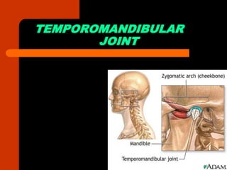
5. The temporomandibular joint is the
joint of the jaw and is frequently
referred to as TMJ.
The TMJs are one of the only synovial
joints in the human body with an
articular disc.
The name is derived from the two
bones which form the joint : the upper
temporal bone which is part of the
cranium and the lower jaw bone
called the mandible.
6. COMPONENTS
There are six main
components of the TMJ.
Mandibular condyles
Articular surface of
the temporal bone
Capsule
Articular disc
Ligaments
Lateral pterygoid
7. CAPSULE and ARTICULAR DISC
The capsule is a fibrous membrane that
surrounds the joint and incorporates the
articular eminence. It attaches to the
articular eminence, the articular disc and
the neck of the mandibular condyle.
The articular disc is a fibrous extension of
the capsule in between the two bones of
the joint. The disc functions as articular
surfaces against both the temporal bone
and the condyles and divides the joint into
two sections, as described in more detail
below. It is biconcave in structure and
attaches to the condyle medially and
laterally.
8. The anterior portion of the disc splits
in the vertical dimension, coincident
with the insertion of the superior head
of the lateral pterygoid.
The posterior portion also splits in the
vertical dimension, and the area
between the split continues
posteriorly and is referred to as the
retrodiscal tissue.
9.
10. LIGAMENTS
The ligaments are-the fibrous capsule,the lateral
ligament,the sphenomandibular
ligament,stylomandibular ligament.
The fibrous capsule- attached above to articular
tubercle,below to mandible,circumference of
mandibular fossa
The lateral ligament- attached above to articular
tubercle,below to posterolateral aspect of neck of
mandible.
The sphenomandibular ligament- attached
superiorly to spine of sphenoid,inferiorly to
lingula.it is accessory ligament.
The Stylomandibular ligament- attached above
to lateral styloid process and below to angle of
mandible.it is also accessory.
11. ARTICULATION
The TMJ is a ginglymoarthrodial
joint, referring to its dual compartment
structure and function (ginglymo-and
arthrodial).
The condyle articulates with the
temporal bone in the mandibular
fossa. The mandibular fossa is a
concave depression in the squamous
portion of the temporal bone.
12. There are two TMJs, one on either side,
working in unison.
The unique feature of the TMJs is the
articular disc. The disc is composed of
fibrocartilagenous tissue which is positioned
between the two bones that form the joint.
The disc divides each joint into two.
The lower joint compartment formed by the
mandible and the articular disc is involved in
rotational movement-this is the initial
movement of the jaw when the mouth opens.
The upper joint compartment formed by
the articular disc and the temporal bone is
involved in translational movements-this is
the secondary gliding motion of the jaw as it
is opened widely.
13.
14. MUSCLE PRODUCING MOVEMENTS
Depression- lateral pterygoid mainly
Elevation- masster,temporalis,medial
petygoid of both sides.
Protrusion- lateral and medial
pterygoid.
Retraction- posterior fibres of
temporalis.
Lateral or side to side movement
eg.turning chin to left side- left lateral
pterygoid and right medial pterygoid.
15.
16.
17. CLINICAL CONSIDERATIONS
The most common disorder of the TMJ is disc
displacement.
The most common cause of TMJ pain is
myofascial pain dysfunction syndrome,
primarily involving the muscles of mastication.
Internal derangements is defined as an abnormal
relationship of the disc to any of the other
components of the TMJ. Disc displacement is an
example of internal derangement.
Degenerative joint disease, otherwise known as
osteoarthritis is the organic degeneration of the
articular surfaces within the TMJ.
TMJ pain remains one of the most reliable
diagnostic criteria for temporal arthritis.
18.
19. Diagnosis and Treatment of TMJ
TMJ is usually determined by exams, such as xray, MRI and CT scan. If the condition is not
serious, a physician will usually recommend
several steps:
• Resting the jaw joint
• Utilizing conventional analgesic pain killers or
nonsteroidal anti-inflammatory drugs to alleviate
the swelling and tight muscles
• Applying heated compresses to the areas
• Avoiding strain of the jaw
• Avoiding tough foods that require heavy chewing