
Recommandé
Contenu connexe
Similaire à Howard university presentation ver3
Similaire à Howard university presentation ver3 (20)
Howard university presentation ver3
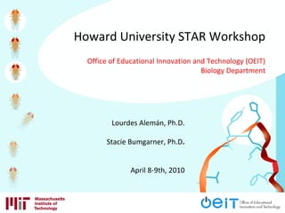
- 1. Lourdes Alemán, Ph.D. Stacie Bumgarner, Ph.D. April 8-9th, 2010 Howard University STAR Workshop Office of Educational Innovation and Technology (OEIT) Biology Department
- 2. 170 liters
- 3. Nobel Prize website Aquaporin structural model
- 4. 10% of what they hear 30% of what they see 60% of what they hear & see 80% of what they hear, see, & do 100% of what they hear, see, do, smell, feel, taste…& purchase on credit By Ronald A. Berk Professors are from Mars. Students are from Snickers. Students learn…
- 5. Challenge Bridging the divide between research & the classroom using interactive technology.
- 8. Who we are MIT Biology Department Office of Educational Innovation & Technology (OEIT) What we are doing Designing interactive teaching software tools to be used IN & OUTSIDE class
- 9. Software Tools for Academics & Researchers (STAR) biology tools 1 Educational goals 2 Design process 3 Access & outreach
- 10. Educational goals Challenge Teaching protein structure in a traditional classroom setting StarBiochem: protein 3D viewer Challenge Teaching experimental design & data interpretation outside of a genetics lab StarGenetics: virtual genetics lab
- 11. Design process Collaboration between MIT faculty & OEIT StarBiochem Professor Graham Walker Introductory Biology Series StarGenetics Professor Chris Kaiser Genetics Graduate Student Bridge Program
- 12. Access & outreach Software & curriculum materials -> freely accessible online StarBiochem http://web.mit.edu/star/biochem StarGenetics http://web.mit.edu/star/genetics
- 13. Create curriculum modules. Conduct workshops. Collaborate with wide range of educational institutions. Assess how software tools impact different populations of students. Access & outreach
- 14. StarBiochem usage beyond MIT Visits 9881 Countries 82
- 15. StarBiochem Workshop – Day 1 1 Introduction to StarBiochem 2 StarBiochem hands-on activity 3 StarBiochem educational applications (Part I) Classroom applications 4 StarBiochem educational applications (Part II) Lab applications 5 Designing & writing new StarBiochem curriculum activities
- 16. StarBiochem Workshop – Day 1 1 Introduction to StarBiochem 2 StarBiochem hands-on activity 3 StarBiochem educational applications (Part I) Classroom applications 4 StarBiochem educational applications (Part II) Lab applications 5 Designing & writing new StarBiochem curriculum activities
- 17. Proteins perform many important biological functions hemoglobin carry oxygen in the blood F1 ATPase generate energy antibody protect against infections aquaporin water transport microtubules cell division Protein Data Bank website
- 18. The function of a protein is determined by its structure structure function
- 19. Challenge Teaching protein structure in a traditional classroom setting.
- 20. Traditional methods for teaching about protein structure and function Cartoons Can introduce misconceptions 2-D representations Limits student exploration Protein Structure & Function (2007)
- 21. Using 3-D protein viewers for teaching about protein structure & function Research PyMol, KING, Rasmol, Chimera, First Glance in Jmol, Webmol Educational Protein Explorer, BioMolecular Explorer, other guided tutorials
- 22. Limitations of existing 3-D proteins viewers for teaching about proteins structure & function Research Educational uses crystallography jargon not user-friendly ability to scale-up not known windows functionality only tutorials with limited views/manipulations
- 23. StarBiochem: protein 3-D viewer
- 24. StarBiochem is a protein 3-D viewer designed for educational purposes Website: http://web.mit.edu/star/biochem/ Platform independent. Interface is designed with students in mind. Parallels how proteins are taught in introductory biology. Can upload >54,000 Protein Data Bank structures.
- 25. StarBiochem hands-on guided tour http://web.mit.edu/star/biochem/
- 26. StarBiochem Workshop – Day 1 1 Introduction to StarBiochem 2 StarBiochem hands-on activity 3 StarBiochem educational applications (Part I) Classroom applications 4 StarBiochem educational applications (Part II) Lab applications 5 Designing & writing new StarBiochem curriculum activities
- 27. How mutations in DNA can result in disease sickle cell anemia: hemoglobin
- 28. Hemoglobin Highly expressed in red blood cells Responsible for oxygen transport Contains four protein subunits -> each one binds to a O2 molecule Protein Data Bank website
- 29. Hemoglobin heme heme Protein Data Bank website Highly expressed in red blood cells Responsible for oxygen transport Contains four protein subunits -> each one binds to a O2 molecule
- 30. severe bone pain & necrosis stroke enlarged heart bloodstream infections and pneumonia www.commercialappeal.com Sickle cell anemia
- 31. A mutation in hemoglobin causes sickle cell anemia University of Utah Learn Genetics website
- 32. How does a mutation in the hemoglobin gene causes the protein to become sticky? normal hemoglobin vs.sickle hemoglobin (2HBS)(1A3N)
- 33. Structure of sickle hemoglobin: DNA sequence A -> T protein hemoglobin becomes sticky amino acid sequence Glu6 -> Val6 disease sickle cell anemia
- 34. StarBiochem Workshop – Day 1 1 Introduction to StarBiochem 2 StarBiochem hands-on activity 3 StarBiochem educational applications (Part I) Classroom applications 4 StarBiochem educational applications (Part II) Lab applications 5 Designing & writing new StarBiochem curriculum activities
- 35. Goal To illustrate how StarBiochem can be implemented into an existing class curriculum.
- 36. StarBiochem educational applications (Part I) Classroom applications 1 Current StarBiochem applications 2 In-class activities 3 Outside-class activities 4 Assessment & testing
- 37. StarBiochem educational applications (Part I) Classroom applications 1 Current StarBiochem applications 2 In-class activities 3 Outside-class activities 4 Assessment & testing
- 38. Current StarBiochem educational applications • Undergraduate setting Introductory Biology (MIT) Introductory Biology Lab (Brandeis) • High school setting MIT Museum/Environmental Health Sciences Outreach Program (MIT) Outreach HS program (Broad Institute) HS fieldtrips (Biology Department, MIT)
- 39. Introductory Biology Courses (7.01 series) StarBiochem modules core topics StarBiochem case study: MIT biochemistry molecular biology recombinant DNA technology signalingimmunology cancer biology neurobiology genetics PAH amyloid GFP BCR-Abl Herceptin DNA pol Ras Hemoglobin PFK crystallin Diviya Sinha
- 40. StarBiochem case study: Brandeis University Introductory Biology Lab Course (Bio118) crystallin protein cataracts • explore the structure of crystallin using StarBiochem • design mutation in crystallin based on structure • test mutant crystallin for in vitro cataract formation Melissa Kosinski-Collins
- 41. StarBiochem educational applications (Part I) Classroom applications 1 Current StarBiochem applications 2 In-class activities 3 Outside-class activities 4 Assessment & testing
- 42. StarBiochem in-class activities Short (2-5 min) Intermediate (10-20 min) Long (40-60 min)
- 43. StarBiochem in-class activities Short Example: snapshot of a structure or a concept use StarBiochem live create video using StarBiochem create images of a particular structural snapshot
- 44. StarBiochem in-class activities Short Example: snapshot of a structure or a concept use StarBiochem live create video using StarBiochem (ex: hemoglobin) create images of a particular structural snapshot
- 45. StarBiochem in-class activities Intermediate Example: interactive building protein activity Protein 1 Protein 2 cell membrane protein cytoplasmic protein
- 46. StarBiochem in-class activities Intermediate Example: interactive building protein activity Protein 1 Protein 2 cell membrane protein cytoplasmic protein 1 Students are asked to consider what would be necessary to build proteins: location & function. 2 Students answers are compared with actual protein structures in StarBiochem.
- 47. StarBiochem in-class activities Long Example: basics of protein structure lecture using StarBiochem (Dr. Chia-Yung Wu) Example 1 Example 2 Potassium channel GFP
- 48. Protein structure lecture using StarBiochem Introductory Biology The live demos using StarBiochem helped me gain a better understanding of the relationship between a protein's structure and its function. The live demos on the potassium channel protein in StarBiochem helped me understand how this channel allows for the passage of potassium ions ONLY. The live demos using StarBiochem helped me understand the four levels of protein structure: primary, secondary, tertiary & quaternary. The live demos using StarBiochem helped me understand what protein structures look like in 3D. I enjoyed today’s lecture on protein structure. Howard University (2009)
- 49. StarBiochem educational applications (Part I) Classroom applications 1 Current StarBiochem applications 2 In-class activities 3 Outside-class activities 4 Assessment & testing
- 51. Review available curriculum modules online (exercises). Modify current modules or develop your own. Students download software from web. Each module contains full step-by-step instructions for students to use software. Outside-class activities
- 52. StarBiochem educational applications (Part I) Classroom applications 1 Current StarBiochem applications 2 In-class activities 3 Outside-class activities 4 Assessment & testing
- 53. Goal Assess effectiveness of the software in illustrating a concept Assess implementation of StarBiochem activity Assessment & testing
- 54. Example: open-ended exam question Introductory Biology (MIT) Aquaporin exercise (homework) •basics of protein structure •aquaporin specificity for passage of water only Aquaporin question (exam) •assess both protein structure basics and aquaporin structure -> function relationships •could not be completed without doing the aquaporin exercise Assessment of software’s ability to illustrate concept
- 55. Assessment of software’s curriculum implementation Use simple sample survey provided 1 Survey Monkey (http://www.surveymonkey.com/) • best for remote assessment • data is automatically analyzed • basic membership (free), Pro membership ($300/year) 2 Paper survey • best for direct assessment • manual data manipulation • free
- 56. StarBiochem educational applications (Part I) Classroom applications 1 Current StarBiochem applications 2 In-class activities 3 Outside-class activities 4 Assessment & testing
- 57. StarBiochem Workshop – Day 1 1 Introduction to StarBiochem 2 StarBiochem hands-on activity 3 StarBiochem educational applications (Part I) Classroom applications 4 StarBiochem educational applications (Part II) Lab applications 5 Designing & writing new StarBiochem curriculum activities
- 58. Goal To illustrate how to implement StarBiochem lab activity.
- 59. 10, 000
- 60. DNA mutations neutral positive -> evolution negative -> disease Effects Consequences of DNA mutations
- 61. Consequences of DNA mutations DNA mutations neutral positive -> evolution negative -> disease Effects
- 62. What causes DNA mutations?
- 63. DNA mutations can be caused by various agents
- 64. DNA mutations can be caused by various agents man-made agents (X-rays, smoking, toxic chemicals) environmental agents (UV rays, toxic chemicals) foreign biological agents (viruses, transposons) cellular processes (DNA replication, cell division, respiration)
- 65. respiration radiation smoking Each agent is associated with a particular type of DNA mutations oxidation agent mutation type
- 66. respiration radiation smoking Each mutation type is fixed by a different cellular mechanism oxidation agent mutation type DNA glycosylase
- 67. How our cells try to fix DNA mutations DNA glycosylase
- 68. StarBiochem educational applications (Part II) Lab activity 1 Pre-assignment 2 Hands-on activity: DNA glycosylase exercise 3 Post-assignment
- 69. StarBiochem educational applications (Part II) Lab activity 1 Pre-assignment 2 Hands-on activity: DNA glycosylase exercise 3 Post-assignment
- 70. Lab activity: pre-assignment Goal 1 Illustrate basic science research -> medical applications 2 Understand relevance of lab activity
- 71. Lab activity: pre-assignment Reading “Mapping the Cancer Genome” by Francis S. Collins and Anna D. Barker Scientific American Magazine, March 2007: 50-5 Questions • usefulness of the genomic cancer atlas for studying & treating cancer. • hallmarks of cancer cells such as abnormalities in self-control and repair mechanisms. • complexity of assembly a genomic cancer atlas.
- 72. StarBiochem educational applications (Part II) Lab activity 1 Pre-assignment 2 Hands-on activity: DNA glycosylase exercise 3 Post-assignment
- 73. Oxidation can cause mutations in DNA
- 74. Oxidation can cause mutations in DNA 1 2
- 75. Oxidation can cause mutations in DNA 1 2 1a 1b 2a 2b
- 76. DNA glycosylase proteins help repair DNA damage caused by oxidation DNA glycosylases 1 2 1a 1b 2a 2b
- 77. How does DNA glycosylase fix DNA oxidation damage? DNA glycosylase: human 8-oxoguanine glycosylase (hOGG1)
- 78. Structure of DNA glycosylase: structure human DNA glycosylase & DNA substrate mechanism rotating DNA bases outside of double helix to check for mutations
- 79. Personalized medicine for treating cancer Rebecca Henretta (2007)
- 80. Personalized medicine for treating cancer Non-responders Responders Rebecca Henretta (2007)
- 81. Personalized medicine for treating cancer Non-responders Responders alternative treatment conventional treatment Rebecca Henretta (2007)
- 82. Personalized medicine for treating cancer: hOGG1 levels & lung cancer low cancer risk high low high hOGG1 levels Cleveland Clinic website
- 83. StarBiochem educational applications (Part II) Lab activity 1 Pre-assignment 2 Hands-on activity: DNA glycosylase exercise 3 Post-assignment
- 84. Lab activity: post-assignment Goal Synthesis of pre-assignment & DNA glycosylase exercise
- 85. Lab activity: post-assignment Questions • understand how the genomic cancer atlas can be used to uncover potential new roles of hOGG1 in cancer. • understand how new cancer treatments can be design based on hOGG1.
- 86. StarBiochem educational applications (Part II) Lab activity 1 Pre-assignment 2 Hands-on activity: DNA glycosylase exercise 3 Post-assignment
- 87. StarBiochem Workshop – Day 1 1 Introduction to StarBiochem 2 StarBiochem hands-on activity 3 StarBiochem educational applications (Part I) Classroom applications 4 StarBiochem educational applications (Part II) Lab applications 5 Designing & writing new StarBiochem curriculum activities
- 88. Goal Empower you to build your own StarBiochem exercises by illustrating the design and building process.
- 89. Designing & writing StarBiochem curriculum activities 1 Design & building process What is the goal of the activity? What type of activity accomplishes goal? How to select a structure that accomplishes goal? How to structure the activity? 2 Interactive exercise creating a StarBiochem activity
- 90. Designing & writing StarBiochem curriculum activities 1 Design & building process What is the goal of the activity? What type of activity accomplishes goal? How to select a structure that accomplishes goal? How to structure the activity? 2 Interactive exercise creating a StarBiochem activity
- 91. What is the goal of the activity? structure -> function structure -> function -> disease structure -> medical therapies protein regulation evolution of protein structure
- 92. What type of activity accomplishes goal? 1 Course • Introductory Biology • Biochemistry • Cell/Molecular Biology • Pharmacology • Etc. 2 Course curriculum • format • duration of activity
- 93. What type of activity accomplishes goal? Example 1 Structure -> function Course Introductory Biology Curriculum central concept of the course Activity StarBiochem protein structure lecture + StarBiochem exercise (homework/lab activity)
- 94. What type of activity accomplishes goal? Example 2 Structure -> medical therapies Course Cell Biology Curriculum minor point within a lecture Activity StarBiochem video or in class-demo illustrating structure –> targeted therapies
- 95. How to select a structure that accomplishes goal? 1 Goal Is the structure suitable for illustrating goal? 2 Course/format of activity Is the structure accessible to your students? Is the activity format suitable for explaining the structure?
- 96. Schneider, T.L. and Linton, B. (2008) Introduction to Protein Structure through Genetics Diseases. Journal of Chemical Education 85: 663-665 How to select a structure that accomplishes goal?
- 97. Schneider, T.L. and Linton, B. (2008) Introduction to Protein Structure through Genetics Diseases. Journal of Chemical Education 85: 663-665 How to select a structure that accomplishes goal?
- 98. Researching & finding the appropriate structure Protein Data Bank http://www.pdb.org/pdb/home/home.do Search: • keyword • four digit PDB ID • title of paper How to select a structure that accomplishes goal?
- 99. Researching & finding the appropriate structure Hemoglobin: exercise with a well defined activity structure -> function -> disease Glu6 -> Val6 in hemoglobin leads to sickle cell anemia DNA glycosylase: exercise with less well defined activity structure -> function illustrate how DNA glycosylase repairs DNA How to select a structure that accomplishes goal?
- 100. How to structure the activity? 1 Format/duration Short activity straight to the the point ex: DNA glycosylase video Long activity framework for building complexity ex: DNA glycosylase exercise
- 101. How to structure the activity? 1 Format/duration Short activity straight to the the point ex: DNA glycosylase video Long activity framework for building complexity ex: DNA glycosylase exercise
- 102. How to structure the activity? 1 Format/duration Short activity straight to the the point ex: DNA glycosylase video Long activity framework for building complexity ex: DNA glycosylase exercise
- 103. How to structure the activity? References Structure: 1EBM Bruner, S. et al. (2000) Structural basis for recognition and repair of the endogenous mutagen 8-oxoguanine in DNA. Nature 403: 859-866. Structures: 1YQK, 1YQR Banerjee, A. et al. (2005) Structure of a repair enzyme interrogating undamaged DNA elucidates recognition of damaged DNA. Nature 434: 612-618.
- 104. How to structure the activity? Structure: 1EBM Bruner, S. et al. (2000) Structural basis for recognition and repair of the endogenous mutagen 8-oxoguanine in DNA. Nature 403: 859-866. How hOGG1 repairs oxidized guanines: 1.Extrusion of oxoG from the double helix 1.Recognition of oxoG by Gly 42
- 105. How hOGG1 repairs oxidized guanines: 1.Extrusion of oxoG from the double helix 1.Recognition of oxoG by Gly 42 “The structure of the hOGG1-DNA complex provides a clear solution to the long-standing puzzle of how the cellular repair machinery recognizes oxoG in DNA amidst the vast excess of guanine, a close structural relative. The oxoG residue is fully extruded from the DNA helix and inserted into an extrahelical active-site pocket on the enzyme, consistent with structure-based predictions made for other members of the HhH- GPD superfamily.”
- 106. “The most characteristic feature of oxoG - its 8-oxo-carbonyl function - is completely devoid of any interacting partner. Instead, the enzyme recognizes the urea system in oxoG by virtue of its N7-H, which donates a hydrogen bond to the main chain carbonyl of Gly 42… Indeed, of all the contacts made to the oxoG base, that involving Gly 42 is the only one that would clearly be different with oxoG versus guanine; thus, the responsibility for discriminating oxoG from guanine appears to be borne by a single hydrogen bond.” How hOGG1 repairs oxidized guanines: 1.Extrusion of oxoG from the double helix 1.Recognition of oxoG by Gly 42
- 107. How to structure the activity? Structures: 1YQK, 1YQR Banerjee, A. et al. (2005) Structure of a repair enzyme interrogating undamaged DNA elucidates recognition of damaged DNA. Nature 434: 612-618. How hOGG1 distinguishes oxidized guanines from undamaged guanines: 1.oxoG:hOGG1 interaction differs from the G:hOGG1 interaction
- 108. How hOGG1 distinguishes oxidized guanines from undamaged guanines: 1.oxoG:hOGG1 interaction differs from the G:hOGG1 interaction “Whereas the oxoG nucleobase inserts itself deeply into the lesion recognition pocket on the enzyme (Figs 2, left, and 3a), G is rejected by the lesion recognition pocket and instead lies against the protein surface at an exo-site some 5A ° outside the pocket (Figs 2, right panel, and 3b). The G base does interact with two active- site residues, Phe 319 and His 270, but the contacts are completely different from those made with oxoG.”
- 109. How hOGG1 distinguishes oxidized guanines from undamaged guanines: 1.oxoG:hOGG1 interaction differs from the G:hOGG1 interaction “…a hydrogen bond is apparent between the carbonyl oxygen of Gly 42 and N7 H of oxoG. This attractive interaction would be replaced in the case of G by a strongly repulsive interaction with the N7 lone pair, if G and Gly 42 assumed the same positions as in the oxoG complex.”
- 110. How to structure the activity? Long activity: framework for building complexity Begin with: simple questions Allow students to become familiar with protein structure basics End with: complex questions Allow students to understand the goal of the activity
- 111. How to structure the activity? Long activity: framework for building complexity Begin with: simple questions Allow students to become familiar with protein structure basics DNA Glycosylase Exercise: questions 1-5 End with: goal of the exercise structure -> function DNA Glycosylase Exercise: questions 6-8
- 112. DNA Glycosylase Exercise: simple questions Question 1: overall structure explore different 3-D models for representing atoms Question 2: amino acid structure explore difference between amino acid backbone and side chain Question 3,4: secondary structure explore how individual amino acids fold to form secondary structures Question 5: tertiary structure explore relationship between a protein’s environment & its tertiary structure
- 113. DNA Glycosylase Exercise: complex questions Question 6: hOGG1 repairs DNA by extruding oxidized bases from double helix location of oxoG with respect to the double helix Question 7: hOGG1 recognizes oxidized bases by contacting the damaged base directly and recognizing a specific modification comparison of secondary structures where amino acids that interact with oxoG are likely to be located Question 8: hOGG1’s specificity for oxidized guanines lies in the inability of guanine to bind to the active site of hOGG1 comparison of the oxoG:hOGG1 interaction with that of the G:hOGG1 interaction
- 114. Designing & writing StarBiochem curriculum activities 1 Design & building process What is the goal of the activity? What type of activity accomplishes goal? How to select a structure that accomplishes goal? How to structure the activity? 2 Interactive exercise creating a StarBiochem activity
- 115. Designing & writing StarBiochem curriculum activities 1 Design & building process What is the goal of the activity? What type of activity accomplishes goal? How to select a structure that accomplishes goal? How to structure the activity? 2 Interactive exercise creating a StarBiochem activity
- 116. Interactive exercise: creating a StarBiochem activity - Goal of the activity you would like to implement - Type of activity (length, structure, specifics) 1. Work alone 2. Discuss with a partner 3. Share ideas with the group
- 117. StarBiochem Workshop – Day 1 1 Introduction to StarBiochem 2 StarBiochem hands-on activity 3 StarBiochem educational applications (Part I) Classroom applications 4 StarBiochem educational applications (Part II) Lab applications 5 Designing & writing new StarBiochem curriculum activities
- 118. Acknowledgements Star Team Sara Bonner Rocklyn Clarke Ivan Ceraj Justin Riley Chuck Shubert Biology Department Chris Kaiser Graham Walker Diviya Sinha OEIT Vikay Kumar Molly Ruggles Collaborators Stacie Bumgarner Melissa Kosinski-Collins Megan Rokop Kathy Vandiver Lourdes Aleman laleman@mit.edu Collaborating Institutions Brandeis University Broad Institute Outreach Program Howard University MIT Museum Suffolk University Tufts University Outside Funding Davis Educational Foundation HHMI MIT’s Institutional Grant
- 119. “The ‘trip’ through the potassium channel protein was AMAZING. I'll explain to Grandma. Refreshing! :)” “I'm not a bio major but found the interphase fun, easy, and educational. I plan to recommend it to my high school bio teacher.” “I have found an appreciation for biochemistry almost entirely due to this program.” “Great Job! Thank you! The StarBiochem program is truly innovative and gives a new perspective on the 2-D images provided by books and overheads.” StarBiochem feedback
- 120. StarGenetics •test new version (Yeast) •add visualizers -C. elegans -bacteria •curriculum modules •tutorials •videos: -illustrate use cases •assessment •human pedigrees •association studies •population genetics Other genetic tools StarCellBio •test new version •curriculum modules •videos: -illustrate use cases StarBiochem Future directions for STAR biology software
- 121. StarBiochem & StarGenetics usage beyond MIT Visits 14,963
Notes de l'éditeur
- Some exciting educational tools that we have been developing to increase student’s understanding of biological concepts. In particular, I am here to tell you about StarBiochem.
- Therefore, faculty and teachers face the challenge of communicating and explaining these 3D relationships in traditional classrooms, where 2D mediums are prevalent.
- Be specific about the last statement
- Why is it that understanding protein structure is an important educational goal in biology classrooms? Proteins are the cells work horses, performing most of the cell’s function. Here are five different proteins that have important functions. Hemoglobin carries oxygen from the lungs to the rest of the body. F1 ATPase produces the energy necessary that allows you to breath, walk, think, etc. Antibodies protect us from current and future infections. Microtubules form the skeleton of the cell and are responsible for properly dividing the DNA content of the cell into two identical halves during cell division. Aquaporins are the proteins that allow the passage of H2O in and out of cells. What is apparent from these examples is that proteins come in many sizes and shapes. Some are globular, others look like long hollow cylinders and others have very unique shapes.
- This diversity in 3D structures has a functional consequence, because the structure of a protein determines its function. In other words, the structure of a protein is directly linked to the specific function that a protein is responsible for. Therefore to understand how a particular protein performs an specific function we turn to its structure.
- Therefore, faculty and teachers face the challenge of communicating and explaining these 3D relationships in traditional classrooms, where 2D mediums are prevalent.
- Neither of these is actually an optimal tool for teaching about protein structure. Cartoons, can introduce huge misconceptions, given that the real structure of proteins is overly simplified and typically proteins are represented by a globular blob, even though proteins are extremely varied in their 3D mensional structure. 2D snapshots of protein structures offer a more realistic representations of proteins, but they limited in that they only offer a single view of the structure and cannot be manipulated.
- StarBiochem resolves limitations of both educational and research based 3D viewers. StarBiochem is designed so that hundreds of students access it without crashing! Interface represents each level of protein structure.
- Now, I will like to turn your attention to the second teaching point that we will be exploring today and for this hands on activity we will be using sickle cell anemia as a case study.
- Sickle cell anemia is disease of the blood that is genetically inherited. It has pretty severe symptoms and medical consequences. Patients with sickle cell anemia have severe bone pain. Sickle cell anemia can lead to stroke and other cardiovascular complications such as an enlarged heart and severe infections.
- The reason sickle cell anemia leads to such severe medical problems is because of a mutation in hemoglobin. Hemoglobin is a protein found in red blood cells that is responsible for binding oxygen in the lungs and distributing it throughout the body. In normal red blood cells hemoglobin is freely floating. Normal red blood cells are shaped are compact and flexible allowing them to squeeze through small capillaries. In patients with sickle cell anemia, hemoglobin is sticky and likes to form long, inflexible chains with other hemoglobin molecules. This long inflexible hemoglobin chains, distore the shape of red blood cells, causing them to become elongated and unflexible. This causes sickle red blood cells to become stuck in small capillaries. This is the reason why patients with this disease often have strokes, enlarged hearts and infections.
- What makes hemoglobin sticky in patients with sickle cell anemia? To understand this issue, we now turn to the structure of sickle hemoglobin in StarBiochem.
- I hope that this small hands-on activity allows you to appreciate how the altered structure sickle hemoglobin, in particular the lysine amino acid at position 6, alters the function of this protein, making it sticky and causing this severe disease.
- Therefore, faculty and teachers face the challenge of communicating and explaining these 3D relationships in traditional classrooms, where 2D mediums are prevalent.
- StarBiochem has been used in both undergraduate and high school settings inside and outside of MIT.
- Hypothesis driven lab project
- StarBiochem has been used in both undergraduate and high school settings inside and outside of MIT.
- StarBiochem has been used in both undergraduate and high school settings inside and outside of MIT.
- StarBiochem has been used in both undergraduate and high school settings inside and outside of MIT.
- StarBiochem has been used in both undergraduate and high school settings inside and outside of MIT.
- StarBiochem has been used in both undergraduate and high school settings inside and outside of MIT.
- StarBiochem has been used in both undergraduate and high school settings inside and outside of MIT.
- We are interest in collecting both direct and indirect assessment tools. This is an example In this particular example, StarBiochem viewer was used as a chore part of a lecture of protein structure fort the introductory biology course at Howard University.
- Put a lot of energy into developing these and
- Therefore, faculty and teachers face the challenge of communicating and explaining these 3D relationships in traditional classrooms, where 2D mediums are prevalent.
- Therefore, faculty and teachers face the challenge of communicating and explaining these 3D relationships in traditional classrooms, where 2D mediums are prevalent.
- Therefore, faculty and teachers face the challenge of communicating and explaining these 3D relationships in traditional classrooms, where 2D mediums are prevalent.
- Therefore, faculty and teachers face the challenge of communicating and explaining these 3D relationships in traditional classrooms, where 2D mediums are prevalent.
- 10, 000 is the number of unintended changes (mutations) that occur to a single cell’s DNA each day. Note: a human body has approximately 10-50 trillion cells. Activity Question: How many mutations per cell would you have after 3 days?
- DNA mutations can be cause by different types of agents: External manmade substances/agents : industrial chemicals, X-rays used for medical purposes (dentist, hospital, etc), chemicals found in cigarettes, etc. - External substances found in the environment: natural chemicals, certain types of viruses (ex: Human Papillomavirus which can cause cervical cancer in women), UV rays, natural radiation present in the soil and in the atmosphere, etc. Internal body processes: respiration (breathing) and DNA replication mistakes. Different agents cause different types of DNA changes. For example, respiration, radadiation and smoking cause a type of damage call DNA oxidation. DNA oxidation is caused by the inappropriate attachment of a reactive oxygen atom (‘radical’) to one of the DNA bases. Activity Question: Which of these type of DNA mutations causing agents can be avoided?
- DNA mutations can be cause by different types of agents: External manmade substances/agents : industrial chemicals, X-rays used for medical purposes (dentist, hospital, etc), chemicals found in cigarettes, etc. - External substances found in the environment: natural chemicals, certain types of viruses (ex: Human Papillomavirus which can cause cervical cancer in women), UV rays, natural radiation present in the soil and in the atmosphere, etc. Internal body processes: respiration (breathing) and DNA replication mistakes. Different agents cause different types of DNA changes. For example, respiration, radadiation and smoking cause a type of damage call DNA oxidation. DNA oxidation is caused by the inappropriate attachment of a reactive oxygen atom (‘radical’) to one of the DNA bases. Activity Question: Which of these type of DNA mutations causing agents can be avoided?
- Now, I will like to turn your attention to the second teaching point that we will be exploring today and for this hands on activity we will be using sickle cell anemia as a case study.
- Attachment of an active (radical) oxygen molecule can occur to any of the four DNA bases (A,T, C & G), but the most commonly modified base due to oxidation is guanine (G). Among, the four DNA bases, guanine is most susceptible to oxidation.
- During the first round of DNA replication, the strand containing the oxidated guanine (G=O) is misread and instead of pairing with ‘C’, the modified guanine pairs with ‘A’, resulting in double helix 1. At the end of the first round of DNA replication, we have one double helix with a G=0 to A pair (1) and a double helix with a G to C pair (2). Let’s follow what happens when each of these double helixes is replicated.
- During the next round of DNA replication, each strand of double helix 1 and 2 serves as a template. Double helix 2 will yield two helices (2a and 2b), each one with a G to C pair. For double helix 1, the strand with the oxidated G (G=O) will again be misspaired to an ‘A’, resulting in double helix 1b. The template strand with an A at that position will be paired to a T, resulting in double helix 1a. Therefore, the result of an oxidated G is the conversion of a G to C pair into a T to A pair.
- DNA glycosylase prevents this conversion of a G to C pair into an T to A pair by fixing the oxidated G (G=O) before DNA replication.
- What makes hemoglobin sticky in patients with sickle cell anemia? To understand this issue, we now turn to the structure of sickle hemoglobin in StarBiochem.
- I hope that this small hands-on activity allows you to appreciate how the structure of this protein in its relationship to DNA allows us to visualize how this protein works to recognize damaged DNA bases.
- Let’s say that you are a cancer doctor and that you have 10 people who all have been diagnosed with lung cancer. You treat them with a strong medication against lung cancer: some patients will respond well to the treatment and some will not (in some cases the treatment is even toxic). In the past we had no way of knowing which patients responded to what type of treatments. Today we can look at the genes in the individual’s cancer cells to see which patients will respond to this particular therapy and therefore only give the therapy to those who will respond to it.
- Let’s say that you are a cancer doctor and that you have 10 people who all have been diagnosed with lung cancer. You treat them with a strong medication against lung cancer: some patients will respond well to the treatment and some will not (in some cases the treatment is even toxic). In the past we had no way of knowing which patients responded to what type of treatments. Today we can look at the genes in the individual’s cancer cells to see which patients will respond to this particular therapy and therefore only give the therapy to those who will respond to it.
- We are currently learning how an individual genes informs us about which treatments are effective and which are not.
- Let’s say you are a doctor and you are having difficult convincing your patient to stop smoking. If you were to find out that this person had low hOGG1 levels, then you can try to convince them to stop smoking because not only are they are risk for smoking but on top of that they have an added risk of developing cancer because of their hOGG1 levels are low.
- Therefore, faculty and teachers face the challenge of communicating and explaining these 3D relationships in traditional classrooms, where 2D mediums are prevalent.
- What concept are you trying to teach with the activity.
- What concept are you trying to teach with the activity.
- What concept are you trying to teach with the activity.
- What concept are you trying to teach with the activity.
- What concept are you trying to teach with the activity.
- What concept are you trying to teach with the activity.
- What concept are you trying to teach with the activity.
- What concept are you trying to teach with the activity.
- What concept are you trying to teach with the activity.
- What concept are you trying to teach with the activity.
- What concept are you trying to teach with the activity.
- What concept are you trying to teach with the activity.
- What concept are you trying to teach with the activity.
- What concept are you trying to teach with the activity.
- What concept are you trying to teach with the activity.
- What concept are you trying to teach with the activity.
- What concept are you trying to teach with the activity.
- What concept are you trying to teach with the activity.
- What concept are you trying to teach with the activity.
- What concept are you trying to teach with the activity.
- What concept are you trying to teach with the activity.
- What concept are you trying to teach with the activity.
- What concept are you trying to teach with the activity.
- As we continue to learn from faculty and student user experience with both software tools, we are moving forward with additional changes and improvements with our current suite of programs and looking forward to addressing new challenges outside of biochemistry and genetics.