SlideShare une entreprise Scribd logo
1  sur  61
Compiled by
V. Magendira Mani
Assistant Professor,
PG & Research Department of Biochemistry,
Islamiah College (Autonomous),
Vaniyambadi,
Vellore District – 6357512,
Tamilnadu, India.
magendiramani@rediffmail.com ; vinayagam magendiramani@academia.edu
DNA REPLICATION
DNA replication is the process of producing two
identical replicas from one original DNA molecule.
This biological process occurs in all living organisms
and is the basis for biological inheritance. DNA is
made up of two strands and each strand of the original
DNA molecule serves as template for the production
of the complementary strand, a process referred to as
semiconservative replication.
REPLICATION FORK
The replication fork is a structure that forms within
the nucleus during DNA replication. It is created by
helicases, which break the hydrogen bonds holding
the two DNA strands together. The resulting
structure has two branching "prongs", each one
made up of a single strand of DNA. These two
strands serve as the template for the leading and
lagging strands, which will be created as DNA
polymerase matches complementary nucleotides to
the templates; the templates may be properly
referred to as the leading strand template and the
lagging strand template.
REPLICATION BUBBLE
It is formed during replication in both
eukaryotic and prokaryotic DNA. It is the place
where replication occurs actively. It is otherwise
known as replication bubble. Formation of the
replication eye provides the theta like structure to
the circular DNA during replication in prokaryotes.
Each replication bubble found to have two
replication forks, each at the corner of an eye.
BIDIRECTIONAL REPLICATION
DNA replication typically proceeds in both
directions simultaneously at any particular
replicon. Note that the leading and lagging
strands at the two replication forks are on
opposite physical strands of the dsDNA starting
at the Origin of Replication. This creates two
replication forks, moving in opposite directions.
Bi-directional nature of replication was identified
by studying the appearance of nucleotides at the
replication forks in connection with the change in
the radioactive material intensity.
REPLICON
Initiation of DNA replication at origins leads to the
concept of a replicon. A replicon is a DNA molecule
or RNA molecule, or a region of DNA or RNA, that
replicates from a single origin of replication. For
eukaryotic chromosomes, there are multiple
replicons per chromosome. The definition of
replicons is somewhat confused with mitochondria,
as they use unidirectional replication with two
separate origins.
SINGLE-STRANDED DNA-BINDING PROTEIN
(SSB)
It was called a DNA-unwinding protein (M.W. 22,000).
Single-stranded DNA-binding protein, or SSB, binds to
single-stranded regions of DNA to prevent premature
annealing, to protect the single-stranded DNA from
being digested by nucleases, and to remove secondary
structure from the DNA to allow other enzymes to
function effectively upon it. Single-stranded DNA is
produced during all aspects of DNA metabolism:
replication, recombination and repair. As well as
stabilizing this single-stranded DNA, SSB proteins bind
to and modulate the function of numerous proteins
involved in all of these processes. SSB proteins have
been identified in both viruses and organisms from
bacteria to humans.
It contains four subunits. The major function of
SSB is to prevent recoiling of DNA strands after
it’s unwinding by helicases. Thus, SSB plays
vital role in replication.
PRIMER
A primer is a strand of nucleic acid that
serves as a starting point for DNA
synthesis. It is required for DNA
replication because the enzymes that
catalyze this process, DNA polymerases,
can only add new nucleotides to an
existing strand of DNA. The polymerase
starts replication at the 3'-end of the
primer, and copies the opposite strand.
In most cases of natural DNA
replication, the primer for DNA
synthesis and replication is a short
strand of RNA.
DNA PRIMASE
DNA primase is an enzyme involved in
the replication of DNA. DNA primase is,
in fact, a type of RNA polymerase which
creates an RNA primer (later this RNA
piece is removed by a 5' to 3' exonuclease);
next, DNA polymerase uses the RNA
primer to replicate ssDNA.
Primase catalyzes the synthesis of a short
RNA (or DNA in some organisms)
segment called a primer complementary to
a ssDNA template. Primase is of key
importance in DNA replication because no
known DNA polymerases can initiate the
synthesis of a DNA strand without an
initial RNA or DNA primer (for temporary
DNA elongation).
PRIMOSOME
It is a complex containing Pri A, Pri B, Pri C, Dna T, Dna
B, Dna C and Primase (Dna G) proteins. Primososme
complex without primase referred as preprimosome
complex. Primosome complex plays a vital role in lagging
strand synthesis by synthesizing the primer at frequent
intervals. Of all the proteins actual primer synthesizing
ability relies with primase molecule. But each protein of
the complex has their own role i.e. each of them necessary
for the function of primosome complex in replication for
example. Pri A, Pri B, Pri C necessary for the binding of
primosome complex to DNA. Dna B responsible for
helicase activity. Dna C aids the binding of Dna B. Dna T
helps the binding of DnaB and Dna C complex to Pri A,
Pri B and Pri C complex.
Enzyme Function in DNA replication
DNA Helicase
Also known as helix destabilizing enzyme.
Unwinds the DNA double helix at
the Replication Fork.
DNA Polymerase
Builds a new duplex DNA strand by adding
nucleotides in the 5' to 3' direction. Also
performs proof-reading and error correction.
DNA clamp
A protein which prevents DNA polymerase
III from dissociating from the DNA parent
strand.
Single-Strand Binding (SSB) Proteins
Bind to ssDNA and prevent the DNA double
helix from re-annealing after DNA helicase
unwinds it, thus maintaining the strand
separation.
Topoisomerase
Relaxes the DNA from its super-coiled
nature.
DNA Gyrase
Relieves strain of unwinding by DNA
helicase; this is a specific type of
topoisomerase
DNA Ligase
Re-anneals the semi-conservative strands
and joins Okazaki Fragments of the lagging
strand.
Primase
Provides a starting point of RNA (or DNA)
for DNA polymerase to begin synthesis of
the new DNA strand.
Telomerase
Lengthens telomeric DNA by adding
repetitive nucleotide sequences to the ends
of eukaryotic chromosomes.
DNA REPLICATION PROTEINS
At the replication fork, many replication enzymes assemble on
the DNA into a complex molecular machine called
the replisome. The following is a list of major DNA replication
enzymes that participate in the replisome:
DNA GYRASE
DNA gyrase, often referred to simply as gyrase, is an
enzyme that relieves strain while double-strand DNA
is being unwound by helicase. This causes negative
supercoiling of the DNA. The ability of gyrase to relax
positive supercoils comes into play during DNA
replication. The right-handed nature of the DNA
double helix causes positive supercoils to accumulate
ahead of a translocating enzyme, in the case of DNA
replication, a DNA polymerase. The ability of gyrase
(and topoisomerase IV) to relax positive supercoils
allows superhelical tension ahead of the polymerase
to be released so that replication can continue.
Negative supercoiling of bacterial DNA by DNA
gyrase influences all metabolic processes involving
DNA and is essential for replication. Gyrase
supercoils DNA by a mechanism called sign
inversion, whereby a positive supercoil is directly
inverted to a negative one by passing a DNA
segment through a transient double-strand break.
Reversal of this scheme relaxes DNA, and this
mechanism also accounts for the ability of gyrase to
catenate and uncatenate DNA rings. Each round of
supercoiling is driven by a conformational change
induced by adenosine triphosphate (ATP) binding:
ATP hydrolysis permits fresh cycles. The A subunit is
particularly associated with the concerted breakage-
and-rejoining of DNA and the B subunit mediates
energy transduction. Gyrase is a prototype for a
growing class of prokaryotic and eukaryotic
topoisomerases that interconvert complex forms by
way of transient double-strand breaks.
DNA HELICASES
Helicases are often used to separate strands of
a DNA double helix or a self-annealed RNA
molecule using the energy from ATP
hydrolysis, a process characterized by the
breaking of hydrogen bonds between
annealed nucleotide bases. They also function
to remove nucleic acid-associated proteins and
catalyze homologous DNA recombination.
They are proteins, which are involved in the
unwinding of DNA molecule. There are four kinds of
helicases namely Dna A, Dna B, Rep Proteins, and
Helicases - II.
Dna-A protein (Mw 48,000) - It binds to 4 of 9 mer
sequence and unwinds a 3 of 13 mer sequence at Ori
C site and forms an open complex during initiation of
replication. It is the first protein which binds to DNA
to initiate DNA replication.
Dna-B proteins (Mw- 3,00,000)- It is a primosome
constituent and consists of six subunits. It unwinds
DNA during replication. It is responsible for the
extension of open complex during replication.
Rep Proteins (Mw 65,000) - It is a helicase consisting
of one subunit. It binds to the 5'-- 3' template and
moves in 3'-- 5' direction. It actively participates in
leading strand synthesis in replication.
DNA Helicase - II (Mw 75,000) - It is helicase
consisting of only one subunit. It binds with
3'--5' template stand and moves along in 5'-- 3'
direction. It is involved in lagging strand
synthesis in replication.
The role of helicases is to unwind the duplex
DNA in order to provide a single-stranded DNA
for replication, transcription, and recombination
for instance.
DNA LIGASE
The DNA ligases are responsible for connecting DNA
segments during replication, repair and recombination.
They are class of enzymes that catalyze the formation of
alpha-phosphodiester bond between two DNA chains.
This enzyme requires the free OH group at the 3' end of
other DNA strand and phosphate group at 5' end of the
other. The formation of a phosphodiester bond between
these groups is an endergonic (energy absorption)
reaction. Hence energy source required for ligation. In
E.Coli and other bacteria NAD+ supplies the energy
whereas in animals i.e. eukaryotes ATP play the role.
It plays a role in repairing single-strand breaks in duplex
DNA in living organisms, but some forms (such as DNA
ligase IV) may specifically repair double-strand breaks
(i.e. a break in both complementary strands of DNA).
The DNA ligase reaction, which proceeds in three steps:
adenylation (addition of AMP) of a lysine residue in the
active center of the enzyme, pyrophosphate is released;
transfer of the AMP to the 5' phosphate of the so-called
donor, formation of a pyrophosphate bond;
formation of a phosphodiester bond between the 5'
phosphate of the donor and the 3' hydroxyl of the acceptor.
In mammals, there are four specific types of ligase.
DNA ligase I: ligates the nascent DNA of the lagging strand
after the DNA polymerase I has removed the RNA primer
from the Okazaki fragments.
DNA ligase II: alternatively spliced form of DNA
ligase III found in non-dividing cells.
DNA ligase III: complexes with DNA repair protein
XRCC1 to aid in sealing DNA during the process of
nucleotide excision repair and recombinant
fragments.
DNA ligase IV: complexes with XRCC4. It catalyzes
the final step in the non-homologous end joining
DNA double-strand break repair pathway. It is also
required for V(D)J recombination, the process that
generates diversity in immunoglobulin and T-cell
receptor loci during immune system development.
TOPOISOMERASES
Topoismerases are a group of enzymes which controls
supercoiling of DNA thereby maintaining it in the proper
topological state or superhelical tension. There are two
classes of topoisomerases. They are type – I topoisomerase
and type – II topoisomerase.
TYPE - I TOPOISOMERASE
Type-I topoisomerase (Nicking-Closing enzymes) are
monomeric 100kd proteins that are widespread in both
prokaryotes and eukaryotes. They can remove negative
supercoils without leaving nicks in the DNA molecule.
MECHANISM
After the enzyme binds to a DNA molecule and
cuts on strand, the free 5' phosphate on the DNA is
covalently attached to a tyrosine residue in the
enzyme in the case of prokaryotes (the free 3'-
phosphate on the DNA is covalently attached in the
case of eukaryotes). The DNA strand that has not
been cleaved is then passed through the single
stranded break. The cleaved strand is then
resealed.
The type -I topoisomerase from E.Coli acts on
negatively supercoiled molecules but not on
positively supercoiled molecules. In contrast, type-
I topoisomerase from eukaryotic cells can remove
both positive and negative supercoils. Type-I
topoisomerase reversibly catenates (interlinks)
single stranded circles.
TYPE-II TOPOISOMERASES
The first type II topoisomerase (Topo II) to be
described was isolated from E. coli. and named DNA
gyrase are 375 kd proteins that consists of two pairs
of subunits designated A and B. Topo II enzymes
have the ability to cut both strands of a double-
stranded DNA molecule, pass another portion of the
duplex through the cut, and reseal the cut in a
process that utilizes ATP. Depending on the DNA
substrate, these movements will have the effect of
changing a positive supercoil into a negative
supercoil or of increasing the number of negative
supercoils by 2.
The Topo II enzymes from mammalian cells cannot,
like E. coli DNA gyrase, increase the superhelical
density at the expense of ATP; presumably no such
activity is required in eukaryotes, since binding of
histones increases the potential superhelicity. All
type II topoisomerases catalyze catenation and
decatenation, that is, the linking and unlinking, of
two different DNA duplexes. DNA gyrase has the
ability to cut a double stranded DNA molecule, pass
another portion of the duplex DNA through the cut,
and reseal the cut. It changes the linking number of
the DNA by 2
DNA POLYMERASES
These enzymes copy DNA sequences by using
one strand as a template. The reaction catalyzed
by DNA polymerases is the addition of
deoxyribonucleotides to a DNA chain by using
dNTPs as substrates.
All DNA polymerases require a template strand,
which is copied. DNA polymerases also require a
primer, which is complementary to the template.
The reaction of DNA polymerases is thus better
understood as the addition of nucleotides to a
primer to make a sequence complementary to a
template.
The best‐studied bacterium, E. coli, has three DNA
polymerase types. DNA polymerase I (Pol I) is
primarily a repair enzyme, although it also has a
function in replication. About 400 Pol I molecules
exist in a single bacterium. DNA polymerase I only
makes an average of 20 phosphodiester bonds
before dissociating from the template. These
properties make good sense for an enzyme that is
going to replace damaged DNA. Damage occurs at
separate locations so the large number of Pol I
molecules means that a repair enzyme is always
close at hand.
DNA polymerase I has nucleolytic
(depolymerizing) activities, which are an intimate
part of their function. The 5′ to 3′ exonuclease
activity removes base‐paired sequences ahead of
the polymerizing activity. During replication, this
can remove primers ahead of the polymerizing
function of the polymerase.
Another intimate function of DNA polymerase I (and of
the other forms of DNA polymerase found in E. coli) is
the 3′ to 5′ exonuclease activity. These activities can
de‐polymerize DNA starting from the newly synthesized
end.
The 3′ to 5′ exonuclease activity serves an editing
function to ensure the fidelity of replication. Suppose
DNA polymerase were to make a mistake and add a T
opposite a G in the template strand. When the enzyme
begins the next step of polymerization, the T is not
properly paired with the template. The 3′ to 5′
exonucleolytic activity of DNA polymerase then removes
the unpaired nucleotide, releasing TMP, until a properly
paired stretch is detected. Then polymerization can
resume. This cycle costs two high‐energy phosphate
bonds because TTP is converted to TMP. While this may
seem wasteful of energy, the editing process does keep
the information store of the cell intact.
DNA polymerase II is a specialized repair enzyme. Like Pol I,
a large number of Pol II molecules reside in the cell (about
100). The enzyme is more processive than Pol I. Pol II has the
same editing (3′ to 5′) activity as Pol I, but not the 5′ to 3′
exonuclease activity.
The actual replication enzyme in E. coli is DNA polymerase
III. Its properties contrast with Pol I and Pol II in several
respects. Pol III is much more processive than the other
enzymes, making about 500,000 phosphodiester bonds on
the average. In other words, it is about 5,000 times more
processive than Pol I and 50 times more processive than Pol
II. Pol III is a multisubunit enzyme. It lacks a 5′ to 3′
exonucleolytic activity, although a subunit of the enzyme
carries out the editing (3′ to 5′) function during replication.
Finally, only about 10 molecules of Pol III reside in each cell.
This remains consistent with the function of Pol III in
replication, because the chromosome only needs to be copied
once per generation.
Therefore, the cell only requires a few molecules of
the enzyme. Pol III synthesizes DNA at least a
hundred times more rapidly than the other
polymerases. It can synthesize half of the bacterial
chromosome in a little more than 20 minutes, which
is the fastest that the bacterium can replicate.
DNA POLYMERASE – EUKARYOTIC
Following are the six important
eukaryotic DNA polymerases. They are
as follows:
i) DNA polymerase α
ii) DNA polymerase β
iii) DNA polymerase γ
iv) DNA polymerase δ
v) DNA polymerase Ɛ
vi) DNA polymerase σ
i) DNA polymerase α
It is composed of four subunits of which one has the
primase activity. The largest subunit of it has
polymerase activity. It is supposed to exist in several
forms like α 1, α 2, and α 3. DNA polymerase a in
association with DNA polymerase δ, is involved in the
replication of nuclear chromosomal DNA. The
replication is also aided by another protein factors called
Accessory Proteins and AP4A. These protein factors
appear to have regulatory function. DNA pol α is
supposed to carryout lagging strand synthesis.
ii) DNA polymerase -β
It is involved in the repair of DNA.
iii) DNA polymerase - γ
This polymerase is involved in the replication of
mitochondria DNA.
iv) DNA polymerase -δ
This polymerase has polymerase function and 3'--
>5’ exonuclease activity. It is involved in leading
strand synthesis.
v) DNA polymerase -Ɛ
It is supposed to replace DNA pol delta in some
situations such as DNA repair.
vi) DNA polymerase - σ
This polymerase seems to be expressed only in the
bone marrow cells. It is the only eukaryotic
polymerase that has a deoxy ribonuclease activity
RNA PRIMERS
RNA primers are single strand
oligoribonucleotides with 40 to 60 base pairs.
For replication RNA primers are required. This
is because DNA polymerases require RNA
primer for their action to start in invivo
condition. In replication RNA primer is
synthesized by two different enzymes namely
RNA polymerase and Primosome complex.
RNA polymerase synthesize RNA primer for
synthesize of leading strand whereas
Primosome synthesize RNA primer for lagging
strand synthesis. In addition to this, RNA
polymerase synthesizes only one primer
whereas Primosome synthesizes many primers.
OKAZAKI FRAGMENTS
Fragments synthesized during lagging strand
formation of replication was identified and
proved by Rejis Okazaki. Hence, by his name
these fragments are called as Okazaki’s
fragments. They are short polynucleotides
with 1000-2000 base pairs in length. These
fragments are synthesized by DNA
polymerases. Even though they are formed
during replication, they are joined to form
larger DNA at the completion of replication
by the action of DNA ligase. The invention of
Okazaki fragments lead to the proposal of
semidiscontinuous replication concept.
RESTRICTION ENDONUCLEASE
A restriction enzyme (or restriction
endonuclease) is an enzyme that cuts DNA at
or near specific recognition nucleotide
sequences known as restriction sites.
Restriction enzymes are commonly classified
into three types, which differ in their structure
and whether they cut their DNA substrate at
their recognition site, or if the recognition and
cleavage sites are separate from one another.
To cut DNA, all restriction enzymes make two
incisions, once through each sugar-phosphate
backbone (i.e. each strand) of the DNA double
helix.
These enzymes are found in bacteria and archaea
and provide a defense mechanism against
invading viruses. Inside a prokaryote, the
restriction enzymes selectively cut up foreign
DNA in a process called restriction; while host
DNA is protected by a modification enzyme (a
methylase) that modifies the prokaryotic DNA
and blocks cleavage. Together, these two
processes form the restriction modification
system.
EXONUCLEASES
Exonucleases are enzymes that work by
cleaving nucleotides one at a time from the
end (exo) of a polynucleotide chain. A
hydrolyzing reaction that breaks
phosphodiester bonds at either the 3’ or the 5’
end occurs. Its close relative is the
endonuclease, which cleaves phosphodiester
bonds in the middle (endo) of a
polynucleotide chain. Eukaryotes and
prokaryotes have three types of exonucleases
involved in the normal turnover of mRNA: 5’
to 3’ exonuclease, which is a dependent
decapping protein, 3’ to 5’ exonuclease, an
independent protein, and poly (A)-specific 3’
to 5’ exonuclease.
TELOMERASE
Telomerase also called telomere terminal
transferase is a ribonucleoprotein that is an
enzyme that adds DNA sequence repeats
("TTAGGG" in all vertebrates) to the 3' end of
DNA strands in the telomere regions, which
are found at the ends of eukaryotic
chromosomes. This region of repeated
nucleotide called telomeres contains
noncoding DNA and hinders the loss of
important DNA from chromosome ends. As a
result, every time the chromosome is copied,
only 100–200 nucleotides are lost, which
causes no damage to the organism's DNA.
Telomerase is a reverse transcriptase that
carries its own RNA molecule, which is used
as a template when it elongates telomeres,
which are shortened after each replication
cycle.
RETROVIRAL REPLICATION
Retroviruses contain viral RNA and several copies of
reverse transcriptase (DNA polymerase). After
infecting a cell, the reverse transcriptase is used to
make the initial copies of viral DNA from viral RNA.
Once a DNA strand has been synthesized, a
complementary viral DNA strand is made. These
double strand copies of viral DNA are inserted into
the host-cell chromosome and host-cell RNA
polymerase is used to make virus-related RNA. These
RNA strands serve as templates for making new
copies of the viral chromosomal RNA and serve also
as mRNA. mRNA is translated into viral proteins that
are used to make the virus envelope. New viral
particles are assembled, bud from the plasma
membrane, and are released.
An example of this process is illustrated in the replication of
the retrovirus, HIV (human immunodeficiency virus).
Retroviruses are enveloped animal ribonucleic acid (RNA)
viruses that replicate via a deoxyribonucleic acid (DNA)
intermediate, which is integrated into the host genome as a
provirus. Interaction of the viral envelope protein with a
target cell receptor triggers entry of the viral nucleoprotein
core by fusion of viral and cellular membranes. After entry,
the viral enzymes reverse transcriptase and integrase mediate
reverse transcription of viral RNA and integration of the
resulting double‐stranded DNA copy of the viral genome,
respectively. Expression of viral RNA and proteins from
proviral DNA utilises the transcription and translation
machinery of the host. Retrovirus particles are assembled
through protein–protein and protein–lipid interactions,
released from the cell by budding, and subsequently matured
by a viral protease. A provirus can be transmitted through the
germline from parents to offspring as an endogenous
retrovirus. Host cell restriction factors target multiple steps of
retroviral replication in a complex interplay of virus–host
interactions.
REVERSE TRANSCRIPTASE
Reverse transcriptase is an enzyme used to generate
complementary DNA (cDNA) from an RNA template, a
process termed reverse transcription. It is mainly
associated with retroviruses.
Retroviral RT has three sequential biochemical
activities:
(a) RNA-dependent DNA polymerase activity,
(b) ribonuclease H, and
(c) DNA-dependent DNA polymerase activity.
These activities are used by the retrovirus to convert
single-stranded genomic RNA into double-stranded
cDNA which can integrate into the host genome,
potentially generating a long-term infection that can be
very difficult to eradicate. The same sequence of
reactions is widely used in the laboratory to convert
RNA to DNA for use in molecular cloning, RNA
sequencing, polymerase chain reaction (PCR), or
genome analysis.
HISTONE PROTEIN
In biology, histones are highly alkaline proteins
found in eukaryotic cell nuclei that package
and order the DNA into structural units called
nucleosomes. They are the chief protein
components of chromatin, acting as spools
around which DNA winds, and play a role in
gene regulation. Without histones, the
unwound DNA in chromosomes would be
very long (a length to width ratio of more than
10 million to 1 in human DNA). For example,
each human cell has about 1.8 meters of DNA,
(~6 ft) but wound on the histones it has about
90 micrometers (0.09 mm) of chromatin, which,
when duplicated and condensed during
mitosis, result in about 120 micrometers of
chromosomes.
Types of Histone proteins
There are 5 families of histones (H1 through H5).
H2A, H2B, H3, and H4 are the core histones, and H1
and H5 are the linker histones. The core histones
form the center of the nucleosome, hence the term
'core'. The linker histones are found at the entrance
and exit sites of the nucleosome and lock the DNA in
place, hence the term 'linker'.
A strand of DNA will wrap around the core histones
1.65 times. Interactions between nucleosomes allow
for higher-order structures to form. These higher-
order structures can condense the chromatin to the
point where chromosomes form. You are probably
familiar with what a chromosome looks like (it's an
'X' shape), and now you know that it is histones that
make this familiar structure possible.
Histones form dimers (2 histones) and
tetramers (4 histones). Each nucleosome has
two identical dimers each comprised of one
H2A and one H2B histone (called an H2A-
H2B dimer). Each nucleosome also has one
tetramer comprised of two H3 and two H4
histones (called an H3-H4 tetramer). Figure 1
shows how the core of a nucleosome is
formed.
TEMPORAL CONTROL OF REPLICATION
In all organisms, the protein machinery responsible for the
replication of DNA, the replisome, is faced with a
directionality problem. The antiparallel nature of duplex
DNA permits the leading-strand polymerase to advance in
a continuous fashion, but forces the lagging-strand
polymerase to synthesize in the opposite direction. By
extending RNA primers, the lagging-strand polymerase
restarts at short intervals and produces Okazaki fragments.
At least in prokaryotic systems, this directionality problem
is solved by the formation of a loop in the lagging strand
of the replication fork to reorient the lagging-strand DNA
polymerase so that it advances in parallel with the leading-
strand polymerase.
The replication loop grows and shrinks during
each cycle of Okazaki fragment synthesis. Here
we use single-molecule techniques to visualize, in
real time, the formation and release of replication
loops by individual replisomes of bacteriophage
T7 supporting coordinated DNA replication.
Analysis of the distributions of loop sizes and lag
times between loops reveals that initiation of
primer synthesis and the completion of an
Okazaki fragment each serve as a trigger for loop
release. The presence of two triggers may
represent a fail-safe mechanism ensuring the
timely reset of the replisome after the synthesis of
every Okazaki fragment.
INHIBITORS OF REPLICATION
Dauromycin and Adriamycin:
They are synthetic chemotherapeutic agents and are
inhibitors of both DNA replication and transcription in
prokaryotes. These are presumably act by interfering
with the passage of both DNA and RNA polymerase.
They have planar aromatic ring system which gets
intercalated between GC pairs of the double helical
structure. Thus, they prevent its replication and
transcription.
Adriamycin is otherwise known as doxorubicin.
Doxorubicin is a cytotoxic anthracycline antibiotic
isolated from cultures of Streptomyces peucetius
var. caesius. Doxorubicin consists of a
naphthacenequinone nucleus linked through a
glycosidic bond at ring atom 7 to an amino sugar,
daunosamine. Doxorubicin binds to nucleic acids,
presumably by specific intercalation of the planar
anthracycline nucleus with the DNA double helix.
The anthracycline ring is lipophilic, but the
saturated end of the ring system contains abundant
hydroxyl groups adjacent to the amino sugar,
producing a hydrophilic center. The molecule is
amphoteric, containing acidic functions in the ring
phenolic groups and a basic function in the sugar
amino group. It binds to cell membranes as well as
plasma proteins.
Actinomycin – D
It is an antibiotic produced by streptomyces and inhibits
replication and transcription. It acts by intercalating its
phenoxazone ring between two successive GC pairs in
duplex DNA. Actinomycin D has two identical
pentapeptides which have unusual composition of D-
Valine and Sarcosine which stabilizes this intercalating
interaction.
It was the first antibiotic shown to have anti-
cancer activity, but is not normally used as such,
as it is highly toxic, causing damage to genetic
material. Actinomycin-D is marketed under the
trade name Dactinomycin. Actinomycin-D is one
of the older chemotherapy drugs which has been
used in therapy for many years. It is a clear,
yellow liquid which is administered
intravenously and most commonly used in
treatment of a variety of cancers.
Ethidium bromide and proflavin
They inhibit both replication and transcription by
intercalation. As with most fluorescent compounds,
it is aromatic. The main portion of the molecule is a
tricyclic structure with aniline (aminobenzene)
groups on either side of a pyridine (a six-atom,
nitrogen-containing, aromatic ring). The
dibenzopyridine structure is known as a
phenanthridine. The reason for ethidium bromide's
intense fluorescence after binding with DNA is
probably not due to rigid stabilization of the phenyl
moiety, because the phenyl ring has been shown to
project outside the intercalated bases. In fact, the
phenyl group is found to be almost perpendicular to
the plane of the ring system, as it rotates about its
single bond to find a position where it will abut the
ring system minimally. Instead, the hydrophobic
environment found between the base pairs is
believed to be responsible.
By moving into this hydrophobic
environment and away from the solvent, the
ethidium cation is forced to shed any water
molecules that were associated with it. As
water is a highly efficient fluorescent
quencher, the removal of these water
molecules allows the ethidium to fluoresce.
This property is used to identify the presence
of DNA in gel und UV light.
Novobiocin and oxolinic Acid
Prokaryotic DNA gyrases are specifically inhibited by
two classes of antibiotics. One of these classes includes
the streptomyces-derived novobiocin and the other
contains the clinically useful synthetic antibacterial
agent oxolinic acid.
Both classes of antibiotics profoundly inhibit bacterial
DNA replication and transcription, thereby
demonstrating the importance of properly supercoiled
DNA in these processes. Studies using antibiotic-
resistant E.Coli mutants have demonstrated that
oxolinic acid associates with DNA gyrase’s A subunit
and novobiocin binds to its B subunit. When DNA
gyrase is incubated with DNA and oxolinic acid, and
subsequently denatured with guanidinium chloride, it’s
A subunits remain covalently linked to the 5’-ends of
both cut strands through phosphotyrosine linkages.
Apparently oxolinic acid interferes with gyrase action
by blocking the strand breaking-rejoining process.
Novobiocin, on the other hand, prevents ATP from
binding to the enzyme.
Aphidicolin
Aphidicolin inhibits DNA pol α of eukaryotes, so it
inhibits eukaryotic replication. Aphidicolin also
inhibits DNA pol δ and DNA pol Ɛ. Thus Aphidicolin
inhibits both leading and lagging strand synthesis in
eukaryotes.
Rifamycin
Rifammycin inhibits RNA polymerase, so RNA primer
for leading strand synthesis is not available. Thus
replication inhibited. The rifamycins are a group of
antibiotics which are synthesized either naturally by the
bacterium Amycolatopsis mediterranei, or artificially.
The rifamycin group includes the "classic" rifamycin
drugs as well as the rifamycin derivatives Rifampicin,
Rifabutin and Rifapentine. The biological activity of
rifamycins relies on the inhibition of DNA-dependent
RNA synthesis. This is due to the high affinity of
rifamycins to prokaryotic RNA polymerase. Crystal
structure data of the antibiotic bound to RNA polymerase
indicates that rifamycin blocks synthesis by causing
strong steric clashes with the growing oligonucleotide.
If rifamycin binds the polymerase after the chain
elongation process has started, no effect is observed
on the biosynthesis, which is consistent with a
model that suggests rifamycin physically blocks the
chain elongation. In addition, rifamycins showed
potency towards tumors. This is due to their
inhibition of the enzyme reverse transcriptase,
which is essential for tumor persistence. However,
rifamycins potency proved to be mild and this
never leads to their introduction to clinical trials.
ALL THE BEST
By
VMM
V. Magendira Mani
Assistant Professor,
PG & Research Department of Biochemistry,
Islamiah College (Autonomous),
Vaniyambadi,
Vellore District – 6357512,
Tamilnadu, India.
magendiramani@rediffmail.com ;
vinayagam magendiramani@academia.edu
https://tvuni.academia.edu/mvinayagam

Contenu connexe

Tendances

DNA repair and recombination
DNA repair and recombinationDNA repair and recombination
DNA repair and recombinationKAUSHAL SAHU
 
Transcription in prokaryotes and eukaryotes
Transcription in prokaryotes and eukaryotesTranscription in prokaryotes and eukaryotes
Transcription in prokaryotes and eukaryotesMicrobiology
 
Yeast Artificial Chromosomes (YACs)
Yeast Artificial Chromosomes (YACs)Yeast Artificial Chromosomes (YACs)
Yeast Artificial Chromosomes (YACs)Amna Jalil
 
Various model of DNA replication
Various model of DNA replicationVarious model of DNA replication
Various model of DNA replicationEmaSushan
 
Sanger sequencing (DNA sequencing by ENZYMATIC METHOD)
Sanger sequencing (DNA sequencing by ENZYMATIC METHOD)Sanger sequencing (DNA sequencing by ENZYMATIC METHOD)
Sanger sequencing (DNA sequencing by ENZYMATIC METHOD)RaihanathusSahdhiyya
 
Transcription in prokaryotes
Transcription in prokaryotesTranscription in prokaryotes
Transcription in prokaryotesKaayathri Devi
 
homologus recombination
homologus recombinationhomologus recombination
homologus recombinationDeepak Rohilla
 
Linker, Adaptor, Homopolymeric Tailing & Terminal Transferase
Linker, Adaptor, Homopolymeric Tailing & Terminal TransferaseLinker, Adaptor, Homopolymeric Tailing & Terminal Transferase
Linker, Adaptor, Homopolymeric Tailing & Terminal TransferaseUtsa Roy
 
Denaturation and renaturation of dna
Denaturation and renaturation of dnaDenaturation and renaturation of dna
Denaturation and renaturation of dnapunya08
 
Eukaryotic Chromosome Organisation
Eukaryotic Chromosome OrganisationEukaryotic Chromosome Organisation
Eukaryotic Chromosome OrganisationMeera C R
 
DNA REPLICATION DAMAGE AND REPAIR
DNA REPLICATION DAMAGE AND REPAIRDNA REPLICATION DAMAGE AND REPAIR
DNA REPLICATION DAMAGE AND REPAIRMSCW Mysore
 

Tendances (20)

DNA repair and recombination
DNA repair and recombinationDNA repair and recombination
DNA repair and recombination
 
Transcription in eukaryotes
Transcription in eukaryotesTranscription in eukaryotes
Transcription in eukaryotes
 
Sos repair mechanism
Sos repair mechanismSos repair mechanism
Sos repair mechanism
 
Transcription in prokaryotes and eukaryotes
Transcription in prokaryotes and eukaryotesTranscription in prokaryotes and eukaryotes
Transcription in prokaryotes and eukaryotes
 
Yeast Artificial Chromosomes (YACs)
Yeast Artificial Chromosomes (YACs)Yeast Artificial Chromosomes (YACs)
Yeast Artificial Chromosomes (YACs)
 
Various model of DNA replication
Various model of DNA replicationVarious model of DNA replication
Various model of DNA replication
 
Sanger sequencing (DNA sequencing by ENZYMATIC METHOD)
Sanger sequencing (DNA sequencing by ENZYMATIC METHOD)Sanger sequencing (DNA sequencing by ENZYMATIC METHOD)
Sanger sequencing (DNA sequencing by ENZYMATIC METHOD)
 
Transcription in prokaryotes
Transcription in prokaryotesTranscription in prokaryotes
Transcription in prokaryotes
 
DNA TOPOLOGY
DNA TOPOLOGYDNA TOPOLOGY
DNA TOPOLOGY
 
DNA footprinting
DNA footprintingDNA footprinting
DNA footprinting
 
homologus recombination
homologus recombinationhomologus recombination
homologus recombination
 
Operon
Operon Operon
Operon
 
Linker, Adaptor, Homopolymeric Tailing & Terminal Transferase
Linker, Adaptor, Homopolymeric Tailing & Terminal TransferaseLinker, Adaptor, Homopolymeric Tailing & Terminal Transferase
Linker, Adaptor, Homopolymeric Tailing & Terminal Transferase
 
C value paradox
C value paradoxC value paradox
C value paradox
 
Denaturation and renaturation of dna
Denaturation and renaturation of dnaDenaturation and renaturation of dna
Denaturation and renaturation of dna
 
Rna polymerase
Rna polymeraseRna polymerase
Rna polymerase
 
Sos repair
Sos repairSos repair
Sos repair
 
Eukaryotic Chromosome Organisation
Eukaryotic Chromosome OrganisationEukaryotic Chromosome Organisation
Eukaryotic Chromosome Organisation
 
Dna damage
Dna damage Dna damage
Dna damage
 
DNA REPLICATION DAMAGE AND REPAIR
DNA REPLICATION DAMAGE AND REPAIRDNA REPLICATION DAMAGE AND REPAIR
DNA REPLICATION DAMAGE AND REPAIR
 

En vedette (8)

Enzymes and proteins in dna replication
Enzymes and proteins in dna replicationEnzymes and proteins in dna replication
Enzymes and proteins in dna replication
 
(20071002)
(20071002)(20071002)
(20071002)
 
Dna replication
Dna replicationDna replication
Dna replication
 
DNA replication and types of DNA
DNA replication and types of DNADNA replication and types of DNA
DNA replication and types of DNA
 
Types of replication, pooling and ha
Types of replication, pooling and haTypes of replication, pooling and ha
Types of replication, pooling and ha
 
Enzymology
Enzymology Enzymology
Enzymology
 
Dna replication
Dna replication Dna replication
Dna replication
 
DNA Replication
 DNA Replication DNA Replication
DNA Replication
 

Similaire à Enzymes of DNA replication

Origin of replication, replication fork, enzymes
Origin of replication, replication fork, enzymesOrigin of replication, replication fork, enzymes
Origin of replication, replication fork, enzymesAnuKiruthika
 
Replication
ReplicationReplication
Replicationammara12
 
DNA replication in prokaryotes
DNA replication in prokaryotes DNA replication in prokaryotes
DNA replication in prokaryotes RanaMuhammadFurqan
 
REPLICATION
REPLICATIONREPLICATION
REPLICATIONYESANNA
 
Replication in prokaryotes
Replication in prokaryotesReplication in prokaryotes
Replication in prokaryotesPraveen Garg
 
dnareplication-151218084840 (1).pdf
dnareplication-151218084840 (1).pdfdnareplication-151218084840 (1).pdf
dnareplication-151218084840 (1).pdfCYBEROCTAPAS
 
DNA Replication in Prokaryotes and Eukaryotes .pptx
DNA Replication in Prokaryotes and Eukaryotes .pptxDNA Replication in Prokaryotes and Eukaryotes .pptx
DNA Replication in Prokaryotes and Eukaryotes .pptxiftikharnarc1
 
molecular biology- Replication in Prokaryotes
molecular biology- Replication in Prokaryotesmolecular biology- Replication in Prokaryotes
molecular biology- Replication in Prokaryotesnazeg8482
 
DNA Replication
DNA ReplicationDNA Replication
DNA ReplicationSalmaAjmal
 
Replication of DNA
Replication of DNAReplication of DNA
Replication of DNAShankar0103
 
DNA replication
DNA replicationDNA replication
DNA replicationEmaSushan
 
Dna replication in prokaryotes
Dna replication in prokaryotesDna replication in prokaryotes
Dna replication in prokaryotesAhmad Raza
 
Proteins involved in DNA replication (25).pptx
Proteins involved in DNA replication (25).pptxProteins involved in DNA replication (25).pptx
Proteins involved in DNA replication (25).pptxunspokenbuddy
 

Similaire à Enzymes of DNA replication (20)

Origin of replication, replication fork, enzymes
Origin of replication, replication fork, enzymesOrigin of replication, replication fork, enzymes
Origin of replication, replication fork, enzymes
 
Replication
ReplicationReplication
Replication
 
DNA replication in prokaryotes
DNA replication in prokaryotes DNA replication in prokaryotes
DNA replication in prokaryotes
 
Dna replication
Dna replication Dna replication
Dna replication
 
REPLICATION
REPLICATIONREPLICATION
REPLICATION
 
dna replication
dna replicationdna replication
dna replication
 
DNA REPLICATION
DNA REPLICATIONDNA REPLICATION
DNA REPLICATION
 
DNA Replication
DNA ReplicationDNA Replication
DNA Replication
 
Replication in prokaryotes
Replication in prokaryotesReplication in prokaryotes
Replication in prokaryotes
 
Replication
ReplicationReplication
Replication
 
dnareplication-151218084840 (1).pdf
dnareplication-151218084840 (1).pdfdnareplication-151218084840 (1).pdf
dnareplication-151218084840 (1).pdf
 
DNA Replication
DNA ReplicationDNA Replication
DNA Replication
 
DNA Replication in Prokaryotes and Eukaryotes .pptx
DNA Replication in Prokaryotes and Eukaryotes .pptxDNA Replication in Prokaryotes and Eukaryotes .pptx
DNA Replication in Prokaryotes and Eukaryotes .pptx
 
molecular biology- Replication in Prokaryotes
molecular biology- Replication in Prokaryotesmolecular biology- Replication in Prokaryotes
molecular biology- Replication in Prokaryotes
 
DNA Replication
DNA ReplicationDNA Replication
DNA Replication
 
Replication of DNA
Replication of DNAReplication of DNA
Replication of DNA
 
DNA replication
DNA replicationDNA replication
DNA replication
 
Dna replication in prokaryotes
Dna replication in prokaryotesDna replication in prokaryotes
Dna replication in prokaryotes
 
Proteins involved in DNA replication (25).pptx
Proteins involved in DNA replication (25).pptxProteins involved in DNA replication (25).pptx
Proteins involved in DNA replication (25).pptx
 
DNA Replication
DNA ReplicationDNA Replication
DNA Replication
 

Plus de Marudhar Kesari Jain College for Women Vaniyambadi - 635 751, Tamil Nadu, INDIA.

Plus de Marudhar Kesari Jain College for Women Vaniyambadi - 635 751, Tamil Nadu, INDIA. (20)

Homocystinuria
HomocystinuriaHomocystinuria
Homocystinuria
 
Maple syrup urine disease (msud)
Maple syrup urine disease (msud)Maple syrup urine disease (msud)
Maple syrup urine disease (msud)
 
Albinism
AlbinismAlbinism
Albinism
 
Phenylketonuria
PhenylketonuriaPhenylketonuria
Phenylketonuria
 
Tyrosinosis
TyrosinosisTyrosinosis
Tyrosinosis
 
Alkaptonuria
AlkaptonuriaAlkaptonuria
Alkaptonuria
 
Immuno deficiency disorder Associated with purine metabolism
Immuno deficiency disorder Associated with purine metabolismImmuno deficiency disorder Associated with purine metabolism
Immuno deficiency disorder Associated with purine metabolism
 
Tumour markers
Tumour markersTumour markers
Tumour markers
 
Free radicals
Free radicalsFree radicals
Free radicals
 
Carcinogenic agents
Carcinogenic agentsCarcinogenic agents
Carcinogenic agents
 
Cancer character and types
Cancer character and typesCancer character and types
Cancer character and types
 
Biosensor
BiosensorBiosensor
Biosensor
 
2 d page
2 d page2 d page
2 d page
 
Inborn errors of metabolism
Inborn errors of metabolismInborn errors of metabolism
Inborn errors of metabolism
 
Hemoglobinopathy & erythrocyte enzyme disorder
Hemoglobinopathy & erythrocyte enzyme disorderHemoglobinopathy & erythrocyte enzyme disorder
Hemoglobinopathy & erythrocyte enzyme disorder
 
Guidelines to scientific writing thesis (1)
Guidelines to scientific writing   thesis (1)Guidelines to scientific writing   thesis (1)
Guidelines to scientific writing thesis (1)
 
Guidelines to scientific paper writing
Guidelines to scientific paper writingGuidelines to scientific paper writing
Guidelines to scientific paper writing
 
Overview of Electrophoresis
Overview of ElectrophoresisOverview of Electrophoresis
Overview of Electrophoresis
 
Agarose gel electrophoresis
Agarose gel electrophoresisAgarose gel electrophoresis
Agarose gel electrophoresis
 
Sodium Dodecyl Poly Acryl AMide Gel electrophoresis (SDS-PAGE)
Sodium Dodecyl Poly Acryl AMide Gel electrophoresis (SDS-PAGE)Sodium Dodecyl Poly Acryl AMide Gel electrophoresis (SDS-PAGE)
Sodium Dodecyl Poly Acryl AMide Gel electrophoresis (SDS-PAGE)
 

Dernier

JAPAN: ORGANISATION OF PMDA, PHARMACEUTICAL LAWS & REGULATIONS, TYPES OF REGI...
JAPAN: ORGANISATION OF PMDA, PHARMACEUTICAL LAWS & REGULATIONS, TYPES OF REGI...JAPAN: ORGANISATION OF PMDA, PHARMACEUTICAL LAWS & REGULATIONS, TYPES OF REGI...
JAPAN: ORGANISATION OF PMDA, PHARMACEUTICAL LAWS & REGULATIONS, TYPES OF REGI...anjaliyadav012327
 
BAG TECHNIQUE Bag technique-a tool making use of public health bag through wh...
BAG TECHNIQUE Bag technique-a tool making use of public health bag through wh...BAG TECHNIQUE Bag technique-a tool making use of public health bag through wh...
BAG TECHNIQUE Bag technique-a tool making use of public health bag through wh...Sapna Thakur
 
Kisan Call Centre - To harness potential of ICT in Agriculture by answer farm...
Kisan Call Centre - To harness potential of ICT in Agriculture by answer farm...Kisan Call Centre - To harness potential of ICT in Agriculture by answer farm...
Kisan Call Centre - To harness potential of ICT in Agriculture by answer farm...Krashi Coaching
 
Organic Name Reactions for the students and aspirants of Chemistry12th.pptx
Organic Name Reactions  for the students and aspirants of Chemistry12th.pptxOrganic Name Reactions  for the students and aspirants of Chemistry12th.pptx
Organic Name Reactions for the students and aspirants of Chemistry12th.pptxVS Mahajan Coaching Centre
 
Measures of Central Tendency: Mean, Median and Mode
Measures of Central Tendency: Mean, Median and ModeMeasures of Central Tendency: Mean, Median and Mode
Measures of Central Tendency: Mean, Median and ModeThiyagu K
 
SOCIAL AND HISTORICAL CONTEXT - LFTVD.pptx
SOCIAL AND HISTORICAL CONTEXT - LFTVD.pptxSOCIAL AND HISTORICAL CONTEXT - LFTVD.pptx
SOCIAL AND HISTORICAL CONTEXT - LFTVD.pptxiammrhaywood
 
1029 - Danh muc Sach Giao Khoa 10 . pdf
1029 -  Danh muc Sach Giao Khoa 10 . pdf1029 -  Danh muc Sach Giao Khoa 10 . pdf
1029 - Danh muc Sach Giao Khoa 10 . pdfQucHHunhnh
 
Ecosystem Interactions Class Discussion Presentation in Blue Green Lined Styl...
Ecosystem Interactions Class Discussion Presentation in Blue Green Lined Styl...Ecosystem Interactions Class Discussion Presentation in Blue Green Lined Styl...
Ecosystem Interactions Class Discussion Presentation in Blue Green Lined Styl...fonyou31
 
Call Girls in Dwarka Mor Delhi Contact Us 9654467111
Call Girls in Dwarka Mor Delhi Contact Us 9654467111Call Girls in Dwarka Mor Delhi Contact Us 9654467111
Call Girls in Dwarka Mor Delhi Contact Us 9654467111Sapana Sha
 
Sanyam Choudhary Chemistry practical.pdf
Sanyam Choudhary Chemistry practical.pdfSanyam Choudhary Chemistry practical.pdf
Sanyam Choudhary Chemistry practical.pdfsanyamsingh5019
 
Software Engineering Methodologies (overview)
Software Engineering Methodologies (overview)Software Engineering Methodologies (overview)
Software Engineering Methodologies (overview)eniolaolutunde
 
Accessible design: Minimum effort, maximum impact
Accessible design: Minimum effort, maximum impactAccessible design: Minimum effort, maximum impact
Accessible design: Minimum effort, maximum impactdawncurless
 
APM Welcome, APM North West Network Conference, Synergies Across Sectors
APM Welcome, APM North West Network Conference, Synergies Across SectorsAPM Welcome, APM North West Network Conference, Synergies Across Sectors
APM Welcome, APM North West Network Conference, Synergies Across SectorsAssociation for Project Management
 
Beyond the EU: DORA and NIS 2 Directive's Global Impact
Beyond the EU: DORA and NIS 2 Directive's Global ImpactBeyond the EU: DORA and NIS 2 Directive's Global Impact
Beyond the EU: DORA and NIS 2 Directive's Global ImpactPECB
 
Separation of Lanthanides/ Lanthanides and Actinides
Separation of Lanthanides/ Lanthanides and ActinidesSeparation of Lanthanides/ Lanthanides and Actinides
Separation of Lanthanides/ Lanthanides and ActinidesFatimaKhan178732
 
Student login on Anyboli platform.helpin
Student login on Anyboli platform.helpinStudent login on Anyboli platform.helpin
Student login on Anyboli platform.helpinRaunakKeshri1
 
Russian Call Girls in Andheri Airport Mumbai WhatsApp 9167673311 💞 Full Nigh...
Russian Call Girls in Andheri Airport Mumbai WhatsApp  9167673311 💞 Full Nigh...Russian Call Girls in Andheri Airport Mumbai WhatsApp  9167673311 💞 Full Nigh...
Russian Call Girls in Andheri Airport Mumbai WhatsApp 9167673311 💞 Full Nigh...Pooja Nehwal
 
POINT- BIOCHEMISTRY SEM 2 ENZYMES UNIT 5.pptx
POINT- BIOCHEMISTRY SEM 2 ENZYMES UNIT 5.pptxPOINT- BIOCHEMISTRY SEM 2 ENZYMES UNIT 5.pptx
POINT- BIOCHEMISTRY SEM 2 ENZYMES UNIT 5.pptxSayali Powar
 

Dernier (20)

INDIA QUIZ 2024 RLAC DELHI UNIVERSITY.pptx
INDIA QUIZ 2024 RLAC DELHI UNIVERSITY.pptxINDIA QUIZ 2024 RLAC DELHI UNIVERSITY.pptx
INDIA QUIZ 2024 RLAC DELHI UNIVERSITY.pptx
 
JAPAN: ORGANISATION OF PMDA, PHARMACEUTICAL LAWS & REGULATIONS, TYPES OF REGI...
JAPAN: ORGANISATION OF PMDA, PHARMACEUTICAL LAWS & REGULATIONS, TYPES OF REGI...JAPAN: ORGANISATION OF PMDA, PHARMACEUTICAL LAWS & REGULATIONS, TYPES OF REGI...
JAPAN: ORGANISATION OF PMDA, PHARMACEUTICAL LAWS & REGULATIONS, TYPES OF REGI...
 
BAG TECHNIQUE Bag technique-a tool making use of public health bag through wh...
BAG TECHNIQUE Bag technique-a tool making use of public health bag through wh...BAG TECHNIQUE Bag technique-a tool making use of public health bag through wh...
BAG TECHNIQUE Bag technique-a tool making use of public health bag through wh...
 
Kisan Call Centre - To harness potential of ICT in Agriculture by answer farm...
Kisan Call Centre - To harness potential of ICT in Agriculture by answer farm...Kisan Call Centre - To harness potential of ICT in Agriculture by answer farm...
Kisan Call Centre - To harness potential of ICT in Agriculture by answer farm...
 
Organic Name Reactions for the students and aspirants of Chemistry12th.pptx
Organic Name Reactions  for the students and aspirants of Chemistry12th.pptxOrganic Name Reactions  for the students and aspirants of Chemistry12th.pptx
Organic Name Reactions for the students and aspirants of Chemistry12th.pptx
 
Measures of Central Tendency: Mean, Median and Mode
Measures of Central Tendency: Mean, Median and ModeMeasures of Central Tendency: Mean, Median and Mode
Measures of Central Tendency: Mean, Median and Mode
 
SOCIAL AND HISTORICAL CONTEXT - LFTVD.pptx
SOCIAL AND HISTORICAL CONTEXT - LFTVD.pptxSOCIAL AND HISTORICAL CONTEXT - LFTVD.pptx
SOCIAL AND HISTORICAL CONTEXT - LFTVD.pptx
 
1029 - Danh muc Sach Giao Khoa 10 . pdf
1029 -  Danh muc Sach Giao Khoa 10 . pdf1029 -  Danh muc Sach Giao Khoa 10 . pdf
1029 - Danh muc Sach Giao Khoa 10 . pdf
 
Ecosystem Interactions Class Discussion Presentation in Blue Green Lined Styl...
Ecosystem Interactions Class Discussion Presentation in Blue Green Lined Styl...Ecosystem Interactions Class Discussion Presentation in Blue Green Lined Styl...
Ecosystem Interactions Class Discussion Presentation in Blue Green Lined Styl...
 
Call Girls in Dwarka Mor Delhi Contact Us 9654467111
Call Girls in Dwarka Mor Delhi Contact Us 9654467111Call Girls in Dwarka Mor Delhi Contact Us 9654467111
Call Girls in Dwarka Mor Delhi Contact Us 9654467111
 
Sanyam Choudhary Chemistry practical.pdf
Sanyam Choudhary Chemistry practical.pdfSanyam Choudhary Chemistry practical.pdf
Sanyam Choudhary Chemistry practical.pdf
 
Software Engineering Methodologies (overview)
Software Engineering Methodologies (overview)Software Engineering Methodologies (overview)
Software Engineering Methodologies (overview)
 
Accessible design: Minimum effort, maximum impact
Accessible design: Minimum effort, maximum impactAccessible design: Minimum effort, maximum impact
Accessible design: Minimum effort, maximum impact
 
APM Welcome, APM North West Network Conference, Synergies Across Sectors
APM Welcome, APM North West Network Conference, Synergies Across SectorsAPM Welcome, APM North West Network Conference, Synergies Across Sectors
APM Welcome, APM North West Network Conference, Synergies Across Sectors
 
Beyond the EU: DORA and NIS 2 Directive's Global Impact
Beyond the EU: DORA and NIS 2 Directive's Global ImpactBeyond the EU: DORA and NIS 2 Directive's Global Impact
Beyond the EU: DORA and NIS 2 Directive's Global Impact
 
Separation of Lanthanides/ Lanthanides and Actinides
Separation of Lanthanides/ Lanthanides and ActinidesSeparation of Lanthanides/ Lanthanides and Actinides
Separation of Lanthanides/ Lanthanides and Actinides
 
Student login on Anyboli platform.helpin
Student login on Anyboli platform.helpinStudent login on Anyboli platform.helpin
Student login on Anyboli platform.helpin
 
Mattingly "AI & Prompt Design: The Basics of Prompt Design"
Mattingly "AI & Prompt Design: The Basics of Prompt Design"Mattingly "AI & Prompt Design: The Basics of Prompt Design"
Mattingly "AI & Prompt Design: The Basics of Prompt Design"
 
Russian Call Girls in Andheri Airport Mumbai WhatsApp 9167673311 💞 Full Nigh...
Russian Call Girls in Andheri Airport Mumbai WhatsApp  9167673311 💞 Full Nigh...Russian Call Girls in Andheri Airport Mumbai WhatsApp  9167673311 💞 Full Nigh...
Russian Call Girls in Andheri Airport Mumbai WhatsApp 9167673311 💞 Full Nigh...
 
POINT- BIOCHEMISTRY SEM 2 ENZYMES UNIT 5.pptx
POINT- BIOCHEMISTRY SEM 2 ENZYMES UNIT 5.pptxPOINT- BIOCHEMISTRY SEM 2 ENZYMES UNIT 5.pptx
POINT- BIOCHEMISTRY SEM 2 ENZYMES UNIT 5.pptx
 

Enzymes of DNA replication

  • 1. Compiled by V. Magendira Mani Assistant Professor, PG & Research Department of Biochemistry, Islamiah College (Autonomous), Vaniyambadi, Vellore District – 6357512, Tamilnadu, India. magendiramani@rediffmail.com ; vinayagam magendiramani@academia.edu
  • 2. DNA REPLICATION DNA replication is the process of producing two identical replicas from one original DNA molecule. This biological process occurs in all living organisms and is the basis for biological inheritance. DNA is made up of two strands and each strand of the original DNA molecule serves as template for the production of the complementary strand, a process referred to as semiconservative replication.
  • 3. REPLICATION FORK The replication fork is a structure that forms within the nucleus during DNA replication. It is created by helicases, which break the hydrogen bonds holding the two DNA strands together. The resulting structure has two branching "prongs", each one made up of a single strand of DNA. These two strands serve as the template for the leading and lagging strands, which will be created as DNA polymerase matches complementary nucleotides to the templates; the templates may be properly referred to as the leading strand template and the lagging strand template.
  • 4. REPLICATION BUBBLE It is formed during replication in both eukaryotic and prokaryotic DNA. It is the place where replication occurs actively. It is otherwise known as replication bubble. Formation of the replication eye provides the theta like structure to the circular DNA during replication in prokaryotes. Each replication bubble found to have two replication forks, each at the corner of an eye.
  • 5. BIDIRECTIONAL REPLICATION DNA replication typically proceeds in both directions simultaneously at any particular replicon. Note that the leading and lagging strands at the two replication forks are on opposite physical strands of the dsDNA starting at the Origin of Replication. This creates two replication forks, moving in opposite directions. Bi-directional nature of replication was identified by studying the appearance of nucleotides at the replication forks in connection with the change in the radioactive material intensity.
  • 6. REPLICON Initiation of DNA replication at origins leads to the concept of a replicon. A replicon is a DNA molecule or RNA molecule, or a region of DNA or RNA, that replicates from a single origin of replication. For eukaryotic chromosomes, there are multiple replicons per chromosome. The definition of replicons is somewhat confused with mitochondria, as they use unidirectional replication with two separate origins.
  • 7. SINGLE-STRANDED DNA-BINDING PROTEIN (SSB) It was called a DNA-unwinding protein (M.W. 22,000). Single-stranded DNA-binding protein, or SSB, binds to single-stranded regions of DNA to prevent premature annealing, to protect the single-stranded DNA from being digested by nucleases, and to remove secondary structure from the DNA to allow other enzymes to function effectively upon it. Single-stranded DNA is produced during all aspects of DNA metabolism: replication, recombination and repair. As well as stabilizing this single-stranded DNA, SSB proteins bind to and modulate the function of numerous proteins involved in all of these processes. SSB proteins have been identified in both viruses and organisms from bacteria to humans.
  • 8. It contains four subunits. The major function of SSB is to prevent recoiling of DNA strands after it’s unwinding by helicases. Thus, SSB plays vital role in replication.
  • 9. PRIMER A primer is a strand of nucleic acid that serves as a starting point for DNA synthesis. It is required for DNA replication because the enzymes that catalyze this process, DNA polymerases, can only add new nucleotides to an existing strand of DNA. The polymerase starts replication at the 3'-end of the primer, and copies the opposite strand. In most cases of natural DNA replication, the primer for DNA synthesis and replication is a short strand of RNA.
  • 10. DNA PRIMASE DNA primase is an enzyme involved in the replication of DNA. DNA primase is, in fact, a type of RNA polymerase which creates an RNA primer (later this RNA piece is removed by a 5' to 3' exonuclease); next, DNA polymerase uses the RNA primer to replicate ssDNA. Primase catalyzes the synthesis of a short RNA (or DNA in some organisms) segment called a primer complementary to a ssDNA template. Primase is of key importance in DNA replication because no known DNA polymerases can initiate the synthesis of a DNA strand without an initial RNA or DNA primer (for temporary DNA elongation).
  • 11. PRIMOSOME It is a complex containing Pri A, Pri B, Pri C, Dna T, Dna B, Dna C and Primase (Dna G) proteins. Primososme complex without primase referred as preprimosome complex. Primosome complex plays a vital role in lagging strand synthesis by synthesizing the primer at frequent intervals. Of all the proteins actual primer synthesizing ability relies with primase molecule. But each protein of the complex has their own role i.e. each of them necessary for the function of primosome complex in replication for example. Pri A, Pri B, Pri C necessary for the binding of primosome complex to DNA. Dna B responsible for helicase activity. Dna C aids the binding of Dna B. Dna T helps the binding of DnaB and Dna C complex to Pri A, Pri B and Pri C complex.
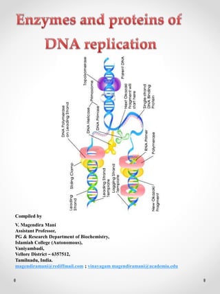
  • 12. Enzyme Function in DNA replication DNA Helicase Also known as helix destabilizing enzyme. Unwinds the DNA double helix at the Replication Fork. DNA Polymerase Builds a new duplex DNA strand by adding nucleotides in the 5' to 3' direction. Also performs proof-reading and error correction. DNA clamp A protein which prevents DNA polymerase III from dissociating from the DNA parent strand. Single-Strand Binding (SSB) Proteins Bind to ssDNA and prevent the DNA double helix from re-annealing after DNA helicase unwinds it, thus maintaining the strand separation. Topoisomerase Relaxes the DNA from its super-coiled nature. DNA Gyrase Relieves strain of unwinding by DNA helicase; this is a specific type of topoisomerase DNA Ligase Re-anneals the semi-conservative strands and joins Okazaki Fragments of the lagging strand. Primase Provides a starting point of RNA (or DNA) for DNA polymerase to begin synthesis of the new DNA strand. Telomerase Lengthens telomeric DNA by adding repetitive nucleotide sequences to the ends of eukaryotic chromosomes. DNA REPLICATION PROTEINS At the replication fork, many replication enzymes assemble on the DNA into a complex molecular machine called the replisome. The following is a list of major DNA replication enzymes that participate in the replisome:
  • 13. DNA GYRASE DNA gyrase, often referred to simply as gyrase, is an enzyme that relieves strain while double-strand DNA is being unwound by helicase. This causes negative supercoiling of the DNA. The ability of gyrase to relax positive supercoils comes into play during DNA replication. The right-handed nature of the DNA double helix causes positive supercoils to accumulate ahead of a translocating enzyme, in the case of DNA replication, a DNA polymerase. The ability of gyrase (and topoisomerase IV) to relax positive supercoils allows superhelical tension ahead of the polymerase to be released so that replication can continue.
  • 14. Negative supercoiling of bacterial DNA by DNA gyrase influences all metabolic processes involving DNA and is essential for replication. Gyrase supercoils DNA by a mechanism called sign inversion, whereby a positive supercoil is directly inverted to a negative one by passing a DNA segment through a transient double-strand break. Reversal of this scheme relaxes DNA, and this mechanism also accounts for the ability of gyrase to catenate and uncatenate DNA rings. Each round of supercoiling is driven by a conformational change induced by adenosine triphosphate (ATP) binding: ATP hydrolysis permits fresh cycles. The A subunit is particularly associated with the concerted breakage- and-rejoining of DNA and the B subunit mediates energy transduction. Gyrase is a prototype for a growing class of prokaryotic and eukaryotic topoisomerases that interconvert complex forms by way of transient double-strand breaks.
  • 15. DNA HELICASES Helicases are often used to separate strands of a DNA double helix or a self-annealed RNA molecule using the energy from ATP hydrolysis, a process characterized by the breaking of hydrogen bonds between annealed nucleotide bases. They also function to remove nucleic acid-associated proteins and catalyze homologous DNA recombination.
  • 16. They are proteins, which are involved in the unwinding of DNA molecule. There are four kinds of helicases namely Dna A, Dna B, Rep Proteins, and Helicases - II. Dna-A protein (Mw 48,000) - It binds to 4 of 9 mer sequence and unwinds a 3 of 13 mer sequence at Ori C site and forms an open complex during initiation of replication. It is the first protein which binds to DNA to initiate DNA replication. Dna-B proteins (Mw- 3,00,000)- It is a primosome constituent and consists of six subunits. It unwinds DNA during replication. It is responsible for the extension of open complex during replication. Rep Proteins (Mw 65,000) - It is a helicase consisting of one subunit. It binds to the 5'-- 3' template and moves in 3'-- 5' direction. It actively participates in leading strand synthesis in replication.
  • 17. DNA Helicase - II (Mw 75,000) - It is helicase consisting of only one subunit. It binds with 3'--5' template stand and moves along in 5'-- 3' direction. It is involved in lagging strand synthesis in replication. The role of helicases is to unwind the duplex DNA in order to provide a single-stranded DNA for replication, transcription, and recombination for instance.
  • 18. DNA LIGASE The DNA ligases are responsible for connecting DNA segments during replication, repair and recombination. They are class of enzymes that catalyze the formation of alpha-phosphodiester bond between two DNA chains. This enzyme requires the free OH group at the 3' end of other DNA strand and phosphate group at 5' end of the other. The formation of a phosphodiester bond between these groups is an endergonic (energy absorption) reaction. Hence energy source required for ligation. In E.Coli and other bacteria NAD+ supplies the energy whereas in animals i.e. eukaryotes ATP play the role. It plays a role in repairing single-strand breaks in duplex DNA in living organisms, but some forms (such as DNA ligase IV) may specifically repair double-strand breaks (i.e. a break in both complementary strands of DNA).
  • 19. The DNA ligase reaction, which proceeds in three steps: adenylation (addition of AMP) of a lysine residue in the active center of the enzyme, pyrophosphate is released; transfer of the AMP to the 5' phosphate of the so-called donor, formation of a pyrophosphate bond; formation of a phosphodiester bond between the 5' phosphate of the donor and the 3' hydroxyl of the acceptor. In mammals, there are four specific types of ligase. DNA ligase I: ligates the nascent DNA of the lagging strand after the DNA polymerase I has removed the RNA primer from the Okazaki fragments.
  • 20. DNA ligase II: alternatively spliced form of DNA ligase III found in non-dividing cells. DNA ligase III: complexes with DNA repair protein XRCC1 to aid in sealing DNA during the process of nucleotide excision repair and recombinant fragments. DNA ligase IV: complexes with XRCC4. It catalyzes the final step in the non-homologous end joining DNA double-strand break repair pathway. It is also required for V(D)J recombination, the process that generates diversity in immunoglobulin and T-cell receptor loci during immune system development.
  • 21. TOPOISOMERASES Topoismerases are a group of enzymes which controls supercoiling of DNA thereby maintaining it in the proper topological state or superhelical tension. There are two classes of topoisomerases. They are type – I topoisomerase and type – II topoisomerase. TYPE - I TOPOISOMERASE Type-I topoisomerase (Nicking-Closing enzymes) are monomeric 100kd proteins that are widespread in both prokaryotes and eukaryotes. They can remove negative supercoils without leaving nicks in the DNA molecule.
  • 22. MECHANISM After the enzyme binds to a DNA molecule and cuts on strand, the free 5' phosphate on the DNA is covalently attached to a tyrosine residue in the enzyme in the case of prokaryotes (the free 3'- phosphate on the DNA is covalently attached in the case of eukaryotes). The DNA strand that has not been cleaved is then passed through the single stranded break. The cleaved strand is then resealed. The type -I topoisomerase from E.Coli acts on negatively supercoiled molecules but not on positively supercoiled molecules. In contrast, type- I topoisomerase from eukaryotic cells can remove both positive and negative supercoils. Type-I topoisomerase reversibly catenates (interlinks) single stranded circles.
  • 23. TYPE-II TOPOISOMERASES The first type II topoisomerase (Topo II) to be described was isolated from E. coli. and named DNA gyrase are 375 kd proteins that consists of two pairs of subunits designated A and B. Topo II enzymes have the ability to cut both strands of a double- stranded DNA molecule, pass another portion of the duplex through the cut, and reseal the cut in a process that utilizes ATP. Depending on the DNA substrate, these movements will have the effect of changing a positive supercoil into a negative supercoil or of increasing the number of negative supercoils by 2.
  • 24. The Topo II enzymes from mammalian cells cannot, like E. coli DNA gyrase, increase the superhelical density at the expense of ATP; presumably no such activity is required in eukaryotes, since binding of histones increases the potential superhelicity. All type II topoisomerases catalyze catenation and decatenation, that is, the linking and unlinking, of two different DNA duplexes. DNA gyrase has the ability to cut a double stranded DNA molecule, pass another portion of the duplex DNA through the cut, and reseal the cut. It changes the linking number of the DNA by 2
  • 25. DNA POLYMERASES These enzymes copy DNA sequences by using one strand as a template. The reaction catalyzed by DNA polymerases is the addition of deoxyribonucleotides to a DNA chain by using dNTPs as substrates. All DNA polymerases require a template strand, which is copied. DNA polymerases also require a primer, which is complementary to the template. The reaction of DNA polymerases is thus better understood as the addition of nucleotides to a primer to make a sequence complementary to a template.
  • 26. The best‐studied bacterium, E. coli, has three DNA polymerase types. DNA polymerase I (Pol I) is primarily a repair enzyme, although it also has a function in replication. About 400 Pol I molecules exist in a single bacterium. DNA polymerase I only makes an average of 20 phosphodiester bonds before dissociating from the template. These properties make good sense for an enzyme that is going to replace damaged DNA. Damage occurs at separate locations so the large number of Pol I molecules means that a repair enzyme is always close at hand. DNA polymerase I has nucleolytic (depolymerizing) activities, which are an intimate part of their function. The 5′ to 3′ exonuclease activity removes base‐paired sequences ahead of the polymerizing activity. During replication, this can remove primers ahead of the polymerizing function of the polymerase.
  • 27. Another intimate function of DNA polymerase I (and of the other forms of DNA polymerase found in E. coli) is the 3′ to 5′ exonuclease activity. These activities can de‐polymerize DNA starting from the newly synthesized end. The 3′ to 5′ exonuclease activity serves an editing function to ensure the fidelity of replication. Suppose DNA polymerase were to make a mistake and add a T opposite a G in the template strand. When the enzyme begins the next step of polymerization, the T is not properly paired with the template. The 3′ to 5′ exonucleolytic activity of DNA polymerase then removes the unpaired nucleotide, releasing TMP, until a properly paired stretch is detected. Then polymerization can resume. This cycle costs two high‐energy phosphate bonds because TTP is converted to TMP. While this may seem wasteful of energy, the editing process does keep the information store of the cell intact.
  • 28. DNA polymerase II is a specialized repair enzyme. Like Pol I, a large number of Pol II molecules reside in the cell (about 100). The enzyme is more processive than Pol I. Pol II has the same editing (3′ to 5′) activity as Pol I, but not the 5′ to 3′ exonuclease activity. The actual replication enzyme in E. coli is DNA polymerase III. Its properties contrast with Pol I and Pol II in several respects. Pol III is much more processive than the other enzymes, making about 500,000 phosphodiester bonds on the average. In other words, it is about 5,000 times more processive than Pol I and 50 times more processive than Pol II. Pol III is a multisubunit enzyme. It lacks a 5′ to 3′ exonucleolytic activity, although a subunit of the enzyme carries out the editing (3′ to 5′) function during replication. Finally, only about 10 molecules of Pol III reside in each cell. This remains consistent with the function of Pol III in replication, because the chromosome only needs to be copied once per generation.
  • 29. Therefore, the cell only requires a few molecules of the enzyme. Pol III synthesizes DNA at least a hundred times more rapidly than the other polymerases. It can synthesize half of the bacterial chromosome in a little more than 20 minutes, which is the fastest that the bacterium can replicate.
  • 30. DNA POLYMERASE – EUKARYOTIC Following are the six important eukaryotic DNA polymerases. They are as follows: i) DNA polymerase α ii) DNA polymerase β iii) DNA polymerase γ iv) DNA polymerase δ v) DNA polymerase Ɛ vi) DNA polymerase σ
  • 31. i) DNA polymerase α It is composed of four subunits of which one has the primase activity. The largest subunit of it has polymerase activity. It is supposed to exist in several forms like α 1, α 2, and α 3. DNA polymerase a in association with DNA polymerase δ, is involved in the replication of nuclear chromosomal DNA. The replication is also aided by another protein factors called Accessory Proteins and AP4A. These protein factors appear to have regulatory function. DNA pol α is supposed to carryout lagging strand synthesis. ii) DNA polymerase -β It is involved in the repair of DNA. iii) DNA polymerase - γ This polymerase is involved in the replication of mitochondria DNA.
  • 32. iv) DNA polymerase -δ This polymerase has polymerase function and 3'-- >5’ exonuclease activity. It is involved in leading strand synthesis. v) DNA polymerase -Ɛ It is supposed to replace DNA pol delta in some situations such as DNA repair. vi) DNA polymerase - σ This polymerase seems to be expressed only in the bone marrow cells. It is the only eukaryotic polymerase that has a deoxy ribonuclease activity
  • 33.
  • 34. RNA PRIMERS RNA primers are single strand oligoribonucleotides with 40 to 60 base pairs. For replication RNA primers are required. This is because DNA polymerases require RNA primer for their action to start in invivo condition. In replication RNA primer is synthesized by two different enzymes namely RNA polymerase and Primosome complex. RNA polymerase synthesize RNA primer for synthesize of leading strand whereas Primosome synthesize RNA primer for lagging strand synthesis. In addition to this, RNA polymerase synthesizes only one primer whereas Primosome synthesizes many primers.
  • 35. OKAZAKI FRAGMENTS Fragments synthesized during lagging strand formation of replication was identified and proved by Rejis Okazaki. Hence, by his name these fragments are called as Okazaki’s fragments. They are short polynucleotides with 1000-2000 base pairs in length. These fragments are synthesized by DNA polymerases. Even though they are formed during replication, they are joined to form larger DNA at the completion of replication by the action of DNA ligase. The invention of Okazaki fragments lead to the proposal of semidiscontinuous replication concept.
  • 36. RESTRICTION ENDONUCLEASE A restriction enzyme (or restriction endonuclease) is an enzyme that cuts DNA at or near specific recognition nucleotide sequences known as restriction sites. Restriction enzymes are commonly classified into three types, which differ in their structure and whether they cut their DNA substrate at their recognition site, or if the recognition and cleavage sites are separate from one another. To cut DNA, all restriction enzymes make two incisions, once through each sugar-phosphate backbone (i.e. each strand) of the DNA double helix.
  • 37. These enzymes are found in bacteria and archaea and provide a defense mechanism against invading viruses. Inside a prokaryote, the restriction enzymes selectively cut up foreign DNA in a process called restriction; while host DNA is protected by a modification enzyme (a methylase) that modifies the prokaryotic DNA and blocks cleavage. Together, these two processes form the restriction modification system.
  • 38. EXONUCLEASES Exonucleases are enzymes that work by cleaving nucleotides one at a time from the end (exo) of a polynucleotide chain. A hydrolyzing reaction that breaks phosphodiester bonds at either the 3’ or the 5’ end occurs. Its close relative is the endonuclease, which cleaves phosphodiester bonds in the middle (endo) of a polynucleotide chain. Eukaryotes and prokaryotes have three types of exonucleases involved in the normal turnover of mRNA: 5’ to 3’ exonuclease, which is a dependent decapping protein, 3’ to 5’ exonuclease, an independent protein, and poly (A)-specific 3’ to 5’ exonuclease.
  • 39. TELOMERASE Telomerase also called telomere terminal transferase is a ribonucleoprotein that is an enzyme that adds DNA sequence repeats ("TTAGGG" in all vertebrates) to the 3' end of DNA strands in the telomere regions, which are found at the ends of eukaryotic chromosomes. This region of repeated nucleotide called telomeres contains noncoding DNA and hinders the loss of important DNA from chromosome ends. As a result, every time the chromosome is copied, only 100–200 nucleotides are lost, which causes no damage to the organism's DNA. Telomerase is a reverse transcriptase that carries its own RNA molecule, which is used as a template when it elongates telomeres, which are shortened after each replication cycle.
  • 40.
  • 41. RETROVIRAL REPLICATION Retroviruses contain viral RNA and several copies of reverse transcriptase (DNA polymerase). After infecting a cell, the reverse transcriptase is used to make the initial copies of viral DNA from viral RNA. Once a DNA strand has been synthesized, a complementary viral DNA strand is made. These double strand copies of viral DNA are inserted into the host-cell chromosome and host-cell RNA polymerase is used to make virus-related RNA. These RNA strands serve as templates for making new copies of the viral chromosomal RNA and serve also as mRNA. mRNA is translated into viral proteins that are used to make the virus envelope. New viral particles are assembled, bud from the plasma membrane, and are released.
  • 42.
  • 43. An example of this process is illustrated in the replication of the retrovirus, HIV (human immunodeficiency virus). Retroviruses are enveloped animal ribonucleic acid (RNA) viruses that replicate via a deoxyribonucleic acid (DNA) intermediate, which is integrated into the host genome as a provirus. Interaction of the viral envelope protein with a target cell receptor triggers entry of the viral nucleoprotein core by fusion of viral and cellular membranes. After entry, the viral enzymes reverse transcriptase and integrase mediate reverse transcription of viral RNA and integration of the resulting double‐stranded DNA copy of the viral genome, respectively. Expression of viral RNA and proteins from proviral DNA utilises the transcription and translation machinery of the host. Retrovirus particles are assembled through protein–protein and protein–lipid interactions, released from the cell by budding, and subsequently matured by a viral protease. A provirus can be transmitted through the germline from parents to offspring as an endogenous retrovirus. Host cell restriction factors target multiple steps of retroviral replication in a complex interplay of virus–host interactions.
  • 44. REVERSE TRANSCRIPTASE Reverse transcriptase is an enzyme used to generate complementary DNA (cDNA) from an RNA template, a process termed reverse transcription. It is mainly associated with retroviruses. Retroviral RT has three sequential biochemical activities: (a) RNA-dependent DNA polymerase activity, (b) ribonuclease H, and (c) DNA-dependent DNA polymerase activity. These activities are used by the retrovirus to convert single-stranded genomic RNA into double-stranded cDNA which can integrate into the host genome, potentially generating a long-term infection that can be very difficult to eradicate. The same sequence of reactions is widely used in the laboratory to convert RNA to DNA for use in molecular cloning, RNA sequencing, polymerase chain reaction (PCR), or genome analysis.
  • 45. HISTONE PROTEIN In biology, histones are highly alkaline proteins found in eukaryotic cell nuclei that package and order the DNA into structural units called nucleosomes. They are the chief protein components of chromatin, acting as spools around which DNA winds, and play a role in gene regulation. Without histones, the unwound DNA in chromosomes would be very long (a length to width ratio of more than 10 million to 1 in human DNA). For example, each human cell has about 1.8 meters of DNA, (~6 ft) but wound on the histones it has about 90 micrometers (0.09 mm) of chromatin, which, when duplicated and condensed during mitosis, result in about 120 micrometers of chromosomes.
  • 46. Types of Histone proteins There are 5 families of histones (H1 through H5). H2A, H2B, H3, and H4 are the core histones, and H1 and H5 are the linker histones. The core histones form the center of the nucleosome, hence the term 'core'. The linker histones are found at the entrance and exit sites of the nucleosome and lock the DNA in place, hence the term 'linker'. A strand of DNA will wrap around the core histones 1.65 times. Interactions between nucleosomes allow for higher-order structures to form. These higher- order structures can condense the chromatin to the point where chromosomes form. You are probably familiar with what a chromosome looks like (it's an 'X' shape), and now you know that it is histones that make this familiar structure possible.
  • 47. Histones form dimers (2 histones) and tetramers (4 histones). Each nucleosome has two identical dimers each comprised of one H2A and one H2B histone (called an H2A- H2B dimer). Each nucleosome also has one tetramer comprised of two H3 and two H4 histones (called an H3-H4 tetramer). Figure 1 shows how the core of a nucleosome is formed.
  • 48. TEMPORAL CONTROL OF REPLICATION In all organisms, the protein machinery responsible for the replication of DNA, the replisome, is faced with a directionality problem. The antiparallel nature of duplex DNA permits the leading-strand polymerase to advance in a continuous fashion, but forces the lagging-strand polymerase to synthesize in the opposite direction. By extending RNA primers, the lagging-strand polymerase restarts at short intervals and produces Okazaki fragments. At least in prokaryotic systems, this directionality problem is solved by the formation of a loop in the lagging strand of the replication fork to reorient the lagging-strand DNA polymerase so that it advances in parallel with the leading- strand polymerase.
  • 49. The replication loop grows and shrinks during each cycle of Okazaki fragment synthesis. Here we use single-molecule techniques to visualize, in real time, the formation and release of replication loops by individual replisomes of bacteriophage T7 supporting coordinated DNA replication. Analysis of the distributions of loop sizes and lag times between loops reveals that initiation of primer synthesis and the completion of an Okazaki fragment each serve as a trigger for loop release. The presence of two triggers may represent a fail-safe mechanism ensuring the timely reset of the replisome after the synthesis of every Okazaki fragment.
  • 50.
  • 51. INHIBITORS OF REPLICATION Dauromycin and Adriamycin: They are synthetic chemotherapeutic agents and are inhibitors of both DNA replication and transcription in prokaryotes. These are presumably act by interfering with the passage of both DNA and RNA polymerase. They have planar aromatic ring system which gets intercalated between GC pairs of the double helical structure. Thus, they prevent its replication and transcription.
  • 52. Adriamycin is otherwise known as doxorubicin. Doxorubicin is a cytotoxic anthracycline antibiotic isolated from cultures of Streptomyces peucetius var. caesius. Doxorubicin consists of a naphthacenequinone nucleus linked through a glycosidic bond at ring atom 7 to an amino sugar, daunosamine. Doxorubicin binds to nucleic acids, presumably by specific intercalation of the planar anthracycline nucleus with the DNA double helix. The anthracycline ring is lipophilic, but the saturated end of the ring system contains abundant hydroxyl groups adjacent to the amino sugar, producing a hydrophilic center. The molecule is amphoteric, containing acidic functions in the ring phenolic groups and a basic function in the sugar amino group. It binds to cell membranes as well as plasma proteins.
  • 53. Actinomycin – D It is an antibiotic produced by streptomyces and inhibits replication and transcription. It acts by intercalating its phenoxazone ring between two successive GC pairs in duplex DNA. Actinomycin D has two identical pentapeptides which have unusual composition of D- Valine and Sarcosine which stabilizes this intercalating interaction.
  • 54. It was the first antibiotic shown to have anti- cancer activity, but is not normally used as such, as it is highly toxic, causing damage to genetic material. Actinomycin-D is marketed under the trade name Dactinomycin. Actinomycin-D is one of the older chemotherapy drugs which has been used in therapy for many years. It is a clear, yellow liquid which is administered intravenously and most commonly used in treatment of a variety of cancers.
  • 55. Ethidium bromide and proflavin They inhibit both replication and transcription by intercalation. As with most fluorescent compounds, it is aromatic. The main portion of the molecule is a tricyclic structure with aniline (aminobenzene) groups on either side of a pyridine (a six-atom, nitrogen-containing, aromatic ring). The dibenzopyridine structure is known as a phenanthridine. The reason for ethidium bromide's intense fluorescence after binding with DNA is probably not due to rigid stabilization of the phenyl moiety, because the phenyl ring has been shown to project outside the intercalated bases. In fact, the phenyl group is found to be almost perpendicular to the plane of the ring system, as it rotates about its single bond to find a position where it will abut the ring system minimally. Instead, the hydrophobic environment found between the base pairs is believed to be responsible.
  • 56. By moving into this hydrophobic environment and away from the solvent, the ethidium cation is forced to shed any water molecules that were associated with it. As water is a highly efficient fluorescent quencher, the removal of these water molecules allows the ethidium to fluoresce. This property is used to identify the presence of DNA in gel und UV light.
  • 57. Novobiocin and oxolinic Acid Prokaryotic DNA gyrases are specifically inhibited by two classes of antibiotics. One of these classes includes the streptomyces-derived novobiocin and the other contains the clinically useful synthetic antibacterial agent oxolinic acid. Both classes of antibiotics profoundly inhibit bacterial DNA replication and transcription, thereby demonstrating the importance of properly supercoiled DNA in these processes. Studies using antibiotic- resistant E.Coli mutants have demonstrated that oxolinic acid associates with DNA gyrase’s A subunit and novobiocin binds to its B subunit. When DNA gyrase is incubated with DNA and oxolinic acid, and subsequently denatured with guanidinium chloride, it’s A subunits remain covalently linked to the 5’-ends of both cut strands through phosphotyrosine linkages. Apparently oxolinic acid interferes with gyrase action by blocking the strand breaking-rejoining process. Novobiocin, on the other hand, prevents ATP from binding to the enzyme.
  • 58. Aphidicolin Aphidicolin inhibits DNA pol α of eukaryotes, so it inhibits eukaryotic replication. Aphidicolin also inhibits DNA pol δ and DNA pol Ɛ. Thus Aphidicolin inhibits both leading and lagging strand synthesis in eukaryotes.
  • 59. Rifamycin Rifammycin inhibits RNA polymerase, so RNA primer for leading strand synthesis is not available. Thus replication inhibited. The rifamycins are a group of antibiotics which are synthesized either naturally by the bacterium Amycolatopsis mediterranei, or artificially. The rifamycin group includes the "classic" rifamycin drugs as well as the rifamycin derivatives Rifampicin, Rifabutin and Rifapentine. The biological activity of rifamycins relies on the inhibition of DNA-dependent RNA synthesis. This is due to the high affinity of rifamycins to prokaryotic RNA polymerase. Crystal structure data of the antibiotic bound to RNA polymerase indicates that rifamycin blocks synthesis by causing strong steric clashes with the growing oligonucleotide.
  • 60. If rifamycin binds the polymerase after the chain elongation process has started, no effect is observed on the biosynthesis, which is consistent with a model that suggests rifamycin physically blocks the chain elongation. In addition, rifamycins showed potency towards tumors. This is due to their inhibition of the enzyme reverse transcriptase, which is essential for tumor persistence. However, rifamycins potency proved to be mild and this never leads to their introduction to clinical trials.
  • 61. ALL THE BEST By VMM V. Magendira Mani Assistant Professor, PG & Research Department of Biochemistry, Islamiah College (Autonomous), Vaniyambadi, Vellore District – 6357512, Tamilnadu, India. magendiramani@rediffmail.com ; vinayagam magendiramani@academia.edu https://tvuni.academia.edu/mvinayagam