Flexor tendon injuries.m
•Télécharger en tant que PPTX, PDF•
28 j'aime•9,808 vues

A 30-year old female presented to the emergency room with a laceration and bleeding in her right hand after falling on glass. She was right hand dominant and worked in telemarketing. Physical examination would focus on the extent of the laceration and potential injury to flexor tendons and nerves. Flexor tendon injuries can lead to loss of finger flexion and grip strength if not repaired properly. The goals of reconstruction are to anatomically repair the tendons with limited motion restrictions and adhere to post-operative rehabilitation to regain function and prevent complications like adhesions.

Recommandé
Contenu connexe
Tendances
Tendances (20)
Similaire à Flexor tendon injuries.m
Similaire à Flexor tendon injuries.m (20)
Plus de Mansoor Khan
Plus de Mansoor Khan (20)
Dernier
Dernier (20)
Flexor tendon injuries.m
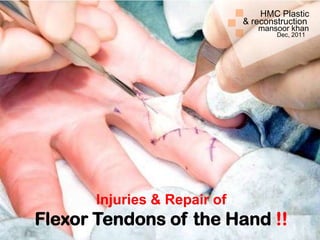
- 1. HMC Plastic & reconstruction mansoor khan Dec, 2011 Injuries & Repair of Flexor Tendons of the Hand !!
- 2. Presentation: Questions to consider: A 30-year old female presents to the 1. What aspects of the physical Emergency room after falling on a piece examination would you focus on? of glass. She complains of 2. What anatomic structures may pain, numbness and bleeding of her have been disrupted given this type right hand. She is right hand dominant of injury? and works for a local telemarketing firm.
- 3. “ glistening structure between muscle & bone which ” transmit force from muscle to the bone
- 4. Tendons tertiary bundles fasciles fibers fibrils endotenon collagen
- 12. PULLEYS
- 18. Skin laceration with loss of normal cascade of the fingerd in resting position!!
- 19. Loss of active flexion at DIP in FDP laceration!!
- 20. Loss of normal tenodesis effect!!
- 21. Passive flexion with forearm squeez!!
- 24. Complain of numbness preceeded by execissive bleed Concider neurovascular insult!!
- 25. Goals of reconstruction: Coaptation of tendons, anatomical repair with a limited accordion effect at the repair site, multiple strand drepair to permit active range of motion rehabilitation Pully reconstruction to minimize bow- stringing, atraumatic surgical technique to minimize adhesionns, strict adherence to rehabilitation protocole.
- 26. Timing of flexor tendon injuries: Primary: repair within 24 hours (contraindicated in case of high grade condtamination i.e. human bites, infection) Delayed Primary: 1-14 days when the wound can be still pulled open without incision Early Secondary: 2-5 weeks. Late Secondary : after 5 weeks i.e. tendon substitution techniques/salvage process.
- 27. Leddy classification of zone I flexor tendon injuries!! Type I: tendon retracted into palm (fullness in palm) Type II: tendon traped in the sheath at PIP (unable to flex PIP) Type III: tendon traped in A4 pully
- 28. Type II injury!!
- 29. Type I injury!!
- 30. Direct repair: if laceration is more than 1 cm from FDP insertion Tendon advancement: if the laceration is less then 1 cm from insertion.
- 32. Wilson One method of attaching tendon to bone. A, Small area of cortex is raised with osteotome. B, Hole is drilled through bone with Kirschner wire in drill. C, Bunnell crisscross stitch is placed in end of tendon, and wire suture is drawn through hole in bone. D, End of tendon is drawn against bone, and suture is tied over button.
- 33. Kleinert method of tendon advancement!!
- 34. Tendon advancement shortens the FDP & completes the grip before the normal fingerd and limit their flexion and thus week grip Quadrigia effect!!
- 35. Laceration during flexion leads to retraction of cut ends of the tendons!!
- 36. Complications: complete disruption, entrapment, triggering. Assess for entrapment, debride if risk of entrapment No drepair if less than <25% laceration, only epitenon repair in 25-50% lacerations, core suture plus epitenon repair when >50% laceration Dorsal blocking splintage for 6-8 weeks as consevative measure Partial lacerations of the tendons!!
- 37. Commonly used incidions for flexor tendon exploration!!
- 41. Because the blood supply to the FDP tendon is jeopardized if the FDS is not also fixed (due to the vinculae anatomy) Repair both tendons:
- 42. Complications: Adhesions & stiffness requiring tenolysis in 18-25% cases Tenolysis is indicated after 3 months if no improvement is noted for 1-2 months extensive physiotherapy.
- 43. Lumbrical muscle bellies usually are not sutured because this can increase the tension of these muscles and result in a “lumbrical plus” finger (paradoxical proximal interphalangeal extension on attempted active finger flexion). Zone 3 injuries
- 44. Tendon repair strength: Core suture: Material, caliber, number of strands, knot location, dorsal vs ventral location Epitendinous suture: Depth, locking, cross hatching, simple
- 48. Silfverskiöld
- 49. Fish-Mouth End-to-End Suture (Pulvertaft)
- 51. Active range of motion rehabilitation Kleinert !!
- 52. Place and hold post-operative exercised!!
- 53. Differential passive exercises for FDP & FDS!!
- 54. Post-operative passive exercises Duran’s
- 55. Lumbrical plus!!
- 56. Risk factors for adhesions: Composite tendon/tissue damage Gap formation Ischaemia due to over mobalizations of tendon ends Immobalization Persistant inflammation Secondary trauma