

Recommandé
Contenu connexe
Tendances
Tendances (20)
Similaire à Becterial
Similaire à Becterial (20)
Plus de Maryam Shakeel
Dernier
Dernier (20)
Becterial
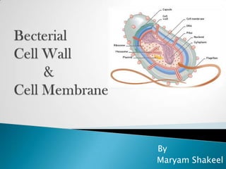
- 2. The cell wall is the tough, usually flexible but sometimes fairly rigid layer that surrounds becteria. It is located outside the cell membrane Mostly in bacteria cell wall chemically composed of peptidoglycan (murein). The cell wall strengthens the plasma membrane and prevents the cell from bursting.
- 3. Gram +ve Gram-ve with the with the Gram stain. Gram G+ cells are stain. G- Purple cells are red.
- 4. Gram Gram - E. coli cells + Staphylococcus cells.
- 5. Gram-positive cell walls consist of many layers of peptidoglycan and also contain teichoic acids. Teichoic acids may: bind and regulate movement of cations into and out of the cell prevent extensive wall breakdown and possible cell lysis during cell growth provide much of the cell wall's antigenicity
- 7. Gram-negative bacteria have a lipopolysaccharide-lipoprotein-phospholipid outer membrane surrounding a thin (sometimes a single) peptidoglycan layer. Gram-negative cell walls have no teichoic acids.
- 9. G +ve G -ve
- 11. • From • Maintain the Osmolysis shape of cell Structural Protection Support Critical Filtering Structure mechanism • Becteria can • Does not allow not live without motion of it large molecules
- 13. Surrounds the cell's cytoplasm and regulates the flow of substances in and out of the cell. The plasma membrane is composed of a phospholipid bilayer (two layers) embedded with proteins and other molecules. The phospholipids are not fixed relative to each other and able to flow past each other, making the membrane fluid.
- 14. Bacteria can have a wide variety of fatty acids within their membranes. Along with typical saturated and unsaturated fatty acids, bacteria can contain fatty acids with additional methyl , hydroxy or even cyclic groups. The relative proportions of these fatty acids can be modulated by the bacterium to maintain the optimum fluidity of the membrane (e.g. following temperature change).
- 15. Phospholipids are molecules composed of two long, hydrocarbon "tails" and a phosphate group "head." The hydrophilic (water-attracted) phosphate group "heads" form the internal and external surface of the plasma membrane. The hydrophobic (water- repelled) tails form the interior of the plasma membrane.
- 16. Embedded in the plasma membrane are proteins that perform multiple functions, including the detection of chemicals in the cell's environment and the transportation of materials into and out of the cell.
- 17. the region between the cytoplasmic and outer membranes. The periplasm contains the peptidoglycan layer and many proteins responsible for substrate binding on hydrolysis and reception of extracellular signals.
- 18. passive transport of many ions, sugar and amino acids across the outer membrane. These molecules are therefore present in the periplasm. acting as a permeability barrier for most molecules and serving as the location for the transport of molecules into the cell.
- 19. prokaryotic membranes also function in energy conservation as the location about which a proton motive force is generated. bacterial membranes (with some exceptions e.g. Mycoplasma and methanotrophs) generally do not contain sterols. bacteria can have a wide variety of fatty acids within their membranes.
- 20. It prevents only prevents toxicity to large molecules the cell whereas cell from entering the membrane prevents cell. entry of smaller Cell wall is molecules. completely cell membrane is permeable semi-permeable.
- 21. The function of the The cell membrane cell wall is to provides support to provide strength the cytoskeleton of and rigidity to the the cell, gives shape cell. to the cell It protects the cell it maintains the against mechanical potential of the cell, forces. helps in It is made up of communication with peptidoglycan. other cells, and act as molecular signals It is made up of lipid and protein.
Notes de l'éditeur
- Electron micrograph of an ultra-thin section of a dividing pair of group A streptococci (20,000X). The cell surface fimbriae (fibrils) are evident. The bacterial cell wall is seen as the light staining region between the fibrils and the dark staining cell interior. Cell division in progress is indicated by the new septum formed between the two cells and by the indentation of the cell wall near the cell equator. The streptococcal cell diameter is equal to approximately one micron. Electron micrograph of Streptococcus pyogenes by Maria Fazio and Vincent A. Fischetti, Ph.D. with permission. The Laboratory of Bacterial Pathogenesis and Immunology, Rockefeller University.For g+