1. EVOLUTIONARY DEVELOPMENT AND ANATOMY OF THE LUNGS
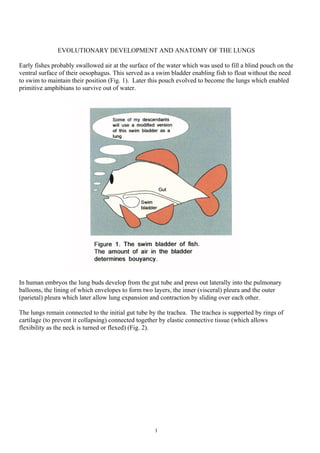
Early fishes probably swallowed air at the surface of the water which was used to fill a blind pouch on the
ventral surface of their oesophagus. This served as a swim bladder enabling fish to float without the need
to swim to maintain their position (Fig. 1). Later this pouch evolved to become the lungs which enabled
primitive amphibians to survive out of water.
In human embryos the lung buds develop from the gut tube and press out laterally into the pulmonary
balloons, the lining of which envelopes to form two layers, the inner (visceral) pleura and the outer
(parietal) pleura which later allow lung expansion and contraction by sliding over each other.
The lungs remain connected to the initial gut tube by the trachea. The trachea is supported by rings of
cartilage (to prevent it collapsing) connected together by elastic connective tissue (which allows
flexibility as the neck is turned or flexed) (Fig. 2).
1
2. The larynx, which was initially a valve that protected the primitive lungs from dehydration is the dividing
line between the upper and lower airways. In the larynx there are two inner folds, the vocal cords (Fig. 3)
which vibrate to give speech. The epiglottis is a cartilaginous valve-like flap which guides air and food
to the respective entrances (Figs. 3 and 4).
2
6. PHYSIOLOGICAL PRINCIPLES
For adequate oxygenation of the tissues it is necessary that the lungs be ventilated, oxygen is able to
diffuse into the blood passing through the lungs, and there is an adequate circulatory system.
Inspiration is an active process achieved by increasing the intrathoracic volume by contraction of the
diaphragm and intercostal muscles (Fig. 8). Expiration, in contrast, is a passive process which relies upon
the natural elastic recoil of the lung. If a more substantial expiration is required the abdominal muscles
can contract and push up the diaphragm. The elasticity of the lung is reduced by processes such as
fibrosis or emphysema.
6
7. Various measurements of ventilatory function (Fig. 9) can be made by getting patients to blow into
various measuring devices.
The peak expiratory flow (PEF) is the peak flow after the largest possible breath in and fastest possible
blow out (Fig. 10). It is reduced by airway narrowing or expiratory muscle weakness. The forced
expiratory volume in the first second (FEV1) and forced vital capacity (FVC) are obtained by getting the
patient to take in the largest possible breath (to total lung capacity) and then blow out as hard, fast and
completely as possible (Fig. 11). Normally the forced expiratory volume in the first second is 70-80
percent of the forced vital capacity. If the airflow is reduced, for example by bronchial narrowing, then
the forced expiratory volume in the first second is reduced proportionally more than the forced vital
capacity and the ratio of FEV1 to FVC is reduced. If the lungs are stiff, a restrictive defect, or the
ventilatory muscles are weak then the forced expiratory volume in the first second and the forced
expiratory volume are both decreased in the same proportion and the FEV1 to FVC ration remains
normal. In general the FEV1 , although less easy to measure, gives a more useful assessment of
ventilatory flow obstruction than the PEF.
7
9. With exercise there is increased extraction of oxygen by skeletal muscles which is met by increases in the
ventilatory rate and tidal volume.
The main business of the lungs is to oxygenate red blood corpuscles (RBCs) in the blood and to blow off
excess carbon dioxide (the major by-product of energy production). To do this the average adult breaths
about 400 mls of air (the tidal volume) in and out about 15-20 times per minute. Air comprises three
gases (oxygen, carbon dioxide, nitrogen) and water vapour. The pressure (p) depends on the
concentration of the gas present in the substance being tested. Gases pass from areas with a high pressure
to areas with a low pressure.
9
10. At rest 250 mls of oxygen per minute are absorbed from the air and 200 mls of carbon dioxide per minute
are exhaled as well as some water vapour. At sea level air is 21 percent oxygen with an arterial blood
oxygen level (pO2 ) of 160mms mercury with minimal carbon dioxide (Fig. 12). The tissues have a pO2
of 40 and an arterial blood carbon dioxide pressure (pCO2 ) of 46 mms mercury. Thus there is a diffusion
gradient across alveolar walls such that oxygen diffuses into the body and carbon dioxide diffuses out
(Fig. 7). As expected alveolar air is intermediate between inspired air and tissue levels (Fig. 12).
Measurement of the systemic arterial pO2 gives a guide as to how well the lungs have oxygenated the
blood that leaves the lungs. The percentage of oxygen saturation of the blood in the peripheries can be
measured by devices which colourmetrically measure the percentage oxygen saturation of the
haemoglobin that is present (usually measured on an earlobe or at a fingertip) and this reflect tissue
oxygenation. If the haemoglobin saturation with oxygen is low but the arterial pO2 is normal then the
tissues are desperate for oxygen. The saturations are not usually affected by mild anaemia but if anaemia
is severe the tissues are oxygen starved and extract what oxygen there is in the haemoglobin and the
saturation will then be low. Thus if the peripheral saturations are normal there is usually no need to
measure the arterial pO2.
Oxygen moves from areas of high partial pressure to areas of low partial pressure (Figs 12 and 13). At a
pO2 of 100 mms mercury only 0.3 mls of oxygen is dissolved in every 100mls of blood and this is
inadequate to sustain aerobic metabolism and an oxygen storage medium is required. This is provided by
the haemoglobin in red blood corpuscles. With a normal haemoglobin level, 20 mls of oxygen is
10
11. contained in every 100 mls of blood. The oxygen-haemoglobin dissociation curve, shows the relevant
pressures and the volumes of oxygen carried by haemoglobin. Once the oxygen carrying power of the
plasma and haemoglobin is saturated hyperventilation of the lungs or giving higher concentrations of
oxygen will not be of significant benefit - the haemoglobin is saturated anyway and the amount or oxygen
carried in the plasma will not alter significantly (unless the pO2 is increased by providing oxygen under
increased pressure - hyperbaric oxygen).
Carbon dioxide can diffuse into and out of blood far more readily than can oxygen. The quantity of
carbon dioxide in the blood can be directly affected by ventilation (hyperventilation causes carbon
dioxide levels to fall whereas hypoventilation causes carbon dioxide levels to rise).
In normal individuals the ventilatory drive is provided by the plasma acidity which usually depends on
the pCO2 - the higher the pCO2 the greater the ventilatory drive.
Why does carbon dioxide and not oxygen normally provide the ventilatory drive? Probably because there
is a need to keep the tissue oxygenation at a near normal level for as long as possible, and this is achieved
11
12. because the oxygen-haemoglobin dissociation curve allows haemoglobin to maintain tissue oxygen levels
at normal levels within a wide range of tissue requirements until “the system” breaks down (this
maintenance of oxygen provision is the main reason why the oxygen-haemoglobin dissociation curve is
as it is). In contrast tissue and blood levels of carbon dioxide rise almost linearly (Fig. 14) and these can
be used as the early warning system to cause increased ventilation before tissue oxygen levels fall.
Aspects of ventilation
If carbon dioxide levels are chronically high the respiratory centre in the brain (link) may downgrade its
sensitivity to carbon dioxide and instead rely on a low oxygen concentration (hypoxia) as the major
ventilatory drive. Patients may become chronically cyanosed – “blue bloaters” (Fig. 15). Giving a high
concentration of oxygen to such patients may cause them to stop breathing until the oxygen returns to a
low level, perhaps to a level dangerously lower than before the start of additional oxygen.
Figure 15.
Oxygen can be given in high concentration to previously well patients with acute onset respiratory
disease and a low pO2 (low oxygen levels and not a high carbon dioxide levels that kill people). In such
circumstances there should be gradual increments in the administered oxygen concentration which will
increase pO2 levels whilst the arterial blood CO2 is being monitored. If the arterial pCO2 goes up then
12
13. too high a concentration of oxygen is being given and the patient’s ventilation will need to be increased
by drug therapy or mechanical ventilation.
A normal or low carbon dioxide level, when combined with a low oxygen level, suggests that blood is not
being exposed to oxygen in the lung, either because a ventilated part of the lung is not receiving blood (a
ventilation-perfusion mismatch) or because the alveolar transmission of oxygen is impaired. The carbon
dioxide in such situations does not rise as the oxygen falls for two main reasons. Firstly, carbon dioxide
diffuses much more easily than oxygen. Secondly, the areas of the lung that are receiving blood are being
hyperventilated because of the low pO2 and thus more carbon dioxide than normal is washed out. The
abnormally low carbon dioxide blood returning from the underperfused part(s) of the lungs results in a
normal or low carbon dioxide content when mixed with the high carbon dioxide blood returning from the
hyperventilated and perfused lung. Oxygen does not have this facility because a mixture of low oxygen
haemoglobin and fully oxygenated haemoglobin can only result in low oxygen levels because
haemoglobin cannot be supersaturated with oxygen. Carbon dioxide does not have a carrier system
analogous to haemoglobin although some carbon dioxide is combined with protein or carried as
bicarbonate (link).
Diffuse airway obstruction is caused by increased bronchial smooth muscle tone (which is normally at a
low level) with increasing tone narrowing the airways. Diffuse narrowing also occurs if:
• there is left sided heart failure with airway narrowing being produced by swollen airway walls
• the bronchial smooth muscle is overdeveloped
• there are copious bronchial secretions.
In diffuse airway narrowing the major difficulty is with expiration - it is easy to suck air into the chest
(an active muscular process), but on attempted expiration (usually a passive process) the airways are
compressed (Fig. 16), and expiration is impaired, leading to overexpansion of the lungs.
13
14. In medical terms oxygen lack is termed anoxia of which there are four types:
1) Anoxic anoxia. There is a low pO2 in arterial blood brought about by:
a) Hypoventilation or
b) Breathing low oxygen concentrations or
c) Breathing oxygen at low pressures such as at high altitudes or
d) Defective transalveolar transport of oxygen or
e) Shunting of blood away from normally ventilated alveoli (ventilation perfusion defects) or
f) An inadequate circulation in shock states.
2) Anaemic anoxia. If a person is anaemic (having a decrease in the number of red blood corpuscles
and/or a less than normal haemoglobin content) or has an abnormal haemoglobin with reduced oxygen
transporting ability then the pO2 will be normal initially - the red blood corpuscles can give up oxygen
but there is a reduced total quantity to give up, but once the depoleted oxygen carrying power is
exhausted then anoxia results. Carbon monoxide poisoning has the same effect as it binds more tightly to
haemoglobin than does oxygen and thus oxygen is displaced.
3) Stagnant anoxia. This occurs when the circulation of blood is so slow that the tissues are starved of
oxygen.
4) Cell damage anoxia. Tissue cells are damaged such that they cannot utilise the available oxygen.
Respiratory failure
Respiratory failure exists when, if a patient is at sea level, awake and breathing air, there is a lung
dysfunction such that 1) arterial pO2 fall below normal or 2) when pCO2 rises above normal There are
two types of respiratory failure.
• Type I respiratory failure occurs when there is a low pO2 with a normal or low pCO2. Causes include
conditions in which the increased carbon dioxide diffusabilty or mixing of high and low carbon
dioxide blood is able to keep the pCO2 at a normal or even low level. Such conditions include acute
asthma or pneumonia. The normal ventilatory drive driven by a (high) carbon dioxide levels is
replaced by a low oxygen drive caused by the pathology.
• Type II respiratory failure occurs when there is a low pO2 with a high pCO2. Type II failure is thus a
ventilatory failure.
Patients with chronic respiratory disease who maintain their pO2 and, unlike blue bloaters, use their pCO2
induced ventilatory drive to prevent their carbon dioxide from rising by hyperventilating are often termed
“pink puffers” whereas patients who do not hyperventilate and rely upon their low pO2 to provide
ventilatory drive gradually accept their low pO2 levels and consequent become cyanosed. Such patients
are often obese and are thus termed “blue bloaters” (Fig. 15). If blue bloaters are given too much oxygen
they will stop breathing and their carbon dioxide rises even further. There are other metabolic effects of
respiratory problems (link).
Figure 17 details the common causes of cough, Figure 18 the common causes of sputum production, and
Figure 19 some common causes of respiratory shortness of breath.
14
16. 16
Some specific conditions
Asthma is diffuse reversible airways obstruction in which there is airway inflammation with eosinophils
and bronchial hyperreactiveness. Treatments include avoidance of provoking factors, use of drugs to
reverse respiratory smooth muscle contraction and to reduce vagal bronchoconstrictor tone, to reduce
inflammation and to reduce secretions. Emphysema is an abnormal and permanent enlargement of air
spaces distal to the terminal bronchiole, accompanied by destruction of their walls and without obvious
fibrosis. In effect there is honeycombing of the lungs. Pneumonia is an accumulation of secretions and
inflammatory cells in the alveolar spaces of the lung, usually caused by infection. Bronchiectasis is
persistent chronic dilatation of the bronchi.