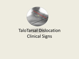
TaloTarsal Dislocation - Radiographic Evidence
•Télécharger en tant que PPTX, PDF•
3 j'aime•3,468 vues

This document discusses taloTarsal dislocation, providing clues to identify it based on clinical signs and radiographic evidence. It notes that taloTarsal dislocation can involve displacement of the talus from the tarsal mechanism in one or more planes (sagittal, transverse, frontal). Left untreated, it can lead to deformity and difficulty weight bearing. The document uses images to illustrate normal versus abnormal taloTarsal alignment and how dislocation in different planes appears radiographically.

Recommandé
Contenu connexe
Tendances
Tendances (20)
Similaire à TaloTarsal Dislocation - Radiographic Evidence
Similaire à TaloTarsal Dislocation - Radiographic Evidence (20)
Plus de GraMedica
Plus de GraMedica (20)
Dernier
Dernier (20)
TaloTarsal Dislocation - Radiographic Evidence
- 2. What are the clues that the talus is not anatomically positioned on the tarsal mechanism?
- 3. Clinical presentation can vary depending on the type of dislocation (incomplete or complete).
- 4. There are external hints indicating internal deformity.
- 5. Sometime these clues are subtle, other times they are very obvious.
- 6. Complete TaloTarsal Dislocation Traumatic injury Patient is not able to bear weight on the foot. Obvious deformity
- 7. Not the biggest bunion, but there is an external observation that something internally is not right.
- 8. Have you developed your x-ray vision? Let’s get started.
- 9. Radiographic Evidence of TaloTarsal Dislocation Michael E. Graham, DPM, FACFAS, FASPS, FAENS
- 10. TaloTarsal Joint is Aligned This is a “normal” example. Talotarsal mechanism (TTM) is neither supinated, nor pronated. Articular facets of the TTM are stable. Weightbearing forces from above are being transferred though the articular facets in an optimum functioning system.
- 11. Is this normal? Yes TTM is balanced, neither supinated nor pronated. TTM will function effortlessly.
- 12. What do you think? Appears that the talus is no longer aligned on the tarsal mechanism. TTM is no longer balanced. Weightbearing forces are not being transmitted through the articular facets in a optimal manner.
- 13. Does this appear to be normal? What is wrong with this foot? Simply that the talus is no longer aligned on the tarsal mechanism. This is a pathologic condition. This will not resolve on its own. It requires physical, physician correction.
- 14. What is the difference between these feet? Talus is not aligned on the tarsal mechanism. Talus is aligned on the tarsal mechanism.
- 15. What is the difference between these feet? Talus is not aligned on the tarsal mechanism. Talus is aligned on the tarsal mechanism.
- 16. Talotarsal dislocation (TTD) can lead to a lowering of the arch. This is due to navicular drop and/or hypermobile/elevated first ray/metatarsal.
- 17. Is this hind-foot normal? No Don’t always see an associated heel valgus (depends on dominate plane of deformity). Transverse plane deformity = forefoot abduction.
- 19. The talus can still displace on the tarsal mechanism leading to other deformity within the foot.
- 21. Don’t be fooled by limiting your observation to stance position alone. Talotarsal dislocation is a dynamic deformity best visualized with the patient walking - gait analysis.
- 22. TaloTarsal Dislocation - Incomplete This is usually a dynamic deformity. Pathologic condition occurs during the walking cycle. Maximum deformity is during walking so when a patient is just standing, you are not “seeing” the maximum talar displacement.
- 23. Gait Analysis Abductory Twist Prolonged period of pronation Calcaneal valgus Pelvic tilt Shoulder tilt
- 24. TaloTarsal Mechanism Relationship of the articular facets of the talus on the calcaneus and navicular. 3 bones, 4 articulations Triplane helicoidal motion Function is to transfer vertical forces from above into horizontal forces below. www.HyProCure.com
- 25. Normal Articular facets are in constant contact Forces are balanced on the articular facets “Normal” amount of joint mechanism motion is available (no more, no less) www.HyProCure.com
- 26. Abnormal – TaloTarsal Mechanism Articular facets are displaced, one on the other. Excessive amount of joint motion is present Excessive forces are placed on supporting tissues Pathologic condition www.HyProCure.com
- 27. Degrees of deformity There are various grading levels of deformities from mild to severe. Regardless of its severity- this is still a pathologic condition that will not resolve on its own; it is progressive and will only get worse with time. Wouldn’t it be better/easier to fix this “sooner than later”? www.HyProCure.com
- 28. TaloTarsal Dislocation Following the same thought process, there are various stages of talotarsal dislocation from mild to severe. This is also a dynamic, progressive deformity that if left undiagnosed or undertreated, will lead to many other secondary pathologic conditions within the foot and also the proximal musculoskeletal chain. www.HyProCure.com
- 29. Normal to abnormal talotarsal alignment www.HyProCure.com
- 30. TaloTarsal Dislocation Incomplete – partial displacement Most common type of TTD Usually dynamic, recurrent No associated fractures Complete – total displacement Least common type of TTD Traumatic etiology Associated with concomitant fractures. www.HyProCure.com
- 31. Complete TaloTarsal Dislocation Total displacement of articular facets. Patients are unable to bear weight Associated fractures Traumatically induced www.HyProCure.com
- 32. TaloTarsal Dislocation Partial This is not normal If there is partial displacement of 1 articular facet to its counter-facet, then the remaining facets will also be displaced. Pathologic condition Results in excessive abnormal motion and excessive abnormal forces acting on the supporting soft tissues Sagittal plane deformity Plantarflexed talus Anterior deviation of Cyma Obliterated Sinus tarsi Increased Talar Declination Angle Diagnosis- TaloTarsal Dislocation (718.37) www.HyProCure.com
- 33. Is this normal? Yes Talus is fully articulated with the tarsal mechanism Weight/force of the body is anatomically and biomechanically correct. No talotarsal displacement Open Sinus Tarsi Normal Cyma www.HyProCure.com
- 34. Is this normal? NO Talus is displaced on the tarsal mechanism. Pathologic event. Deformity is above the bottom of the foot. Sagittal plane deformity (Increased Talar Declination Angle) Plantarflexed Talus Anterior deviation of Cyma Obliterated Sinus Tarsi Diagnosis: Chronic TaloTarsal Dislocation (718.37) www.HyProCure.com
- 35. Is this normal? NO Talus is displaced on the tarsal mechanism Not anatomically aligned (triplane deformity) Pathologic biomechanical mechanism Plantarflexed talus (sagittal plane deformity) Anterior Cyma deviation Fully obliterated sinus tarsi Diagnosis: Chronic TaloTarsal Dislocation (718.37) www.HyProCure.com
- 36. Is this normal? No Pathologic alignment Notice that this is not a “flat foot.” Take a look at the calcaneal inclination angle. Obliterated sinus tarsi Sagittal plane deformity Anterior deviation of Cyma ICD-9 TaloTarsal Dislocation (718.37) www.HyProCure.com
- 37. Is this normal? Yes Articular facets of the talus are anatomically aligned with their counterparts on the tarsal mechanism. No sagittal plane deformity Normal Cyma “Open” sinus tarsi CPT: 28585 with HyProCure. www.HyProCure.com
- 38. Is this foot normal? Yes Talus is properly aligned on the tarsal mechanism. Articular facets are aligned and in constant contact. Normal Cyma Normal TaloNavicular facet alignment No transverse plane deformity Assumed open sinus tarsi on lateral Normal TaloTarsal Alignment www.HyProCure.com
- 39. Is this normal? NO Medial displacement of the head of the talus on the navicular. Displacement of 1 of the 4 articular facets results in partial displacement of the remaining 3 facets. Transverse plane deformity Anterior deviation Cyma TaloNavicular displacement Assumed obliterated sinus tarsi on lateral radiograph. Diagnosis- TaloTarsal Dislocation (718.37) www.HyProCure.com
- 40. Is this Normal? Yes No Transverse plane deformity No displacement of the talus on the tarsal mechanism Normal Cyma Normal TaloNavicular Articulation No transverse plane deformity S/p TaloTarsal Stabilization with Internal Fixation- HyProCure (CPT- 28585) www.HyProCure.com
- 41. Point of Observation- Notice the displacement of the talus on the tarsal mechanism. This particular case has a sagittal and transverse plane deformity with minimal anterior displacement. www.HyProCure.com
- 42. Cardinal Planes of Deformity www.HyProCure.com
- 43. TaloTarsal Dislocation- Sagittal Plane Plantarflexion of the talus on the tarsal mechanism indicates a sagittal plane deformity. This leads to an anterior pelvic tilt. www.HyProCure.com
- 44. Medial displacement of the head of the talus with the navicular. This leads to excessive strain on the knee and hip. Compare the bisection of the talus to the 2nd metatarsal (Talar Second Metatarsal Angle) TaloTarsal Dislocation- Transverse Plane www.HyProCure.com
- 45. What is the easiest way to see a frontal plane deformity on a lateral view?
- 46. Sustentaculumtali is a great indicator of frontal plane deformity. Normal TaloTarsal Mechanism sustentaculum is angled superiorly. In a frontal plane TTD deformity it drops plantarly. TaloTarsal Dislocation- Frontal Plane www.HyProCure.com
- 47. TaloTarsal Dislocation There can be a single dominant plane of deformity: Sagittal > Transverse/Frontal Transverse > Sagittal/Frontal Frontal > Transverse/Sagittal Single Transverse Plane Deformity Single Transverse Plane Deformity www.HyProCure.com
- 48. TaloTarsal Dislocation Two dominate planes of deformity: Sagittal & Transverse > Frontal Sagittal & Frontal > Transverse Frontal & Transverse > Sagittal Transverse & Sagittal www.HyProCure.com
- 49. TaloTarsal Dislocation All three planes are involved. www.HyProCure.com
- 50. TaloTarsal Dislocation before/after “Broken” TaloTarsal Mechanism (718.37) “Fixed” TaloTarsal Mechanism (28585 with HyProCure) www.HyProCure.com
- 51. “Changing Lives, One Step at a Time” www.hyprocure.com View our on-line training www.hyprocuredoctors.com