SlideShare une entreprise Scribd logo
1  sur  65
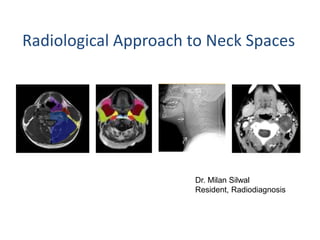
Radiological Approach to Neck Spaces
Dr. Milan Silwal
Resident, Radiodiagnosis
Background:
• Head and neck lesions are a very challenging subject
due to the complex anatomy and wide ranging
pathology.
• Cross sectional imaging is commonly used for clinical
evaluation of head and neck lesions.
• The anatomical location and borders of head and
neck lesions are vital in narrowing radiological
differential diagnosis and allowing formation of an
appropriate clinical management plan.
Neck Spaces:
Described in relation to the hyoid
• Suprahyoid (SHN)
• Infrahyoid (IHN)
Fascia of the neck
• Key to understanding
neck spaces is fascia.
• 2 major fascial layers of
the neck: superficial and
deep cervical fascia.
• Deep cervical fascia comprises three distinct layers:
• Superficial layer splits to enclose a number of muscles (including
sternomastoid and trapezius) and contributes to formation of the carotid
sheath.
• Middle layer envelops the anterior infrahyoid strap muscles, and splits to
enclose the contents of the visceral space. It forms the anterior wall of the
retropharyngeal space and contributing to formation of the carotid
sheath.
• Deep layer encircles the paraspinous and prevertebral muscles and
associated structures. Attaching to the transverse processes, the deep
layer subdivides the perivertebral space into the prevertebral space and
the paraspinal space
• PPS lies in a central location in the
deep face
• PMS is medial to the PPS
• MS is anterior to the PPS
• PS is lateral of the PPS
• CS is posterior to the PPS
• RPS is posterior to PPS
• Perivertebral space is posterior to
the RPS
• Axial graphic depicting the fascia and spaces of the infrahyoid neck. The three
layers of deep cervical fascia are present in the suprahyoid and infrahyoid
neck. The carotid sheath is made up of all 3 layers of deep cervical fascia (tri-
color line around carotid space). Notice the deep layer completely circles the
perivertebral space, diving in laterally to divide it into prevertebral and a
paraspinal components.
Coronal graphic of parapharyngeal space. The parapharyngeal spaces are paired fat-
filled spaces in the more lateral aspect of the suprahyoid neck. This space abuts the
skull base between the masticator and pharyngeal mucosal spaces. There are no
important structures at the point of intersection between the PPS and the skull base.
Inferiorly the PPS communicates inferiorly with the posterior submandibular space.
The masticator space has the largest area of abutment with the skull base, including
CNV3. The pharyngeal mucosal space abuts the basisphenoid and foramen lacerum.
Deep neck spaces
• suprahyoid neck
– parapharyngeal space
– parotid space
– pharyngeal mucosal space
– masticator space
– buccal space
• infrahyoid neck
– anterior cervical space
– posterior cervical space
– visceral space
• supra- and infrahyoid neck
– carotid space
– retropharyngeal space
– perivertebral space
Parapharyngeal space
• PPS: Central, fat-filled spaces in lateral suprahyoid neck (SHN)
around which most of important spaces are located.
• Is shaped like a pyramid, inverted with its base at the skull
base, with its apex inferiorly pointing to the greater cornu of
the hyoid bone.
• Importance of PPS is its conspicuity on CT and MR as well as
its direction of displacement by mass lesions of surrounding
spaces PPS displacement pattern helps define actual space
of origin
– PMS mass lesion pushes PPS laterally
– MS mass lesion pushes PPS posteriorly
– PS mass lesion pushes PPS medially
– CS mass lesion pushes PPS anteriorly
• Boundaries
• superior margin: base of skull
• inferior margin: greater cornu of hyoid bone
• medial margin: pretracheal layer of the deep cervical
fascia
• lateral margin: investing fascia of the deep cervical
fascia covering the deep lobe of the parotid
• anterior margin: investing fascia of the deep cervical
fascia covering the medial pterygoid muscle
• posterior margin: prevertebral layer of the deep
cervical fascia
• Contents
• fat (main component)
• internal maxillary artery
• ascending pharyngeal artery
• pterygoid venous plexus only small portion;
mainly within masticator space
• lymph nodes
This image at the level of the nasopharynx shows the four key spaces
surrounding the parapharyngeal space, the pharyngeal mucosal, masticator,
parotid and carotid spaces.
• Related pathiologies in PPS:
– benign mixed tumor (from minor salivary gland rests in PPS),
– lipoma
– atypical second branchial cleft cyst.
– parapharyngeal cellulitis / parapharyngeal abscess
– trigeminal schwannoma
• To say lesion is primary to PPS, it must be completely
surrounded by PPS fat
• In most cases where lesion is thought to be primary to PPS,
careful observation will find connection to one of surrounding
spaces (usually PS)
Pharyngeal mucosal space
• Anatomy Relationships
• Airway side of PMS has
no fascial border
• Posterior to PMS is
retropharyngeal space
RPS
• Lateral to PMS is PPS
Internal Contents
• Mucosal surface of pharynx
• PMS lymphatic ring: Lymphatic ring of tissue of PMS (Waldeyer ring)
– Nasopharynx: Adenoids
– Oropharynx, lateral wall: Palatine (faucial) tonsil
– Oropharynx, base of tongue: Lingual tonsil
• Minor salivary glands
– Soft palate mucosa has highest concentration
• Pharyngobasilar fascia
– Tough aponeurosis that connects superior constrictor muscle to skull
base
– Posterosuperior margin notch = sinus of Morgagni
• Levator palatini muscle & eustachian tube pass through this notch on way
from skull base to PMS
Lesion is designated primary to PMS under following
circumstances
• Lesion center is medial to parapharyngeal space
• mass pushes PPS fat from medial to lateral
• mass disrupts normal PMS mucosal & submucosal
architecture
MASTICATOR SPACE
• MS is large SHN space spanning area from
high parietal calvarium (suprazygomatic MS)
above to mandibular angle below
• Suprazygomatic MS: Contains only belly of
temporalis muscle
• Infrazygomatic MS: MS "proper"; containing
masseter, medial & lateral pterygoids, CNV3 &
ramus/posterior body of mandible
• Craniocaudal extent of MS is more extensive than
commonly recognized
• On its cephalad margin MS reaches high on parietal
calvarium at top of suprazygomatic MS
• Abuts skull base with foramen ovale & foramen
spinosum included
• MS lesions displace PPS from anterior to posterior
• Masticator space malignancy can spread perineurally via the
mandibular division of the trigeminal nerve into the middle
cranial fossa.
BUCCAL SPACE
• BS is pyramidal fat-filled space of midface; forms
padding of cheeks
• No defined fascial boundaries
• BS predominantly fat & small nodes
– Parotid duct traverses BS; pierces buccinator at level
of upper 2nd molar
– Accessory parotid gland found in ≈ 20%
• Majority of BS pathology is nodal disease or
extension of pathology from adjacent structures
& spaces
• Related pathology
• parotid duct calculi
• odontogenic infection
• tumours: minor salivary gland tumours,
vascular lesions (eg haemangiomas)
PAROTID SPACE
• Paired lateral suprahyoid neck spaces enclosed by superficial
layer of deep cervical fascia containing parotid glands, nodes
& extracranial facial nerve branches
• PS extends from external auditory canal (EAC) & mastoid tip
superiorly to angle of mandible (parotid tail) below
• features to define a primary parotid space lesion:
– Center of lesion is within parotid gland
– If larger mass lesion of deep lobe, mass displaces PPS from
lateral to medial with widening of stylomandibular gap
• Boundaries
• superior margin:
external auditory canal;
apex of the mastoid
process
• inferior margin: inferior
mandibular margin
• anterior margin:
masticator space
• Contents
• parotid glands
• intraparotid lymph
nodes
• intraparotid facial nerve
(CN VII)
• external carotid
artery (ECA)
• retromandibular vein
Related pathology
• congenital
– agenesis
– first branchial cleft cyst
– hemangioma
– cystic hygroma/lymphangioma
• salivary gland tumours-benign and primary malignant tumour
• metastatic adenopathy
• lymphoma
• parotid cysts
• inflammatory
– sialadenitis
– abscess
– Sjogren's syndrome
CAROTID SPACE
• Paired, tubular spaces surrounded by carotid
sheath that contain carotid arteries, internal
jugular veins, cranial nerves (CN) 9-12 (SHN) &
CN10 (IHN)
• extends from skull base (carotid canal and
jugular foramen) to aortic arch below
• divided into its major segments
– Nasopharyngeal, oropharyngeal, cervical and
mediastinal segments
• Boundaries
• superior margin: lower border of jugular
foramen
• inferior margin: aortic arch
• anterolateral margin: sternocleidomastoid
muscle
CAROTID SPACE
The suprahyoid carotid space contains CN9-12, the internal carotid artery & the
internal jugular vein. The carotid sheath is made up of components of all 3
layers of deep cervical fascia (tri-color line around carotid space). In the
suprahyoid neck the carotid sheath is less substantial than in the infrahyoid
neck. The sympathetic trunk runs just medial to the carotid space.
• CS mass displacement pattern
– Nasopharyngeal CS mass: PPS displaced anteriorly,
styloid process displaced anterolaterally
– Oropharyngeal CS mass: PPS displaced anteriorly,
posterior belly of digastric displaced laterally
– Infrahyoid neck CS mass: Surrounds CCA
• May separate CCA from IJV
RETROPHARYNGEAL SPACE
• RPS is fat-filled space in posterior midline of
neck that can be identified on imaging from
skull base to upper mediastinum
• Upper-most RPS (nasopharyngeal portion) is
“narrow"
– In RPS abscess path of least resistance is inferiorly
• RPS nodes only found in suprahyoid neck
• Pharyngeal mucosal space (PMS) is anterior
• Danger space (DS) is directly posterior to RPS
• Carotid space is lateral to RPS
• Fat is primary occupant of SHN RPS
• RPS lymph nodes
– Lateral group:Also called nodes of Rouviere
– Medial group: Less often visible on imaging
• radiologic findings to define a lesion as
primary to RPS:
– Centered posteromedial to parapharyngeal space
(PPS) & directly medial to carotid space
– Encroaches on PPS from posteromedial to
anterolateral
• Retropharyngeal space (RPS) is immediately posterior
to pharynx & anterior to prevertebral muscles
• Distension of space results in biconvex, bow tie, or oval
configurations
• Plain film cannot distinguish RPS from prevertebral
space process
• Consider abscess 1st in differential of low density RPS
mass
– Delay in diagnosis and treatment can have grave
consequences
• Airway compromise
• Abscess spreads to danger space and then mediastinum
Danger space
• Danger space is a potential space located behind the
true retropharyngeal space, which connects the
deep cervical spaces to the mediastinum.
Boundaries:
• anteriorly - alar fascia
• posteriorly - prevertebral fascia
• superiorly - extends from the clivus
• inferiorly - posterior mediastinum at the level of
the diaphragm
• It is a potential path for
spread of infections from
the pharynx to the
mediastinum.
• In healthy patients, it is
indistinguishable from
the RPS.
• It is only visible when
distended by fluid or pus,
below the level of T1-T6,
since the retropharyngeal
space variably ends at this
level.
Expected location of the danger
space (green)
Perivertebral space
• Cylindrical space surrounding vertebral column
extending from skull base to upper mediastinum
bounded by deep layer, deep cervical fascia subdivided
into prevertebral & paraspinal components
• Prevertebral-PVS sits directly behind retropharyngeal
space
• Paraspinal-PVS is deep to posterior cervical space &
posterior to transverse processes of cervical spine
• Imaging findings to define a mass lesion as
primary to prevertebral-PVS:
– Mass is centered within prevertebral muscles or
corpus of vertebral body
– Mass lifts prevertebral muscles anteriorly (RPS
mass pushes them posteriorly)
• Most PVS lesions originate in vertebral body
(infection or metastatic tumor) or Vertebral
body is usu. Involved when PVS lesion is found
• Prevertebral-PVS disease may involve epidural space
• 1st barrier to spread is deep layer of deep cervical
fascia
• Path of least resistance of spreading pus or tumor is
deep through neural foramen into epidural space
• When prevertebral PVS disease is found on imaging,
always check for epidural space extension!
Infrahyoid neck space
• The infrahyoid neck is
divided into 6 anatomical
compartments or spaces
by the various layers of the
cervical fascia.
• These spaces are well
recognized in the axial
plane and therefore suited
for analysis on axial CT or
MR.
Visceral space
• Location: anterior, within
the middle layer of deep
cervical fascia.
• Contents: Thyroid &
parathyroids, paratracheal
nodes, esophagus, trachea,
recurrent laryngeal nerve
• Importance: Trachea &
esophagus traverse
Visceral space
Multinodular goiter
• The swelling is adjacent
to the left lamina of the
thyroid cartilage.
• The strap musculature
seems to be draped over
the lesion (blue arrow).
• Therefore this lesion lies
within the visceral space.
• Anterior cervical space and posterior cervical spaces:
– They are predominantly fat filled, so these spaces typically provide
symmetric imaging landmarks on axial imaging. Posterior cervical
space contain CN XI.
– Posterior cervical space contains the spinal accessory lymph node
chain also, so it is commonly affected by both inflammatory and
malignant nodal disease
Anterior cervical space
• small infrahyoid compartment of the head and
neck. It is not enclosed by fascia.
• Contents-areolar fat
• Related pathology
• lipoma(most common)
• second branchial cleft cysts
The mass has the signal intensity of fat on a T1-
weighted image and the signal is completely suppressed
with fat suppression.
To conclude:
• Head and neck anatomy is described reflecting the
importance of fascia lined spaces in confining various
pathologies.
• Neck has been divided into a number of 'deep spaces‘;
suprahyoid and infrahyoid with some of the spaces having
continuation in both the region.
• Knowledge of neck spaces allows for better communication
and aids in diagnosis as each space has a distinct group of
pathologies.
References:
• Textbook of Radiology and Imaging-David sutton
• Head and neck spaces: Where and what? By K. J. Au
Yong et al; doi: 10.1594/ecr2009/C-468
• Radiology Assistant
Thank You!

Contenu connexe

Tendances

Presentation1.pptx, radiological anatomy of the naso, oro and hypopharynx.
Presentation1.pptx, radiological anatomy of the naso, oro and hypopharynx.Presentation1.pptx, radiological anatomy of the naso, oro and hypopharynx.
Presentation1.pptx, radiological anatomy of the naso, oro and hypopharynx.Abdellah Nazeer
 
Imaging of the neck part i
Imaging of the neck part iImaging of the neck part i
Imaging of the neck part iWafik Ebrahim
 
Larynx anatomy ct and mri
Larynx anatomy ct and mriLarynx anatomy ct and mri
Larynx anatomy ct and mriAnish Choudhary
 
Anatomy of neck spaces
Anatomy of neck spacesAnatomy of neck spaces
Anatomy of neck spacesWalid Rezk
 
Cross sectional anatomy of neck spaces
Cross sectional anatomy of neck spacesCross sectional anatomy of neck spaces
Cross sectional anatomy of neck spacesRamanGhimire3
 
Vascular brain anatomy for Radiology by Dr Soumitra Halder
Vascular brain anatomy for Radiology by Dr Soumitra HalderVascular brain anatomy for Radiology by Dr Soumitra Halder
Vascular brain anatomy for Radiology by Dr Soumitra HalderSoumitra Halder
 
CT ANATOMY OF NORMAL MEDIASTINUM MADE EASY
CT ANATOMY OF NORMAL MEDIASTINUM MADE EASYCT ANATOMY OF NORMAL MEDIASTINUM MADE EASY
CT ANATOMY OF NORMAL MEDIASTINUM MADE EASYDrNikrish Hegde
 
Temporal bone radiology
Temporal bone radiologyTemporal bone radiology
Temporal bone radiologySatish Naga
 
Radiological anatomy of neck
Radiological anatomy of neckRadiological anatomy of neck
Radiological anatomy of neckRuhul Mridul
 
Diagnostic Imaging of Cerebellopontine Angle Masses
Diagnostic Imaging of Cerebellopontine Angle MassesDiagnostic Imaging of Cerebellopontine Angle Masses
Diagnostic Imaging of Cerebellopontine Angle MassesMohamed M.A. Zaitoun
 
Diagnostic Imaging of Deep Neck Spaces
Diagnostic Imaging of Deep Neck SpacesDiagnostic Imaging of Deep Neck Spaces
Diagnostic Imaging of Deep Neck SpacesMohamed M.A. Zaitoun
 
radiological anatomy of retroperitoneum powerpoint
radiological anatomy of  retroperitoneum powerpointradiological anatomy of  retroperitoneum powerpoint
radiological anatomy of retroperitoneum powerpointDactarAdhikari
 
CT procedure of neck
CT procedure of neckCT procedure of neck
CT procedure of neckSabitaMandal1
 
Ct ANATOMY HEAD AND NECK
Ct ANATOMY HEAD AND NECKCt ANATOMY HEAD AND NECK
Ct ANATOMY HEAD AND NECKMUNEER khalam
 
Sulcal and gyral neuroanatomy
Sulcal and gyral neuroanatomySulcal and gyral neuroanatomy
Sulcal and gyral neuroanatomyNavni Garg
 
Presentation1.pptx, radiological imaging of pediatric neck masses.
Presentation1.pptx, radiological imaging of pediatric neck masses.Presentation1.pptx, radiological imaging of pediatric neck masses.
Presentation1.pptx, radiological imaging of pediatric neck masses.Abdellah Nazeer
 

Tendances (20)

Presentation1.pptx, radiological anatomy of the naso, oro and hypopharynx.
Presentation1.pptx, radiological anatomy of the naso, oro and hypopharynx.Presentation1.pptx, radiological anatomy of the naso, oro and hypopharynx.
Presentation1.pptx, radiological anatomy of the naso, oro and hypopharynx.
 
Imaging of the neck part i
Imaging of the neck part iImaging of the neck part i
Imaging of the neck part i
 
Larynx anatomy ct and mri
Larynx anatomy ct and mriLarynx anatomy ct and mri
Larynx anatomy ct and mri
 
Anatomy of neck spaces
Anatomy of neck spacesAnatomy of neck spaces
Anatomy of neck spaces
 
Ct of the larynx
Ct of the larynxCt of the larynx
Ct of the larynx
 
Cross sectional anatomy of neck spaces
Cross sectional anatomy of neck spacesCross sectional anatomy of neck spaces
Cross sectional anatomy of neck spaces
 
Vascular brain anatomy for Radiology by Dr Soumitra Halder
Vascular brain anatomy for Radiology by Dr Soumitra HalderVascular brain anatomy for Radiology by Dr Soumitra Halder
Vascular brain anatomy for Radiology by Dr Soumitra Halder
 
CT ANATOMY OF NORMAL MEDIASTINUM MADE EASY
CT ANATOMY OF NORMAL MEDIASTINUM MADE EASYCT ANATOMY OF NORMAL MEDIASTINUM MADE EASY
CT ANATOMY OF NORMAL MEDIASTINUM MADE EASY
 
Radiological anatomy of the brain
Radiological anatomy of the brainRadiological anatomy of the brain
Radiological anatomy of the brain
 
Temporal bone radiology
Temporal bone radiologyTemporal bone radiology
Temporal bone radiology
 
Radiological anatomy of neck
Radiological anatomy of neckRadiological anatomy of neck
Radiological anatomy of neck
 
Diagnostic Imaging of Cerebellopontine Angle Masses
Diagnostic Imaging of Cerebellopontine Angle MassesDiagnostic Imaging of Cerebellopontine Angle Masses
Diagnostic Imaging of Cerebellopontine Angle Masses
 
Diagnostic Imaging of Deep Neck Spaces
Diagnostic Imaging of Deep Neck SpacesDiagnostic Imaging of Deep Neck Spaces
Diagnostic Imaging of Deep Neck Spaces
 
radiological anatomy of retroperitoneum powerpoint
radiological anatomy of  retroperitoneum powerpointradiological anatomy of  retroperitoneum powerpoint
radiological anatomy of retroperitoneum powerpoint
 
CT procedure of neck
CT procedure of neckCT procedure of neck
CT procedure of neck
 
SKULL BASE IMAGING
SKULL BASE IMAGINGSKULL BASE IMAGING
SKULL BASE IMAGING
 
Ct ANATOMY HEAD AND NECK
Ct ANATOMY HEAD AND NECKCt ANATOMY HEAD AND NECK
Ct ANATOMY HEAD AND NECK
 
Sulcal and gyral neuroanatomy
Sulcal and gyral neuroanatomySulcal and gyral neuroanatomy
Sulcal and gyral neuroanatomy
 
Orbit imaging anatomy
Orbit imaging anatomyOrbit imaging anatomy
Orbit imaging anatomy
 
Presentation1.pptx, radiological imaging of pediatric neck masses.
Presentation1.pptx, radiological imaging of pediatric neck masses.Presentation1.pptx, radiological imaging of pediatric neck masses.
Presentation1.pptx, radiological imaging of pediatric neck masses.
 

Similaire à Radiological approach to neck spaces

Anatomy of Deep Neck spaces Radiology Pictorial representation
Anatomy of Deep Neck spaces Radiology Pictorial representationAnatomy of Deep Neck spaces Radiology Pictorial representation
Anatomy of Deep Neck spaces Radiology Pictorial representationsudhanracki
 
Anatomy of neck, deep neck spaces
Anatomy of neck, deep neck spacesAnatomy of neck, deep neck spaces
Anatomy of neck, deep neck spacesDr Krishna Koirala
 
7. anatomy of neck incuding deep neck spaces
7. anatomy of neck incuding deep neck spaces7. anatomy of neck incuding deep neck spaces
7. anatomy of neck incuding deep neck spaceskrishnakoirala4
 
Surgical anatomy of deep neck spaces
Surgical anatomy of deep neck spacesSurgical anatomy of deep neck spaces
Surgical anatomy of deep neck spacesgoogle
 
DEEP NECK SPACES.pptx
DEEP NECK SPACES.pptxDEEP NECK SPACES.pptx
DEEP NECK SPACES.pptxANKIT PRAKASH
 
Fascial Spaces in the Head & Neck
Fascial Spaces in the Head & NeckFascial Spaces in the Head & Neck
Fascial Spaces in the Head & NeckDr Rayan Malick
 
Deep neck spaces and surgical anatomy
Deep neck spaces and surgical anatomyDeep neck spaces and surgical anatomy
Deep neck spaces and surgical anatomyHimanshu Mishra
 
DEEP NECK SPACES ANATOMY.pptx
DEEP NECK SPACES ANATOMY.pptxDEEP NECK SPACES ANATOMY.pptx
DEEP NECK SPACES ANATOMY.pptxegodoc222
 
Cross sectional anatomy of the neck
Cross sectional anatomy of the neckCross sectional anatomy of the neck
Cross sectional anatomy of the neckSahil Chaudhry
 
Neck spaces .pptx
Neck spaces .pptxNeck spaces .pptx
Neck spaces .pptxIslah Raoof
 
Imaging of salivary gland tumours
Imaging of salivary gland tumoursImaging of salivary gland tumours
Imaging of salivary gland tumoursSindhu Gowdar
 
ANATOMY OF NECK.pptx
ANATOMY OF NECK.pptxANATOMY OF NECK.pptx
ANATOMY OF NECK.pptxaasthamoza
 
Deepneck space space infections.undar.pptx
Deepneck space  space infections.undar.pptxDeepneck space  space infections.undar.pptx
Deepneck space space infections.undar.pptxsangamadhikari9
 

Similaire à Radiological approach to neck spaces (20)

Anatomy of Deep Neck spaces Radiology Pictorial representation
Anatomy of Deep Neck spaces Radiology Pictorial representationAnatomy of Deep Neck spaces Radiology Pictorial representation
Anatomy of Deep Neck spaces Radiology Pictorial representation
 
Anatomy of deep neck spaces
Anatomy of deep neck spacesAnatomy of deep neck spaces
Anatomy of deep neck spaces
 
Anatomy of neck, deep neck spaces
Anatomy of neck, deep neck spacesAnatomy of neck, deep neck spaces
Anatomy of neck, deep neck spaces
 
7. anatomy of neck
7. anatomy of neck7. anatomy of neck
7. anatomy of neck
 
7. anatomy of neck incuding deep neck spaces
7. anatomy of neck incuding deep neck spaces7. anatomy of neck incuding deep neck spaces
7. anatomy of neck incuding deep neck spaces
 
Surgical anatomy of deep neck spaces
Surgical anatomy of deep neck spacesSurgical anatomy of deep neck spaces
Surgical anatomy of deep neck spaces
 
DEEP NECK SPACES.pptx
DEEP NECK SPACES.pptxDEEP NECK SPACES.pptx
DEEP NECK SPACES.pptx
 
Fascial Spaces in the Head & Neck
Fascial Spaces in the Head & NeckFascial Spaces in the Head & Neck
Fascial Spaces in the Head & Neck
 
Deep neck spaces and surgical anatomy
Deep neck spaces and surgical anatomyDeep neck spaces and surgical anatomy
Deep neck spaces and surgical anatomy
 
Cervical fascia
Cervical fasciaCervical fascia
Cervical fascia
 
DEEP NECK SPACES ANATOMY.pptx
DEEP NECK SPACES ANATOMY.pptxDEEP NECK SPACES ANATOMY.pptx
DEEP NECK SPACES ANATOMY.pptx
 
Cross sectional anatomy of the neck
Cross sectional anatomy of the neckCross sectional anatomy of the neck
Cross sectional anatomy of the neck
 
surgical anat neck akku (1).pptx
surgical anat neck akku (1).pptxsurgical anat neck akku (1).pptx
surgical anat neck akku (1).pptx
 
Neck spaces .pptx
Neck spaces .pptxNeck spaces .pptx
Neck spaces .pptx
 
Neck spaces (1).pptx
Neck spaces (1).pptxNeck spaces (1).pptx
Neck spaces (1).pptx
 
Imaging of salivary gland tumours
Imaging of salivary gland tumoursImaging of salivary gland tumours
Imaging of salivary gland tumours
 
3 seminar parotid gland
3 seminar   parotid gland3 seminar   parotid gland
3 seminar parotid gland
 
ANATOMY OF NECK.pptx
ANATOMY OF NECK.pptxANATOMY OF NECK.pptx
ANATOMY OF NECK.pptx
 
fascia.ppt
fascia.pptfascia.ppt
fascia.ppt
 
Deepneck space space infections.undar.pptx
Deepneck space  space infections.undar.pptxDeepneck space  space infections.undar.pptx
Deepneck space space infections.undar.pptx
 

Plus de Milan Silwal

Urinary tract infections
Urinary tract infectionsUrinary tract infections
Urinary tract infectionsMilan Silwal
 
Retroperitoneal masses
Retroperitoneal masses Retroperitoneal masses
Retroperitoneal masses Milan Silwal
 
Renal artery Doppler and renal transplant
Renal artery Doppler and renal transplantRenal artery Doppler and renal transplant
Renal artery Doppler and renal transplantMilan Silwal
 
RGU (Retrograde urethrogram), MCU (Micturating cystourethrogram) and its inte...
RGU (Retrograde urethrogram), MCU (Micturating cystourethrogram) and its inte...RGU (Retrograde urethrogram), MCU (Micturating cystourethrogram) and its inte...
RGU (Retrograde urethrogram), MCU (Micturating cystourethrogram) and its inte...Milan Silwal
 
Intravenous urography
Intravenous urographyIntravenous urography
Intravenous urographyMilan Silwal
 
Imaging of urinary bladder carcinoma
Imaging of urinary bladder carcinomaImaging of urinary bladder carcinoma
Imaging of urinary bladder carcinomaMilan Silwal
 
Imaging in prostatic patholgy
Imaging in prostatic patholgyImaging in prostatic patholgy
Imaging in prostatic patholgyMilan Silwal
 
Genito-urinary trauma
Genito-urinary traumaGenito-urinary trauma
Genito-urinary traumaMilan Silwal
 
Endometrial abnormalities
Endometrial abnormalitiesEndometrial abnormalities
Endometrial abnormalitiesMilan Silwal
 
Embryology of genitourinary system
Embryology of genitourinary systemEmbryology of genitourinary system
Embryology of genitourinary systemMilan Silwal
 
Diseases of ureters
Diseases of uretersDiseases of ureters
Diseases of uretersMilan Silwal
 
An apporach to ovarian pathology
An apporach to ovarian pathologyAn apporach to ovarian pathology
An apporach to ovarian pathologyMilan Silwal
 
Non traumatic Subarachnoid hemorrhage (SAH)
Non traumatic Subarachnoid hemorrhage (SAH)Non traumatic Subarachnoid hemorrhage (SAH)
Non traumatic Subarachnoid hemorrhage (SAH)Milan Silwal
 
Imaging in orbital pathology
Imaging in orbital pathologyImaging in orbital pathology
Imaging in orbital pathologyMilan Silwal
 
Neoplastic disorders of spinal cord
Neoplastic disorders of spinal cordNeoplastic disorders of spinal cord
Neoplastic disorders of spinal cordMilan Silwal
 
Neoplastic disorders of spinal cord
Neoplastic disorders of spinal cordNeoplastic disorders of spinal cord
Neoplastic disorders of spinal cordMilan Silwal
 
Neonatal transcranial USG
Neonatal transcranial USGNeonatal transcranial USG
Neonatal transcranial USGMilan Silwal
 
Mastoid diseases imaging
Mastoid diseases imagingMastoid diseases imaging
Mastoid diseases imagingMilan Silwal
 

Plus de Milan Silwal (20)

Urinary tract infections
Urinary tract infectionsUrinary tract infections
Urinary tract infections
 
Retroperitoneal masses
Retroperitoneal masses Retroperitoneal masses
Retroperitoneal masses
 
Renal artery Doppler and renal transplant
Renal artery Doppler and renal transplantRenal artery Doppler and renal transplant
Renal artery Doppler and renal transplant
 
RGU (Retrograde urethrogram), MCU (Micturating cystourethrogram) and its inte...
RGU (Retrograde urethrogram), MCU (Micturating cystourethrogram) and its inte...RGU (Retrograde urethrogram), MCU (Micturating cystourethrogram) and its inte...
RGU (Retrograde urethrogram), MCU (Micturating cystourethrogram) and its inte...
 
Mammography
MammographyMammography
Mammography
 
Intravenous urography
Intravenous urographyIntravenous urography
Intravenous urography
 
Imaging of urinary bladder carcinoma
Imaging of urinary bladder carcinomaImaging of urinary bladder carcinoma
Imaging of urinary bladder carcinoma
 
Imaging in prostatic patholgy
Imaging in prostatic patholgyImaging in prostatic patholgy
Imaging in prostatic patholgy
 
Genito-urinary trauma
Genito-urinary traumaGenito-urinary trauma
Genito-urinary trauma
 
Endometrial abnormalities
Endometrial abnormalitiesEndometrial abnormalities
Endometrial abnormalities
 
Embryology of genitourinary system
Embryology of genitourinary systemEmbryology of genitourinary system
Embryology of genitourinary system
 
Diseases of ureters
Diseases of uretersDiseases of ureters
Diseases of ureters
 
An apporach to ovarian pathology
An apporach to ovarian pathologyAn apporach to ovarian pathology
An apporach to ovarian pathology
 
X ray c-spine
X ray c-spineX ray c-spine
X ray c-spine
 
Non traumatic Subarachnoid hemorrhage (SAH)
Non traumatic Subarachnoid hemorrhage (SAH)Non traumatic Subarachnoid hemorrhage (SAH)
Non traumatic Subarachnoid hemorrhage (SAH)
 
Imaging in orbital pathology
Imaging in orbital pathologyImaging in orbital pathology
Imaging in orbital pathology
 
Neoplastic disorders of spinal cord
Neoplastic disorders of spinal cordNeoplastic disorders of spinal cord
Neoplastic disorders of spinal cord
 
Neoplastic disorders of spinal cord
Neoplastic disorders of spinal cordNeoplastic disorders of spinal cord
Neoplastic disorders of spinal cord
 
Neonatal transcranial USG
Neonatal transcranial USGNeonatal transcranial USG
Neonatal transcranial USG
 
Mastoid diseases imaging
Mastoid diseases imagingMastoid diseases imaging
Mastoid diseases imaging
 

Dernier

High Profile Call Girls Jaipur Vani 8445551418 Independent Escort Service Jaipur
High Profile Call Girls Jaipur Vani 8445551418 Independent Escort Service JaipurHigh Profile Call Girls Jaipur Vani 8445551418 Independent Escort Service Jaipur
High Profile Call Girls Jaipur Vani 8445551418 Independent Escort Service Jaipurparulsinha
 
Russian Call Girls Chickpet - 7001305949 Booking and charges genuine rate for...
Russian Call Girls Chickpet - 7001305949 Booking and charges genuine rate for...Russian Call Girls Chickpet - 7001305949 Booking and charges genuine rate for...
Russian Call Girls Chickpet - 7001305949 Booking and charges genuine rate for...narwatsonia7
 
97111 47426 Call Girls In Delhi MUNIRKAA
97111 47426 Call Girls In Delhi MUNIRKAA97111 47426 Call Girls In Delhi MUNIRKAA
97111 47426 Call Girls In Delhi MUNIRKAAjennyeacort
 
Low Rate Call Girls Mumbai Suman 9910780858 Independent Escort Service Mumbai
Low Rate Call Girls Mumbai Suman 9910780858 Independent Escort Service MumbaiLow Rate Call Girls Mumbai Suman 9910780858 Independent Escort Service Mumbai
Low Rate Call Girls Mumbai Suman 9910780858 Independent Escort Service Mumbaisonalikaur4
 
VIP Call Girls Mumbai Arpita 9910780858 Independent Escort Service Mumbai
VIP Call Girls Mumbai Arpita 9910780858 Independent Escort Service MumbaiVIP Call Girls Mumbai Arpita 9910780858 Independent Escort Service Mumbai
VIP Call Girls Mumbai Arpita 9910780858 Independent Escort Service Mumbaisonalikaur4
 
Call Girls Kanakapura Road Just Call 7001305949 Top Class Call Girl Service A...
Call Girls Kanakapura Road Just Call 7001305949 Top Class Call Girl Service A...Call Girls Kanakapura Road Just Call 7001305949 Top Class Call Girl Service A...
Call Girls Kanakapura Road Just Call 7001305949 Top Class Call Girl Service A...narwatsonia7
 
Call Girl Lucknow Mallika 7001305949 Independent Escort Service Lucknow
Call Girl Lucknow Mallika 7001305949 Independent Escort Service LucknowCall Girl Lucknow Mallika 7001305949 Independent Escort Service Lucknow
Call Girl Lucknow Mallika 7001305949 Independent Escort Service Lucknownarwatsonia7
 
Housewife Call Girls Bangalore - Call 7001305949 Rs-3500 with A/C Room Cash o...
Housewife Call Girls Bangalore - Call 7001305949 Rs-3500 with A/C Room Cash o...Housewife Call Girls Bangalore - Call 7001305949 Rs-3500 with A/C Room Cash o...
Housewife Call Girls Bangalore - Call 7001305949 Rs-3500 with A/C Room Cash o...narwatsonia7
 
Call Girls In Andheri East Call 9920874524 Book Hot And Sexy Girls
Call Girls In Andheri East Call 9920874524 Book Hot And Sexy GirlsCall Girls In Andheri East Call 9920874524 Book Hot And Sexy Girls
Call Girls In Andheri East Call 9920874524 Book Hot And Sexy Girlsnehamumbai
 
Call Girls Service Chennai Jiya 7001305949 Independent Escort Service Chennai
Call Girls Service Chennai Jiya 7001305949 Independent Escort Service ChennaiCall Girls Service Chennai Jiya 7001305949 Independent Escort Service Chennai
Call Girls Service Chennai Jiya 7001305949 Independent Escort Service ChennaiNehru place Escorts
 
Call Girls Service Noida Maya 9711199012 Independent Escort Service Noida
Call Girls Service Noida Maya 9711199012 Independent Escort Service NoidaCall Girls Service Noida Maya 9711199012 Independent Escort Service Noida
Call Girls Service Noida Maya 9711199012 Independent Escort Service NoidaPooja Gupta
 
Call Girls Hebbal Just Call 7001305949 Top Class Call Girl Service Available
Call Girls Hebbal Just Call 7001305949 Top Class Call Girl Service AvailableCall Girls Hebbal Just Call 7001305949 Top Class Call Girl Service Available
Call Girls Hebbal Just Call 7001305949 Top Class Call Girl Service Availablenarwatsonia7
 
College Call Girls Pune Mira 9907093804 Short 1500 Night 6000 Best call girls...
College Call Girls Pune Mira 9907093804 Short 1500 Night 6000 Best call girls...College Call Girls Pune Mira 9907093804 Short 1500 Night 6000 Best call girls...
College Call Girls Pune Mira 9907093804 Short 1500 Night 6000 Best call girls...Miss joya
 
Call Girls Service In Shyam Nagar Whatsapp 8445551418 Independent Escort Service
Call Girls Service In Shyam Nagar Whatsapp 8445551418 Independent Escort ServiceCall Girls Service In Shyam Nagar Whatsapp 8445551418 Independent Escort Service
Call Girls Service In Shyam Nagar Whatsapp 8445551418 Independent Escort Serviceparulsinha
 
College Call Girls Vyasarpadi Whatsapp 7001305949 Independent Escort Service
College Call Girls Vyasarpadi Whatsapp 7001305949 Independent Escort ServiceCollege Call Girls Vyasarpadi Whatsapp 7001305949 Independent Escort Service
College Call Girls Vyasarpadi Whatsapp 7001305949 Independent Escort ServiceNehru place Escorts
 
VIP Call Girls Lucknow Nandini 7001305949 Independent Escort Service Lucknow
VIP Call Girls Lucknow Nandini 7001305949 Independent Escort Service LucknowVIP Call Girls Lucknow Nandini 7001305949 Independent Escort Service Lucknow
VIP Call Girls Lucknow Nandini 7001305949 Independent Escort Service Lucknownarwatsonia7
 
Call Girls Service in Bommanahalli - 7001305949 with real photos and phone nu...
Call Girls Service in Bommanahalli - 7001305949 with real photos and phone nu...Call Girls Service in Bommanahalli - 7001305949 with real photos and phone nu...
Call Girls Service in Bommanahalli - 7001305949 with real photos and phone nu...narwatsonia7
 
call girls in green park DELHI 🔝 >༒9540349809 🔝 genuine Escort Service 🔝✔️✔️
call girls in green park  DELHI 🔝 >༒9540349809 🔝 genuine Escort Service 🔝✔️✔️call girls in green park  DELHI 🔝 >༒9540349809 🔝 genuine Escort Service 🔝✔️✔️
call girls in green park DELHI 🔝 >༒9540349809 🔝 genuine Escort Service 🔝✔️✔️saminamagar
 
Call Girls Thane Just Call 9910780858 Get High Class Call Girls Service
Call Girls Thane Just Call 9910780858 Get High Class Call Girls ServiceCall Girls Thane Just Call 9910780858 Get High Class Call Girls Service
Call Girls Thane Just Call 9910780858 Get High Class Call Girls Servicesonalikaur4
 
Housewife Call Girls Hsr Layout - Call 7001305949 Rs-3500 with A/C Room Cash ...
Housewife Call Girls Hsr Layout - Call 7001305949 Rs-3500 with A/C Room Cash ...Housewife Call Girls Hsr Layout - Call 7001305949 Rs-3500 with A/C Room Cash ...
Housewife Call Girls Hsr Layout - Call 7001305949 Rs-3500 with A/C Room Cash ...narwatsonia7
 

Dernier (20)

High Profile Call Girls Jaipur Vani 8445551418 Independent Escort Service Jaipur
High Profile Call Girls Jaipur Vani 8445551418 Independent Escort Service JaipurHigh Profile Call Girls Jaipur Vani 8445551418 Independent Escort Service Jaipur
High Profile Call Girls Jaipur Vani 8445551418 Independent Escort Service Jaipur
 
Russian Call Girls Chickpet - 7001305949 Booking and charges genuine rate for...
Russian Call Girls Chickpet - 7001305949 Booking and charges genuine rate for...Russian Call Girls Chickpet - 7001305949 Booking and charges genuine rate for...
Russian Call Girls Chickpet - 7001305949 Booking and charges genuine rate for...
 
97111 47426 Call Girls In Delhi MUNIRKAA
97111 47426 Call Girls In Delhi MUNIRKAA97111 47426 Call Girls In Delhi MUNIRKAA
97111 47426 Call Girls In Delhi MUNIRKAA
 
Low Rate Call Girls Mumbai Suman 9910780858 Independent Escort Service Mumbai
Low Rate Call Girls Mumbai Suman 9910780858 Independent Escort Service MumbaiLow Rate Call Girls Mumbai Suman 9910780858 Independent Escort Service Mumbai
Low Rate Call Girls Mumbai Suman 9910780858 Independent Escort Service Mumbai
 
VIP Call Girls Mumbai Arpita 9910780858 Independent Escort Service Mumbai
VIP Call Girls Mumbai Arpita 9910780858 Independent Escort Service MumbaiVIP Call Girls Mumbai Arpita 9910780858 Independent Escort Service Mumbai
VIP Call Girls Mumbai Arpita 9910780858 Independent Escort Service Mumbai
 
Call Girls Kanakapura Road Just Call 7001305949 Top Class Call Girl Service A...
Call Girls Kanakapura Road Just Call 7001305949 Top Class Call Girl Service A...Call Girls Kanakapura Road Just Call 7001305949 Top Class Call Girl Service A...
Call Girls Kanakapura Road Just Call 7001305949 Top Class Call Girl Service A...
 
Call Girl Lucknow Mallika 7001305949 Independent Escort Service Lucknow
Call Girl Lucknow Mallika 7001305949 Independent Escort Service LucknowCall Girl Lucknow Mallika 7001305949 Independent Escort Service Lucknow
Call Girl Lucknow Mallika 7001305949 Independent Escort Service Lucknow
 
Housewife Call Girls Bangalore - Call 7001305949 Rs-3500 with A/C Room Cash o...
Housewife Call Girls Bangalore - Call 7001305949 Rs-3500 with A/C Room Cash o...Housewife Call Girls Bangalore - Call 7001305949 Rs-3500 with A/C Room Cash o...
Housewife Call Girls Bangalore - Call 7001305949 Rs-3500 with A/C Room Cash o...
 
Call Girls In Andheri East Call 9920874524 Book Hot And Sexy Girls
Call Girls In Andheri East Call 9920874524 Book Hot And Sexy GirlsCall Girls In Andheri East Call 9920874524 Book Hot And Sexy Girls
Call Girls In Andheri East Call 9920874524 Book Hot And Sexy Girls
 
Call Girls Service Chennai Jiya 7001305949 Independent Escort Service Chennai
Call Girls Service Chennai Jiya 7001305949 Independent Escort Service ChennaiCall Girls Service Chennai Jiya 7001305949 Independent Escort Service Chennai
Call Girls Service Chennai Jiya 7001305949 Independent Escort Service Chennai
 
Call Girls Service Noida Maya 9711199012 Independent Escort Service Noida
Call Girls Service Noida Maya 9711199012 Independent Escort Service NoidaCall Girls Service Noida Maya 9711199012 Independent Escort Service Noida
Call Girls Service Noida Maya 9711199012 Independent Escort Service Noida
 
Call Girls Hebbal Just Call 7001305949 Top Class Call Girl Service Available
Call Girls Hebbal Just Call 7001305949 Top Class Call Girl Service AvailableCall Girls Hebbal Just Call 7001305949 Top Class Call Girl Service Available
Call Girls Hebbal Just Call 7001305949 Top Class Call Girl Service Available
 
College Call Girls Pune Mira 9907093804 Short 1500 Night 6000 Best call girls...
College Call Girls Pune Mira 9907093804 Short 1500 Night 6000 Best call girls...College Call Girls Pune Mira 9907093804 Short 1500 Night 6000 Best call girls...
College Call Girls Pune Mira 9907093804 Short 1500 Night 6000 Best call girls...
 
Call Girls Service In Shyam Nagar Whatsapp 8445551418 Independent Escort Service
Call Girls Service In Shyam Nagar Whatsapp 8445551418 Independent Escort ServiceCall Girls Service In Shyam Nagar Whatsapp 8445551418 Independent Escort Service
Call Girls Service In Shyam Nagar Whatsapp 8445551418 Independent Escort Service
 
College Call Girls Vyasarpadi Whatsapp 7001305949 Independent Escort Service
College Call Girls Vyasarpadi Whatsapp 7001305949 Independent Escort ServiceCollege Call Girls Vyasarpadi Whatsapp 7001305949 Independent Escort Service
College Call Girls Vyasarpadi Whatsapp 7001305949 Independent Escort Service
 
VIP Call Girls Lucknow Nandini 7001305949 Independent Escort Service Lucknow
VIP Call Girls Lucknow Nandini 7001305949 Independent Escort Service LucknowVIP Call Girls Lucknow Nandini 7001305949 Independent Escort Service Lucknow
VIP Call Girls Lucknow Nandini 7001305949 Independent Escort Service Lucknow
 
Call Girls Service in Bommanahalli - 7001305949 with real photos and phone nu...
Call Girls Service in Bommanahalli - 7001305949 with real photos and phone nu...Call Girls Service in Bommanahalli - 7001305949 with real photos and phone nu...
Call Girls Service in Bommanahalli - 7001305949 with real photos and phone nu...
 
call girls in green park DELHI 🔝 >༒9540349809 🔝 genuine Escort Service 🔝✔️✔️
call girls in green park  DELHI 🔝 >༒9540349809 🔝 genuine Escort Service 🔝✔️✔️call girls in green park  DELHI 🔝 >༒9540349809 🔝 genuine Escort Service 🔝✔️✔️
call girls in green park DELHI 🔝 >༒9540349809 🔝 genuine Escort Service 🔝✔️✔️
 
Call Girls Thane Just Call 9910780858 Get High Class Call Girls Service
Call Girls Thane Just Call 9910780858 Get High Class Call Girls ServiceCall Girls Thane Just Call 9910780858 Get High Class Call Girls Service
Call Girls Thane Just Call 9910780858 Get High Class Call Girls Service
 
Housewife Call Girls Hsr Layout - Call 7001305949 Rs-3500 with A/C Room Cash ...
Housewife Call Girls Hsr Layout - Call 7001305949 Rs-3500 with A/C Room Cash ...Housewife Call Girls Hsr Layout - Call 7001305949 Rs-3500 with A/C Room Cash ...
Housewife Call Girls Hsr Layout - Call 7001305949 Rs-3500 with A/C Room Cash ...
 

Radiological approach to neck spaces

  • 1. Radiological Approach to Neck Spaces Dr. Milan Silwal Resident, Radiodiagnosis
  • 2. Background: • Head and neck lesions are a very challenging subject due to the complex anatomy and wide ranging pathology. • Cross sectional imaging is commonly used for clinical evaluation of head and neck lesions. • The anatomical location and borders of head and neck lesions are vital in narrowing radiological differential diagnosis and allowing formation of an appropriate clinical management plan.
  • 3. Neck Spaces: Described in relation to the hyoid • Suprahyoid (SHN) • Infrahyoid (IHN)
  • 4. Fascia of the neck • Key to understanding neck spaces is fascia. • 2 major fascial layers of the neck: superficial and deep cervical fascia.
  • 5. • Deep cervical fascia comprises three distinct layers: • Superficial layer splits to enclose a number of muscles (including sternomastoid and trapezius) and contributes to formation of the carotid sheath. • Middle layer envelops the anterior infrahyoid strap muscles, and splits to enclose the contents of the visceral space. It forms the anterior wall of the retropharyngeal space and contributing to formation of the carotid sheath. • Deep layer encircles the paraspinous and prevertebral muscles and associated structures. Attaching to the transverse processes, the deep layer subdivides the perivertebral space into the prevertebral space and the paraspinal space
  • 6. • PPS lies in a central location in the deep face • PMS is medial to the PPS • MS is anterior to the PPS • PS is lateral of the PPS • CS is posterior to the PPS • RPS is posterior to PPS • Perivertebral space is posterior to the RPS
  • 7. • Axial graphic depicting the fascia and spaces of the infrahyoid neck. The three layers of deep cervical fascia are present in the suprahyoid and infrahyoid neck. The carotid sheath is made up of all 3 layers of deep cervical fascia (tri- color line around carotid space). Notice the deep layer completely circles the perivertebral space, diving in laterally to divide it into prevertebral and a paraspinal components.
  • 8. Coronal graphic of parapharyngeal space. The parapharyngeal spaces are paired fat- filled spaces in the more lateral aspect of the suprahyoid neck. This space abuts the skull base between the masticator and pharyngeal mucosal spaces. There are no important structures at the point of intersection between the PPS and the skull base. Inferiorly the PPS communicates inferiorly with the posterior submandibular space. The masticator space has the largest area of abutment with the skull base, including CNV3. The pharyngeal mucosal space abuts the basisphenoid and foramen lacerum.
  • 9. Deep neck spaces • suprahyoid neck – parapharyngeal space – parotid space – pharyngeal mucosal space – masticator space – buccal space • infrahyoid neck – anterior cervical space – posterior cervical space – visceral space • supra- and infrahyoid neck – carotid space – retropharyngeal space – perivertebral space
  • 10.
  • 11. Parapharyngeal space • PPS: Central, fat-filled spaces in lateral suprahyoid neck (SHN) around which most of important spaces are located. • Is shaped like a pyramid, inverted with its base at the skull base, with its apex inferiorly pointing to the greater cornu of the hyoid bone.
  • 12. • Importance of PPS is its conspicuity on CT and MR as well as its direction of displacement by mass lesions of surrounding spaces PPS displacement pattern helps define actual space of origin – PMS mass lesion pushes PPS laterally – MS mass lesion pushes PPS posteriorly – PS mass lesion pushes PPS medially – CS mass lesion pushes PPS anteriorly
  • 13. • Boundaries • superior margin: base of skull • inferior margin: greater cornu of hyoid bone • medial margin: pretracheal layer of the deep cervical fascia • lateral margin: investing fascia of the deep cervical fascia covering the deep lobe of the parotid • anterior margin: investing fascia of the deep cervical fascia covering the medial pterygoid muscle • posterior margin: prevertebral layer of the deep cervical fascia
  • 14. • Contents • fat (main component) • internal maxillary artery • ascending pharyngeal artery • pterygoid venous plexus only small portion; mainly within masticator space • lymph nodes
  • 15.
  • 16. This image at the level of the nasopharynx shows the four key spaces surrounding the parapharyngeal space, the pharyngeal mucosal, masticator, parotid and carotid spaces.
  • 17. • Related pathiologies in PPS: – benign mixed tumor (from minor salivary gland rests in PPS), – lipoma – atypical second branchial cleft cyst. – parapharyngeal cellulitis / parapharyngeal abscess – trigeminal schwannoma • To say lesion is primary to PPS, it must be completely surrounded by PPS fat • In most cases where lesion is thought to be primary to PPS, careful observation will find connection to one of surrounding spaces (usually PS)
  • 18. Pharyngeal mucosal space • Anatomy Relationships • Airway side of PMS has no fascial border • Posterior to PMS is retropharyngeal space RPS • Lateral to PMS is PPS
  • 19. Internal Contents • Mucosal surface of pharynx • PMS lymphatic ring: Lymphatic ring of tissue of PMS (Waldeyer ring) – Nasopharynx: Adenoids – Oropharynx, lateral wall: Palatine (faucial) tonsil – Oropharynx, base of tongue: Lingual tonsil • Minor salivary glands – Soft palate mucosa has highest concentration • Pharyngobasilar fascia – Tough aponeurosis that connects superior constrictor muscle to skull base – Posterosuperior margin notch = sinus of Morgagni • Levator palatini muscle & eustachian tube pass through this notch on way from skull base to PMS
  • 20. Lesion is designated primary to PMS under following circumstances • Lesion center is medial to parapharyngeal space • mass pushes PPS fat from medial to lateral • mass disrupts normal PMS mucosal & submucosal architecture
  • 21.
  • 22. MASTICATOR SPACE • MS is large SHN space spanning area from high parietal calvarium (suprazygomatic MS) above to mandibular angle below • Suprazygomatic MS: Contains only belly of temporalis muscle • Infrazygomatic MS: MS "proper"; containing masseter, medial & lateral pterygoids, CNV3 & ramus/posterior body of mandible
  • 23. • Craniocaudal extent of MS is more extensive than commonly recognized • On its cephalad margin MS reaches high on parietal calvarium at top of suprazygomatic MS • Abuts skull base with foramen ovale & foramen spinosum included • MS lesions displace PPS from anterior to posterior • Masticator space malignancy can spread perineurally via the mandibular division of the trigeminal nerve into the middle cranial fossa.
  • 24.
  • 25. BUCCAL SPACE • BS is pyramidal fat-filled space of midface; forms padding of cheeks • No defined fascial boundaries • BS predominantly fat & small nodes – Parotid duct traverses BS; pierces buccinator at level of upper 2nd molar – Accessory parotid gland found in ≈ 20% • Majority of BS pathology is nodal disease or extension of pathology from adjacent structures & spaces
  • 26. • Related pathology • parotid duct calculi • odontogenic infection • tumours: minor salivary gland tumours, vascular lesions (eg haemangiomas)
  • 27.
  • 28. PAROTID SPACE • Paired lateral suprahyoid neck spaces enclosed by superficial layer of deep cervical fascia containing parotid glands, nodes & extracranial facial nerve branches • PS extends from external auditory canal (EAC) & mastoid tip superiorly to angle of mandible (parotid tail) below • features to define a primary parotid space lesion: – Center of lesion is within parotid gland – If larger mass lesion of deep lobe, mass displaces PPS from lateral to medial with widening of stylomandibular gap
  • 29. • Boundaries • superior margin: external auditory canal; apex of the mastoid process • inferior margin: inferior mandibular margin • anterior margin: masticator space • Contents • parotid glands • intraparotid lymph nodes • intraparotid facial nerve (CN VII) • external carotid artery (ECA) • retromandibular vein
  • 30. Related pathology • congenital – agenesis – first branchial cleft cyst – hemangioma – cystic hygroma/lymphangioma • salivary gland tumours-benign and primary malignant tumour • metastatic adenopathy • lymphoma • parotid cysts • inflammatory – sialadenitis – abscess – Sjogren's syndrome
  • 31.
  • 32. CAROTID SPACE • Paired, tubular spaces surrounded by carotid sheath that contain carotid arteries, internal jugular veins, cranial nerves (CN) 9-12 (SHN) & CN10 (IHN) • extends from skull base (carotid canal and jugular foramen) to aortic arch below • divided into its major segments – Nasopharyngeal, oropharyngeal, cervical and mediastinal segments
  • 33. • Boundaries • superior margin: lower border of jugular foramen • inferior margin: aortic arch • anterolateral margin: sternocleidomastoid muscle
  • 34. CAROTID SPACE The suprahyoid carotid space contains CN9-12, the internal carotid artery & the internal jugular vein. The carotid sheath is made up of components of all 3 layers of deep cervical fascia (tri-color line around carotid space). In the suprahyoid neck the carotid sheath is less substantial than in the infrahyoid neck. The sympathetic trunk runs just medial to the carotid space.
  • 35.
  • 36.
  • 37.
  • 38. • CS mass displacement pattern – Nasopharyngeal CS mass: PPS displaced anteriorly, styloid process displaced anterolaterally – Oropharyngeal CS mass: PPS displaced anteriorly, posterior belly of digastric displaced laterally – Infrahyoid neck CS mass: Surrounds CCA • May separate CCA from IJV
  • 39. RETROPHARYNGEAL SPACE • RPS is fat-filled space in posterior midline of neck that can be identified on imaging from skull base to upper mediastinum • Upper-most RPS (nasopharyngeal portion) is “narrow" – In RPS abscess path of least resistance is inferiorly • RPS nodes only found in suprahyoid neck
  • 40.
  • 41. • Pharyngeal mucosal space (PMS) is anterior • Danger space (DS) is directly posterior to RPS • Carotid space is lateral to RPS • Fat is primary occupant of SHN RPS • RPS lymph nodes – Lateral group:Also called nodes of Rouviere – Medial group: Less often visible on imaging
  • 42. • radiologic findings to define a lesion as primary to RPS: – Centered posteromedial to parapharyngeal space (PPS) & directly medial to carotid space – Encroaches on PPS from posteromedial to anterolateral
  • 43. • Retropharyngeal space (RPS) is immediately posterior to pharynx & anterior to prevertebral muscles • Distension of space results in biconvex, bow tie, or oval configurations • Plain film cannot distinguish RPS from prevertebral space process • Consider abscess 1st in differential of low density RPS mass – Delay in diagnosis and treatment can have grave consequences • Airway compromise • Abscess spreads to danger space and then mediastinum
  • 44.
  • 45. Danger space • Danger space is a potential space located behind the true retropharyngeal space, which connects the deep cervical spaces to the mediastinum. Boundaries: • anteriorly - alar fascia • posteriorly - prevertebral fascia • superiorly - extends from the clivus • inferiorly - posterior mediastinum at the level of the diaphragm
  • 46. • It is a potential path for spread of infections from the pharynx to the mediastinum. • In healthy patients, it is indistinguishable from the RPS. • It is only visible when distended by fluid or pus, below the level of T1-T6, since the retropharyngeal space variably ends at this level. Expected location of the danger space (green)
  • 47. Perivertebral space • Cylindrical space surrounding vertebral column extending from skull base to upper mediastinum bounded by deep layer, deep cervical fascia subdivided into prevertebral & paraspinal components • Prevertebral-PVS sits directly behind retropharyngeal space • Paraspinal-PVS is deep to posterior cervical space & posterior to transverse processes of cervical spine
  • 48.
  • 49. • Imaging findings to define a mass lesion as primary to prevertebral-PVS: – Mass is centered within prevertebral muscles or corpus of vertebral body – Mass lifts prevertebral muscles anteriorly (RPS mass pushes them posteriorly) • Most PVS lesions originate in vertebral body (infection or metastatic tumor) or Vertebral body is usu. Involved when PVS lesion is found
  • 50. • Prevertebral-PVS disease may involve epidural space • 1st barrier to spread is deep layer of deep cervical fascia • Path of least resistance of spreading pus or tumor is deep through neural foramen into epidural space • When prevertebral PVS disease is found on imaging, always check for epidural space extension!
  • 51.
  • 52.
  • 54. Infrahyoid neck space • The infrahyoid neck is divided into 6 anatomical compartments or spaces by the various layers of the cervical fascia. • These spaces are well recognized in the axial plane and therefore suited for analysis on axial CT or MR.
  • 55. Visceral space • Location: anterior, within the middle layer of deep cervical fascia. • Contents: Thyroid & parathyroids, paratracheal nodes, esophagus, trachea, recurrent laryngeal nerve • Importance: Trachea & esophagus traverse Visceral space
  • 56.
  • 57.
  • 58. Multinodular goiter • The swelling is adjacent to the left lamina of the thyroid cartilage. • The strap musculature seems to be draped over the lesion (blue arrow). • Therefore this lesion lies within the visceral space.
  • 59. • Anterior cervical space and posterior cervical spaces: – They are predominantly fat filled, so these spaces typically provide symmetric imaging landmarks on axial imaging. Posterior cervical space contain CN XI. – Posterior cervical space contains the spinal accessory lymph node chain also, so it is commonly affected by both inflammatory and malignant nodal disease
  • 60. Anterior cervical space • small infrahyoid compartment of the head and neck. It is not enclosed by fascia. • Contents-areolar fat • Related pathology • lipoma(most common) • second branchial cleft cysts
  • 61.
  • 62. The mass has the signal intensity of fat on a T1- weighted image and the signal is completely suppressed with fat suppression.
  • 63. To conclude: • Head and neck anatomy is described reflecting the importance of fascia lined spaces in confining various pathologies. • Neck has been divided into a number of 'deep spaces‘; suprahyoid and infrahyoid with some of the spaces having continuation in both the region. • Knowledge of neck spaces allows for better communication and aids in diagnosis as each space has a distinct group of pathologies.
  • 64. References: • Textbook of Radiology and Imaging-David sutton • Head and neck spaces: Where and what? By K. J. Au Yong et al; doi: 10.1594/ecr2009/C-468 • Radiology Assistant

Notes de l'éditeur

  1. Superficial cervical fascia Cutaneous nerves, blood and lymph vessels, superficial lymph nodes, continuous anterolaterally with the platysma
  2. The parapharyngeal space has complex fascial margins occupying the space between the muscles of mastication and the muscles of deglutition
  3. is inflammation of a salivary gland.