SlideShare une entreprise Scribd logo
1  sur  34
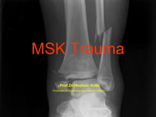
MSK Trauma
.Prof.Dr.Hesham Kotb
.Professor of Radiodiagnosis, Alex. University
Osseous Trauma
1) Acute Osseous Trauma:
1- Complete Fractures.
2- Incomplete Fractures
3- Radiologically Occult Fractures:
a) Bone contusions
b) Avulsion Fractures.

2) Chronic Osseous Trauma:
a) Insufficiency Fractures.
b) Fatigue Fractures.
Radiologically Occult Fractures
Imaging Modalities:
1)
2)

3)
•
•

Conventional Radiography: Mostly negative in many acute
and chronic osseous injuries ( Occult injuries)‫ز‬
Radioisotope Scanning :
It has Limitations
- Can be falsely negative foe 24-72 hours after injury.
- Positive scan is non-specific.
- Examination requires 4-6 hours , so delay diagnosis.
MRI: is highly sensitive. A normal MRI excludes the presence
of an osseous injury.
Linear low signal T1 with low signal edema.
High signal (edema) T2 with linear low signal (fracture line)
Acute Osseous Trauma
A) Impaction Injuries:
1- Contusion: bone contusion or bruises.
STIR or fat Sat T2 == focal areas of
increased signal- easily missed on non-fat
Sat spin echo T2 as edema and
surrounding fat display similar intensities.
• Sites: ACL tear –Patellar Dislocation
Acute Osseous Trauma
• 2- Avulsion Injuries: Occur when excessive
tensile forces result in a piece of bone or cartilage being
pulled away from the host bone by ligament , tendon or
capsular structures.

• Common sites:
• Knee , femur , humerus , elbow ,Ankle & Foot.
:Chronic Osseous Trauma
• Fatigue fractures –abnormal stress
across normal bone.
• Insufficiency fractures—normal stress
across abnormal bone.
- Femoral neck
- Sacrum
- Supraacetabular
- Pubic bones, superior and inferior
pubic rami
Recognizing Fractures
((And describing them
 A disruption in all
or part of the
cortex of a bone.
• All = Complete.
• Part =
Incomplete.
Incomplete Fractures
 GreenstickFacture through one
cortex

 Buckled – Fracture
with buckling of the
cortex.
Description of Fractures







By direction of fractures line.
By the relationship of the fragments.
By the number of the fragments.
By communication with the atmosphere.
Age of fracture (recent-healing- healed (
Type of union ( normally alignedmalaligned(.
Description of Fractures
 By direction of fractures line.
= Transverse.
= Oblique.
= Longitudinal.
= Spiral
Description of Fractures
 By the relationship of the fragments.
- Displacement.
- Shortening.
- Angulation.
- Rotation.
 Most fractures display more than one of
these abnormalities of position.
Description of Fractures
 Number of fragments:
1- Simple: Two fragments.
2- Comminuted : More than two
fragments.
Description of Fractures
 Open Or closed
fracture.
 Gas lucency at the
soft tissue.
??Diagnosis Please
Common Fracture Eponyms
-Colle's Fracture.

-Smith's Fracture.
-Jones Fracture.
-Boxer's Fracture.
Common Fracture Eponyms
• Colle's Fracture.
Fracture of the
distal radius with a
dorsal angulation
Common Fracture Eponyms
• Smith's Fracture.
- Fracture of the
distal radius with a
palmar angulation.
- Fall on a flexed
hand.
Common Fracture Eponyms
• Jones Fracture.
Fracture base of the
fifth metatarsal
Common Fracture Eponyms
 Boxer's Fracture.
Fracture head of the
fifth metacarpal with
palmar angulation.
Fracture Healing
1. Indistinctness of Fracture line.
2. Bony callus formation.
3. Bridging of fracture and obliteration of
fracture line.
4. Remodeling of bone.
Fracture Healing
Msk trauma
Msk trauma

Contenu connexe

Tendances

Imaging of musculo skeletal system
Imaging of musculo skeletal systemImaging of musculo skeletal system
Imaging of musculo skeletal system
orthoprince
 
Dr.salah.radiology.radiological approach to bone diseases
Dr.salah.radiology.radiological approach to bone diseasesDr.salah.radiology.radiological approach to bone diseases
Dr.salah.radiology.radiological approach to bone diseases
abas_lb
 
Foundations of Diagnostic Imaging for Physical Therapist
Foundations of Diagnostic Imaging for Physical TherapistFoundations of Diagnostic Imaging for Physical Therapist
Foundations of Diagnostic Imaging for Physical Therapist
Dana Tew
 
Diagnostic imaging in acl tear
Diagnostic imaging in acl tearDiagnostic imaging in acl tear
Diagnostic imaging in acl tear
Julie Price
 
Introduction to musculoskeletal radiology
Introduction to musculoskeletal radiologyIntroduction to musculoskeletal radiology
Introduction to musculoskeletal radiology
Subhanjan Das
 
radiology.Spine 1.(dr.ameer)
radiology.Spine 1.(dr.ameer)radiology.Spine 1.(dr.ameer)
radiology.Spine 1.(dr.ameer)
student
 

Tendances (20)

Bone lesion approach
Bone lesion approachBone lesion approach
Bone lesion approach
 
PATHOLOGICAL INVESTIGATIONS AND IMAGING TECHNIQUES IN NEUROMUSCULAR DISORDERS...
PATHOLOGICAL INVESTIGATIONS AND IMAGING TECHNIQUES IN NEUROMUSCULAR DISORDERS...PATHOLOGICAL INVESTIGATIONS AND IMAGING TECHNIQUES IN NEUROMUSCULAR DISORDERS...
PATHOLOGICAL INVESTIGATIONS AND IMAGING TECHNIQUES IN NEUROMUSCULAR DISORDERS...
 
Imaging of musculo skeletal system
Imaging of musculo skeletal systemImaging of musculo skeletal system
Imaging of musculo skeletal system
 
Dr.salah.radiology.radiological approach to bone diseases
Dr.salah.radiology.radiological approach to bone diseasesDr.salah.radiology.radiological approach to bone diseases
Dr.salah.radiology.radiological approach to bone diseases
 
MRI IN FOOT PAIN
MRI IN FOOT PAINMRI IN FOOT PAIN
MRI IN FOOT PAIN
 
Foundations of Diagnostic Imaging for Physical Therapist
Foundations of Diagnostic Imaging for Physical TherapistFoundations of Diagnostic Imaging for Physical Therapist
Foundations of Diagnostic Imaging for Physical Therapist
 
Spine mri bir 2 copy
Spine mri bir 2 copySpine mri bir 2 copy
Spine mri bir 2 copy
 
MRI kolano cz.1
MRI kolano cz.1MRI kolano cz.1
MRI kolano cz.1
 
Disorder of the_bones_and_joints
Disorder of the_bones_and_jointsDisorder of the_bones_and_joints
Disorder of the_bones_and_joints
 
Bone scan
Bone scanBone scan
Bone scan
 
Orthopaedics - Bone tumor (compiled cases)
Orthopaedics - Bone tumor (compiled cases)Orthopaedics - Bone tumor (compiled cases)
Orthopaedics - Bone tumor (compiled cases)
 
Bone tumor
Bone tumorBone tumor
Bone tumor
 
Diagnostic imaging of bones and joints (1)
Diagnostic imaging of bones and joints (1)Diagnostic imaging of bones and joints (1)
Diagnostic imaging of bones and joints (1)
 
Ct&Mri
Ct&MriCt&Mri
Ct&Mri
 
Diagnostic imaging in acl tear
Diagnostic imaging in acl tearDiagnostic imaging in acl tear
Diagnostic imaging in acl tear
 
Limb salvage: futile or auspicious
Limb salvage: futile or auspiciousLimb salvage: futile or auspicious
Limb salvage: futile or auspicious
 
Introduction to musculoskeletal radiology
Introduction to musculoskeletal radiologyIntroduction to musculoskeletal radiology
Introduction to musculoskeletal radiology
 
radiology.Spine 1.(dr.ameer)
radiology.Spine 1.(dr.ameer)radiology.Spine 1.(dr.ameer)
radiology.Spine 1.(dr.ameer)
 
Bone tumours
Bone tumoursBone tumours
Bone tumours
 
Pathological fractures
Pathological fracturesPathological fractures
Pathological fractures
 

En vedette

Msk Lecture3 1st Hospital
Msk Lecture3 1st HospitalMsk Lecture3 1st Hospital
Msk Lecture3 1st Hospital
Sumit Prajapati
 
Dr. Suzuki, Dr. Aronowitz, MD
Dr. Suzuki, Dr. Aronowitz, MDDr. Suzuki, Dr. Aronowitz, MD
Dr. Suzuki, Dr. Aronowitz, MD
SDG
 
Nuclear medicine in nerphology
Nuclear medicine in nerphologyNuclear medicine in nerphology
Nuclear medicine in nerphology
Lokender Yadav
 
Skeletal anatomy part 1
Skeletal anatomy part 1Skeletal anatomy part 1
Skeletal anatomy part 1
MissReith
 
Musculoskeletal Imaging 09
Musculoskeletal Imaging 09Musculoskeletal Imaging 09
Musculoskeletal Imaging 09
Steven Sager
 
Presentation1.pptx, radiological anatomy of the upper limb joint.
Presentation1.pptx, radiological anatomy of the upper limb joint.Presentation1.pptx, radiological anatomy of the upper limb joint.
Presentation1.pptx, radiological anatomy of the upper limb joint.
Abdellah Nazeer
 
Kienbock’s disease
Kienbock’s diseaseKienbock’s disease
Kienbock’s disease
orthoprince
 
Presentation1.pptx. ultrasound examination of the ankle joint.
Presentation1.pptx. ultrasound examination of the ankle joint.Presentation1.pptx. ultrasound examination of the ankle joint.
Presentation1.pptx. ultrasound examination of the ankle joint.
Abdellah Nazeer
 

En vedette (20)

Msk 2010 B
Msk 2010 BMsk 2010 B
Msk 2010 B
 
Radiology in Saudi Arabia
Radiology in Saudi ArabiaRadiology in Saudi Arabia
Radiology in Saudi Arabia
 
Radiographic anatomy of upper limb
Radiographic anatomy of upper limbRadiographic anatomy of upper limb
Radiographic anatomy of upper limb
 
Msk Lecture3 1st Hospital
Msk Lecture3 1st HospitalMsk Lecture3 1st Hospital
Msk Lecture3 1st Hospital
 
Dr. Suzuki, Dr. Aronowitz, MD
Dr. Suzuki, Dr. Aronowitz, MDDr. Suzuki, Dr. Aronowitz, MD
Dr. Suzuki, Dr. Aronowitz, MD
 
Nuclear medicine in nerphology
Nuclear medicine in nerphologyNuclear medicine in nerphology
Nuclear medicine in nerphology
 
Skull inside and some separate bones
Skull   inside and some separate bonesSkull   inside and some separate bones
Skull inside and some separate bones
 
Facilitated MSK US examination
Facilitated MSK US examination Facilitated MSK US examination
Facilitated MSK US examination
 
Post-graduate Certifcate Musculoskeletal Ultrasound - The Shoulder
Post-graduate Certifcate Musculoskeletal Ultrasound - The ShoulderPost-graduate Certifcate Musculoskeletal Ultrasound - The Shoulder
Post-graduate Certifcate Musculoskeletal Ultrasound - The Shoulder
 
Nmt631 msk lower extremity pathology
Nmt631 msk lower extremity pathologyNmt631 msk lower extremity pathology
Nmt631 msk lower extremity pathology
 
Skeletal anatomy part 1
Skeletal anatomy part 1Skeletal anatomy part 1
Skeletal anatomy part 1
 
Musculoskeletal Imaging 09
Musculoskeletal Imaging 09Musculoskeletal Imaging 09
Musculoskeletal Imaging 09
 
Presentation1.pptx, radiological anatomy of the upper limb joint.
Presentation1.pptx, radiological anatomy of the upper limb joint.Presentation1.pptx, radiological anatomy of the upper limb joint.
Presentation1.pptx, radiological anatomy of the upper limb joint.
 
Ultrasound Guided Injections - Upper Extremity
Ultrasound Guided Injections - Upper ExtremityUltrasound Guided Injections - Upper Extremity
Ultrasound Guided Injections - Upper Extremity
 
20140913 basic musculoskeletal ultrasound abnormalities kailen tsai更正版
20140913 basic musculoskeletal ultrasound abnormalities kailen tsai更正版20140913 basic musculoskeletal ultrasound abnormalities kailen tsai更正版
20140913 basic musculoskeletal ultrasound abnormalities kailen tsai更正版
 
MRI slides, second yr students 2008
MRI slides, second yr students 2008MRI slides, second yr students 2008
MRI slides, second yr students 2008
 
USG in gall bladder pathologies
USG in gall bladder pathologiesUSG in gall bladder pathologies
USG in gall bladder pathologies
 
Kienbock’s disease
Kienbock’s diseaseKienbock’s disease
Kienbock’s disease
 
Musculoskeletal radiology mocks 2016
Musculoskeletal radiology mocks 2016Musculoskeletal radiology mocks 2016
Musculoskeletal radiology mocks 2016
 
Presentation1.pptx. ultrasound examination of the ankle joint.
Presentation1.pptx. ultrasound examination of the ankle joint.Presentation1.pptx. ultrasound examination of the ankle joint.
Presentation1.pptx. ultrasound examination of the ankle joint.
 

Similaire à Msk trauma

9- BASICS OF ORTHOPEDIC RADIOLOGY.ppt
9- BASICS OF ORTHOPEDIC RADIOLOGY.ppt9- BASICS OF ORTHOPEDIC RADIOLOGY.ppt
9- BASICS OF ORTHOPEDIC RADIOLOGY.ppt
Asgraf
 

Similaire à Msk trauma (20)

clinics.pptx
clinics.pptxclinics.pptx
clinics.pptx
 
Traumatic injuries of the face and jaws
Traumatic injuries of the face and jawsTraumatic injuries of the face and jaws
Traumatic injuries of the face and jaws
 
Special types of trauma
Special types of traumaSpecial types of trauma
Special types of trauma
 
Pelvic fractures.pptx
Pelvic fractures.pptxPelvic fractures.pptx
Pelvic fractures.pptx
 
9- BASICS OF ORTHOPEDIC RADIOLOGY.ppt
9- BASICS OF ORTHOPEDIC RADIOLOGY.ppt9- BASICS OF ORTHOPEDIC RADIOLOGY.ppt
9- BASICS OF ORTHOPEDIC RADIOLOGY.ppt
 
SIêu âm xương sườn, sụn sườn, Bs Tài
SIêu âm xương sườn, sụn sườn, Bs TàiSIêu âm xương sườn, sụn sườn, Bs Tài
SIêu âm xương sườn, sụn sườn, Bs Tài
 
Cervical spine trauma
Cervical spine traumaCervical spine trauma
Cervical spine trauma
 
shanmugham karthick raja ppt.pptx
shanmugham karthick raja ppt.pptxshanmugham karthick raja ppt.pptx
shanmugham karthick raja ppt.pptx
 
basics of orthopedic radiology.ppt
basics of orthopedic radiology.pptbasics of orthopedic radiology.ppt
basics of orthopedic radiology.ppt
 
fracture and dislocation.ppt
fracture and dislocation.pptfracture and dislocation.ppt
fracture and dislocation.ppt
 
Musculoskeletal trauma - dr. Hendy .ppt
Musculoskeletal trauma - dr. Hendy .pptMusculoskeletal trauma - dr. Hendy .ppt
Musculoskeletal trauma - dr. Hendy .ppt
 
Principles of fractures on orthopedic field
Principles of fractures on orthopedic fieldPrinciples of fractures on orthopedic field
Principles of fractures on orthopedic field
 
SKELET RAD.ppt
SKELET RAD.pptSKELET RAD.ppt
SKELET RAD.ppt
 
Musculoskeletal Radiology
Musculoskeletal RadiologyMusculoskeletal Radiology
Musculoskeletal Radiology
 
BONE FRACTURES.pptx
BONE FRACTURES.pptxBONE FRACTURES.pptx
BONE FRACTURES.pptx
 
BONE FRACTURES.pptx
BONE FRACTURES.pptxBONE FRACTURES.pptx
BONE FRACTURES.pptx
 
imaging in skeletal trauma.ppt
imaging in skeletal trauma.pptimaging in skeletal trauma.ppt
imaging in skeletal trauma.ppt
 
Imaging In Orthopaedics.pptx
Imaging In Orthopaedics.pptxImaging In Orthopaedics.pptx
Imaging In Orthopaedics.pptx
 
X-ray spine
X-ray spineX-ray spine
X-ray spine
 
Bone forming tumors rabins
Bone forming tumors rabinsBone forming tumors rabins
Bone forming tumors rabins
 

Plus de Amr Mansour Hassan

Plus de Amr Mansour Hassan (20)

Thoracolumbar fractures classification
Thoracolumbar fractures classificationThoracolumbar fractures classification
Thoracolumbar fractures classification
 
Scoliosis
ScoliosisScoliosis
Scoliosis
 
Idiopathic scoliosis
Idiopathic scoliosisIdiopathic scoliosis
Idiopathic scoliosis
 
Idiopathic scoliosis
Idiopathic scoliosisIdiopathic scoliosis
Idiopathic scoliosis
 
Chiari pelvic osteotomy
Chiari pelvic osteotomyChiari pelvic osteotomy
Chiari pelvic osteotomy
 
Acetabuloplasty indications
Acetabuloplasty   indicationsAcetabuloplasty   indications
Acetabuloplasty indications
 
Salter osteotomy workshop
Salter osteotomy workshopSalter osteotomy workshop
Salter osteotomy workshop
 
Pelvic osteotomy in pediatric
Pelvic osteotomy in pediatricPelvic osteotomy in pediatric
Pelvic osteotomy in pediatric
 
Dega osteotomy, workshop
Dega osteotomy, workshopDega osteotomy, workshop
Dega osteotomy, workshop
 
Pediatric foot deformities
Pediatric foot deformitiesPediatric foot deformities
Pediatric foot deformities
 
Introduction to pediatric foot
Introduction to pediatric footIntroduction to pediatric foot
Introduction to pediatric foot
 
Closed vs. open reduction in lateral condylar fractures of humerus in childern
Closed vs. open reduction in lateral condylar fractures of humerus in childernClosed vs. open reduction in lateral condylar fractures of humerus in childern
Closed vs. open reduction in lateral condylar fractures of humerus in childern
 
Brachial plexus birth palsy
Brachial plexus birth palsy  Brachial plexus birth palsy
Brachial plexus birth palsy
 
Appraoch to child with hip pain
Appraoch to child with hip painAppraoch to child with hip pain
Appraoch to child with hip pain
 
The role of arthography guided closed reductionin reducing the incidence of a...
The role of arthography guided closed reductionin reducing the incidence of a...The role of arthography guided closed reductionin reducing the incidence of a...
The role of arthography guided closed reductionin reducing the incidence of a...
 
Slipped capital femoral epiphysis
Slipped capital femoral epiphysisSlipped capital femoral epiphysis
Slipped capital femoral epiphysis
 
Skeletal dysplasia grand round
Skeletal dysplasia   grand round Skeletal dysplasia   grand round
Skeletal dysplasia grand round
 
Principles of cerebral palsy
Principles of cerebral palsyPrinciples of cerebral palsy
Principles of cerebral palsy
 
Physiological & pathological tibia vara
Physiological & pathological tibia varaPhysiological & pathological tibia vara
Physiological & pathological tibia vara
 
Pelvis osteotomies in ddh patients
Pelvis osteotomies in ddh patientsPelvis osteotomies in ddh patients
Pelvis osteotomies in ddh patients
 

Dernier

Circulation through Special Regions -characteristics and regulation
Circulation through Special Regions -characteristics and regulationCirculation through Special Regions -characteristics and regulation
Circulation through Special Regions -characteristics and regulation
MedicoseAcademics
 
Cardiac Impulse: Rhythmical Excitation and Conduction in the Heart
Cardiac Impulse: Rhythmical Excitation and Conduction in the HeartCardiac Impulse: Rhythmical Excitation and Conduction in the Heart
Cardiac Impulse: Rhythmical Excitation and Conduction in the Heart
MedicoseAcademics
 

Dernier (20)

Video capsule endoscopy (VCE ) in children
Video capsule endoscopy (VCE ) in childrenVideo capsule endoscopy (VCE ) in children
Video capsule endoscopy (VCE ) in children
 
Creating Accessible Public Health Communications
Creating Accessible Public Health CommunicationsCreating Accessible Public Health Communications
Creating Accessible Public Health Communications
 
The Orbit & its contents by Dr. Rabia I. Gandapore.pptx
The Orbit & its contents by Dr. Rabia I. Gandapore.pptxThe Orbit & its contents by Dr. Rabia I. Gandapore.pptx
The Orbit & its contents by Dr. Rabia I. Gandapore.pptx
 
Effects of vaping e-cigarettes on arterial health
Effects of vaping e-cigarettes on arterial healthEffects of vaping e-cigarettes on arterial health
Effects of vaping e-cigarettes on arterial health
 
Anuman- An inference for helpful in diagnosis and treatment
Anuman- An inference for helpful in diagnosis and treatmentAnuman- An inference for helpful in diagnosis and treatment
Anuman- An inference for helpful in diagnosis and treatment
 
Introducing VarSeq Dx as a Medical Device in the European Union
Introducing VarSeq Dx as a Medical Device in the European UnionIntroducing VarSeq Dx as a Medical Device in the European Union
Introducing VarSeq Dx as a Medical Device in the European Union
 
Scientificity and feasibility study of non-invasive central arterial pressure...
Scientificity and feasibility study of non-invasive central arterial pressure...Scientificity and feasibility study of non-invasive central arterial pressure...
Scientificity and feasibility study of non-invasive central arterial pressure...
 
Retinal consideration in cataract surgery
Retinal consideration in cataract surgeryRetinal consideration in cataract surgery
Retinal consideration in cataract surgery
 
180-hour Power Capsules For Men In Ghana
180-hour Power Capsules For Men In Ghana180-hour Power Capsules For Men In Ghana
180-hour Power Capsules For Men In Ghana
 
Relationship between vascular system disfunction, neurofluid flow and Alzheim...
Relationship between vascular system disfunction, neurofluid flow and Alzheim...Relationship between vascular system disfunction, neurofluid flow and Alzheim...
Relationship between vascular system disfunction, neurofluid flow and Alzheim...
 
Muscle Energy Technique (MET) with variant and techniques.
Muscle Energy Technique (MET) with variant and techniques.Muscle Energy Technique (MET) with variant and techniques.
Muscle Energy Technique (MET) with variant and techniques.
 
Circulation through Special Regions -characteristics and regulation
Circulation through Special Regions -characteristics and regulationCirculation through Special Regions -characteristics and regulation
Circulation through Special Regions -characteristics and regulation
 
Scleroderma: Treatment Options and a Look to the Future - Dr. Macklin
Scleroderma: Treatment Options and a Look to the Future - Dr. MacklinScleroderma: Treatment Options and a Look to the Future - Dr. Macklin
Scleroderma: Treatment Options and a Look to the Future - Dr. Macklin
 
Compare home pulse pressure components collected directly from home
Compare home pulse pressure components collected directly from homeCompare home pulse pressure components collected directly from home
Compare home pulse pressure components collected directly from home
 
Integrated Neuromuscular Inhibition Technique (INIT)
Integrated Neuromuscular Inhibition Technique (INIT)Integrated Neuromuscular Inhibition Technique (INIT)
Integrated Neuromuscular Inhibition Technique (INIT)
 
Cardiac Impulse: Rhythmical Excitation and Conduction in the Heart
Cardiac Impulse: Rhythmical Excitation and Conduction in the HeartCardiac Impulse: Rhythmical Excitation and Conduction in the Heart
Cardiac Impulse: Rhythmical Excitation and Conduction in the Heart
 
End Feel -joint end feel - Normal and Abnormal end feel
End Feel -joint end feel - Normal and Abnormal end feelEnd Feel -joint end feel - Normal and Abnormal end feel
End Feel -joint end feel - Normal and Abnormal end feel
 
รายการตํารับยาแผนไทยแห่งชาติ ฉบับ พ.ศ. 2564.pdf
รายการตํารับยาแผนไทยแห่งชาติ ฉบับ พ.ศ. 2564.pdfรายการตํารับยาแผนไทยแห่งชาติ ฉบับ พ.ศ. 2564.pdf
รายการตํารับยาแผนไทยแห่งชาติ ฉบับ พ.ศ. 2564.pdf
 
Hemodialysis: Chapter 2, Extracorporeal Blood Circuit - Dr.Gawad
Hemodialysis: Chapter 2, Extracorporeal Blood Circuit - Dr.GawadHemodialysis: Chapter 2, Extracorporeal Blood Circuit - Dr.Gawad
Hemodialysis: Chapter 2, Extracorporeal Blood Circuit - Dr.Gawad
 
Factors Affecting child behavior in Pediatric Dentistry
Factors Affecting child behavior in Pediatric DentistryFactors Affecting child behavior in Pediatric Dentistry
Factors Affecting child behavior in Pediatric Dentistry
 

Msk trauma

  • 1. MSK Trauma .Prof.Dr.Hesham Kotb .Professor of Radiodiagnosis, Alex. University
  • 2. Osseous Trauma 1) Acute Osseous Trauma: 1- Complete Fractures. 2- Incomplete Fractures 3- Radiologically Occult Fractures: a) Bone contusions b) Avulsion Fractures. 2) Chronic Osseous Trauma: a) Insufficiency Fractures. b) Fatigue Fractures.
  • 3. Radiologically Occult Fractures Imaging Modalities: 1) 2) 3) • • Conventional Radiography: Mostly negative in many acute and chronic osseous injuries ( Occult injuries)‫ز‬ Radioisotope Scanning : It has Limitations - Can be falsely negative foe 24-72 hours after injury. - Positive scan is non-specific. - Examination requires 4-6 hours , so delay diagnosis. MRI: is highly sensitive. A normal MRI excludes the presence of an osseous injury. Linear low signal T1 with low signal edema. High signal (edema) T2 with linear low signal (fracture line)
  • 4. Acute Osseous Trauma A) Impaction Injuries: 1- Contusion: bone contusion or bruises. STIR or fat Sat T2 == focal areas of increased signal- easily missed on non-fat Sat spin echo T2 as edema and surrounding fat display similar intensities. • Sites: ACL tear –Patellar Dislocation
  • 5. Acute Osseous Trauma • 2- Avulsion Injuries: Occur when excessive tensile forces result in a piece of bone or cartilage being pulled away from the host bone by ligament , tendon or capsular structures. • Common sites: • Knee , femur , humerus , elbow ,Ankle & Foot.
  • 6. :Chronic Osseous Trauma • Fatigue fractures –abnormal stress across normal bone. • Insufficiency fractures—normal stress across abnormal bone. - Femoral neck - Sacrum - Supraacetabular - Pubic bones, superior and inferior pubic rami
  • 7.
  • 8.
  • 9.
  • 10. Recognizing Fractures ((And describing them  A disruption in all or part of the cortex of a bone. • All = Complete. • Part = Incomplete.
  • 11. Incomplete Fractures  GreenstickFacture through one cortex  Buckled – Fracture with buckling of the cortex.
  • 12. Description of Fractures       By direction of fractures line. By the relationship of the fragments. By the number of the fragments. By communication with the atmosphere. Age of fracture (recent-healing- healed ( Type of union ( normally alignedmalaligned(.
  • 13. Description of Fractures  By direction of fractures line. = Transverse. = Oblique. = Longitudinal. = Spiral
  • 14.
  • 15.
  • 16.
  • 17. Description of Fractures  By the relationship of the fragments. - Displacement. - Shortening. - Angulation. - Rotation.  Most fractures display more than one of these abnormalities of position.
  • 18.
  • 19.
  • 20. Description of Fractures  Number of fragments: 1- Simple: Two fragments. 2- Comminuted : More than two fragments.
  • 21.
  • 22.
  • 23. Description of Fractures  Open Or closed fracture.  Gas lucency at the soft tissue.
  • 24.
  • 26. Common Fracture Eponyms -Colle's Fracture. -Smith's Fracture. -Jones Fracture. -Boxer's Fracture.
  • 27. Common Fracture Eponyms • Colle's Fracture. Fracture of the distal radius with a dorsal angulation
  • 28. Common Fracture Eponyms • Smith's Fracture. - Fracture of the distal radius with a palmar angulation. - Fall on a flexed hand.
  • 29. Common Fracture Eponyms • Jones Fracture. Fracture base of the fifth metatarsal
  • 30. Common Fracture Eponyms  Boxer's Fracture. Fracture head of the fifth metacarpal with palmar angulation.
  • 31. Fracture Healing 1. Indistinctness of Fracture line. 2. Bony callus formation. 3. Bridging of fracture and obliteration of fracture line. 4. Remodeling of bone.