Contenu connexe
Similaire à cgt201624a (20)
cgt201624a
- 1. ORIGINAL ARTICLE
Physalis alkekengi and Alhagi maurorum ameliorate the side
effect of cisplatin-induced nephrotoxicity
S Changizi-Ashtiyani1
, M Alizadeh2
, H Najafi3
, S Babaei4
, M Khazaei2
, M Jafari2
, N Hossaini5
, A Avan6
and B Bastani7
Cisplatin is frequently being used for the treatment of different tumors, although the application of this agent is associated with
nephrotoxicity. Here, we explored the antioxidant and anti-inflammatory activities of Physalis alkekengi and Alhagi maurorum;
400 mg kg− 1
per day P. alkekengi and 100 mg kg− 1
per day A. maurorum were administered in rats, orally for 10 days after a single
dose of 7 mg kg− 1
intraperitoneal cisplatin. The concentrations of creatinine, urea-nitrogen, and relative and absolute excretion of
sodium/potassium were evaluated before/after therapy. Levels of malondialdehyde (MDA) and ferric-reducing antioxidant power
(FRAP) were measured to assess the oxidative stress induced by cisplatin. Moreover, tissues sections were used for histological
analyses and evaluation of the degree of tissue damage. Cisplatin increased serum levels of creatinine and urea-nitrogen, relative/
absolute excretion of sodium/potassium, and MDA, whereas decreased FRAP level. Interestingly, P. alkekengi or A. maurorum were
able to reduce the level of the renal function markers as well as the levels of sodium/potassium. This effect was more pronounced
by P. alkekengi. Moreover, cisplatin induced pathological damage in kidney, whereas treatment with these agents improved this
condition. Our findings demonstrate the potential therapeutic impact of P. alkekengi and A. maurorum for improving cisplatin-
induced nephrotoxicity, supporting further investigations on the novel potential clinical application of these agents for patients
being treated with cisplatin to ameliorate cisplatin-induced nephrotoxicity.
Cancer Gene Therapy advance online publication, 3 June 2016; doi:10.1038/cgt.2016.24
INTRODUCTION
Cisplatin is an anti-neoplastic drug that is being used in the
treatment of different tumors.1–2
Although the increased dose
could result in a remarkable increase in the therapeutic effect of
cisplatin, it is associated with nephrotoxicity.3–5
It has been
reported that approximately 20–30% of patients receiving
cisplatin have the signs of nephrotoxicity.6,7
Several studies have
been performed on the molecular mechanisms behind cisplatin-
induced nephrotoxicity; indicating the key role of inflammation
and oxidative stress in this condition.6,8–10
There is a growing body of evidence showing antioxidant and anti-
inflammatory activities of Physalis alkekengi in a variety of human
diseases. It has been suggested that alkaloids, glucocorticoids, lycopene
and vitamin C are among its active ingredients.11
Moreover, several
studies have been shown its antioxidant12
and anti-inflammatory
properties.13–15
Another study suggested the antioxidant activity of
Alhagi maurorum, which is enriched in flavonoids.16
Increasing evidence
has shown the antioxidant activity of A. maurorum.17–20
Therefore, in
the present study, we explored the anti-inflammatory and antioxidants
effects of oral administration of P. alkekengi and A. maurorum extracts
on cisplatin-induced nephrotoxicity in an in vivo model.
MATERIALS AND METHODS
Animals
In this study, 28 male Sprague–Dawley rats (weighing 250–300 g) were
used and kept in the central animal house of Arak University of Medical
Sciences. The animals were housed under standard laboratory conditions
and 12 h light/dark cycles at the temperature of 23 ± 2 °C with free access
to food and water throughout the experiment. All tests and procedures
were conducted according to the internationally accepted guidelines for
the care and use of laboratory animals. The animal experiment was
approved by the Ethics Committee at Arak University of Medical Sciences
(AUMS) and performed according to a protocol approved by the AUMS,
Iran and the Declaration of Helsinki.
Extraction of P. alkekengi and A. maurorum
P. alkekengi and A. maurorum were purchased from Arak University center
and were confirmed by a botanist. P. alkekengi (#771230) and A. maurorum
(#78549) was deposited in the herbarium of agriculture and natural
resources research center of Arak, Iran. The aerial parts of the plants were
dried in shade and used for extraction by maceration. Five hundred grams
of dried plant powder was dissolved in 70% ethyl alcohol and was kept for
72 h at room temperature. The mixture was centrifuged and the solution
was carefully isolated. This procedure was repeated three times and the
resulting solution was concentrated in a vacuum evaporator (model
R-1001-VN, Seoul, Korea) at 40 °C, and stored until use at − 20 °C.
Treatment of animal
The animals were randomly divided into four groups (n = 7, in each group).
The sham group received a single intraperitoneal normal saline injection
(1 ml), followed by daily normal saline gavages for 10 consecutive days.
The second group received a single dose of cisplatin (intraperitoneal; 7 mg
kg− 1
), followed by 10 daily oral gavages of normal saline. The third group
received a single dose of cisplatin (intraperitoneal; 7 mg kg− 1
), followed by
treatment with P. alkekengi (400 mg kg− 1
per day) orally for 10 days. The
1
Department of Physiology, Arak University of Medical Sciences, Arak, Iran; 2
Student Research Committee, Arak University of Medical Sciences, Arak, Iran; 3
Medical Biology
Research Center, Kermanshah University of Medical Sciences, Kermanshah, Iran; 4
Department of Histology, Arak University of Medical Sciences, Arak, Iran; 5
Department of
Medicinal Plants, University of Arak, Arak, Iran; 6
Molecular Medicine Group, Department of Modern Sciences and Technologies, School of Medicine, Mashhad University
of Medical Sciences, Mashhad, Iran and 7
Division of Nephrology, School of Medicine, Saint Louis University, Saint Louis, MO, USA. Correspondence: Dr B Bastani, Division of
Nephrology, School of Medicine, Saint Louis University, 3635 Vista Avenue, Saint Louis, MO 63110, USA.
E-mail: bastanib@slu.edu
Received 2 April 2016; accepted 9 May 2016
Cancer Gene Therapy (2016), 1–6
© 2016 Nature America, Inc. All rights reserved 0929-1903/16
www.nature.com/cgt
- 2. fourth group received the same protocol as the third group, but the extract
was A. maurorum at a dose of 100 mg kg− 1
per day. At the end of the
10-day period, rats were kept in metabolic cages for 6 h. Urine and blood
samples were collected from all the animals. Right kidneys were removed
and fixed in 10% formaldehyde to be stained with hematoxylin and eosin
for histological study. The left kidneys were frozen in liquid nitrogen for the
assessment of oxidative stress.
Biochemical analyses
The concentrations of plasma creatinine and urea-nitrogen were measured
using an autoanalyzer (Technicon, RA-1000, Bayer, Tarrytown, NY, USA).
The concentrations of sodium and potassium were evaluated in plasma
and urine samples. Creatinine clearance and the relative and absolute
excretion of sodium and potassium were calculated. For the assessment of
oxidative stress, malondialdehyde (MDA) and ferric-reducing antioxidant
power values were measured in kidney tissue using the methods by
Ohkawa et al.21
and Benzie and Strain,22
as described in our previous
studies.23–24
Histological analysis
The degree of tissue damage was assessed by hematoxylin and eosin-
stained tissue sections. In particular, renal histopathologic damages in
at least 10 microscopic fields (magnification × 400) were quantified
for evaluation of Bowman space, red blood cells in glomerular capillaries,
tubular cell necrosis and their exfoliation into the tubular lumen,
intracellular vacuolization, vascular congestion and proteinaceous casts.
The Bowman space widening and reduced number of red blood cells in
glomerular capillaries in rats showed the highest rate of changes,
compared with the control group. Other changes such as cellular necrosis
and exfoliation, intracellular vacuolization, vascular congestion and intra-
tubular proteinaceous casts were calculated as a percentage of the total
area. The degree of histological damages was scored as zero for no
damage, 1 for 1–20% damage, 2 for 21–40%, 3 for 41–60%, 4 for 61–80%
and 5 for 81–100%. The total histopathological score was calculated, which
was equal to all scores of different damages in each group.23–26
Statistical analysis
Data were analyzed by using SPSS-16 software and expressed as mean ±
s.e.m. To compare the functional parameters as well as the data related to
renal oxidative stress, one-way analysis of variance and Duncan's post hoc
test were used; and the LSD test was used. Non-parametric Kruskal–Wallis
and Mann–Whitney tests were carried out to compare histopathologic
damages. Po0.05 was considered as significant.
RESULTS
The effects of P. alkekengi and A. maurorum on cisplatin-induced
renal dysfunction
In the present study, we first sought to explore the effect of
P. alkekengi and A. maurorum on cisplatin-induced nephrotoxicity.
Thus, we evaluated the effect of these extracts in an in vivo model.
Our data showed that a single dose of cisplatin significantly
increased the plasma creatinine and urea-nitrogen concentrations,
compared with the sham group (Po0.05), whereas treatment
with P. alkekengi or A. maurorum significantly reduced their levels,
compared with the cisplatin group. Of note, the reduction in
serum creatinine was greater in the group receiving P. alkekengi
(Figures 1a and b). Moreover, the decreased creatinine clearance
(Po0.01) and increased absolute and relative excretion of sodium
and potassium caused by cisplatin were improved by the
application of both A. maurorum and P. alkekengi (Table 1).
0
0.5
1
1.5
2
2.5
3
Sham Cisplatin Cis+Physalis Cis+Alhagi
Experimental groups
PlasmaCreatinineConcentration
(mg/dl)
***
††
†††
0
5
10
15
20
25
30
35
40
45
50
Sham Cisplatin Cis+Physalis Cis+Alhagi
Experimental groups
PlasmaNitrogen-UreaConcentration
(mg/dl)
††
**
***
††
**
Figure 1. Effects of oral administration of P. alkekengi or A. maurorum
extracts on plasma creatinine (a) and urea-nitrogen (b) concentra-
tions in rats with cisplatin-induced nephrotoxicity. *Po0.05,
**Po0.01, ***Po0.001 in comparison with the sham group.
†
Po0.05, ††
Po0.01, †††
Po0.001 for comparison of Cisplatin group
with Cis+Physal or Cis+Alhagi group.
Table 1. The effects of oral administration of Physalis alkekengi or Alhagi maurorum on renal functional parameters induced by cisplatin
Functional parameters Experimental groups
Sham Cisplatin Cis+Physal Cis+Alhagi
CCr (μl min− 1
KgW) 1128.8 ± 74.4 375.5 ± 36.4*** 751.5 ± 61.6**†† 734.9 ± 56.5**††
UNaVº
(μmol min− 1
KgW) 4.6 ± 0.8 19.1 ± 2.9*** 6.0 ± 0.9††† 6.4 ± 0.9†††
FENa (%) 2.3 ± 0.3 12.5 ± 1.4** 2.8 ± 0.5†† 5.7 ± 1.6††
UKVº
(μmol min− 1
KgW) 1.8 ± 0.3 5.4 ± 1.7*** 1.9 ± 0.5††† 1.6 ± 0.3†††
FEK (%) 26.7 ± 2.6 48.3 ± 3.4** 28.2 ± 2.1††† 34.5 ± 2.5††
Abbreviation: KgW, Kilogram body weight. Values are represented as mean ± s.e. for creatinine clearance (CCr), absolute excretion of sodium (UNaVº
) and
potassium (UKVº
), and fractional excretion of sodium (FENa) and potassium (FEK) in rats receiving normal saline (Sham), Cisplatin, Cisplatin plus Physalis alkekengi
(Cis+Physal) or Cisplatin plus Alhagi maurorum (Cis+Alhagi) extract. *Po0.05, **Po0.01, ***Po0.001 in comparison with the sham group. †
Po0.05, ††
Po0.01,
†††
Po0.001 for comparison of Cisplatin group with Cis+Physal or Cis+Alhagi group.
Protection from cisplatin-induced nephrotoxicity
S Changizi-Ashtiyani et al
2
Cancer Gene Therapy (2016), 1 – 6 © 2016 Nature America, Inc.
- 3. The effect of P. alkekengi and A. maurorum extracts on oxidative
stress induced by cisplatin
We further evaluated the effect of P. alkekengi and A. maurorum
on the oxidative stress parameter by analyzing MDA and ferric-
reducing antioxidant power. As shown in Figures 2a and b, MDA
level in kidney tissue of the cisplatin group was significantly
increased, while administration of P. alkekengi and A. maurorum
extracts significantly (Po0.05) reduced the level of MDA.
Interestingly, MDA levels in P. alkekengi and A. maurorum groups
were still higher than that in the sham group. On the other hand,
cisplatin decreased the ferric-reducing antioxidant power level
that was partially improved by P. alkekengi and A. maurorum
(Figures 2a and b).
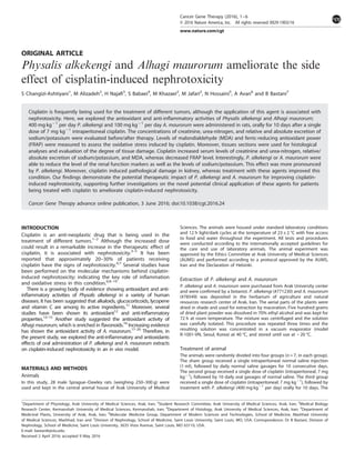
The effects of P. alkekengi and A. maurorum on cisplatin-induced
tissue damages
Tissue damage in rats treated with cisplatin was determined by
histological analyses. As shown in Table 2 and Figure 3, cisplatin
resulted in the enlargement of Bowman space, necrotic
epithelial cells in proximal tubule and thick ascending limb of
loop of Henle and their exfoliation into the lumen, reduced
number of red blood cells in the glomerular capillaries and
vacuolization of proximal tubules cells. In the outer medulla,
epithelial cells of pars recta and thick ascending limb of Henle's
loop showed cellular necrosis and exfoliation, increased vascular
congestion and intra-tubular proteinaceous casts (Table 2 and
Figure 4). In the inner medulla, the degree of vascular congestion
and intra-tubular proteinaceous casts was increased in compar-
ison with the sham group. In particular, histopathologic score in
the cisplatin group was 40, which was significantly greater than
sham group.
The administration of both P. alkekengi and A. maurorum
extracts reduced the severity of the damages; in the P. alkekengi
group, the total histopathologic score was reduced to 21.2. Also,
in the A. maurorum group, the total histopathologic score was
decreased compared with the cisplatin group. However, in both
groups, the total histopathologic scores were significantly higher
than the sham group (Table 2).
DISCUSSION
To the best of our knowledge, this is the first study showing the
effects of oral administration of P. alkekengi and A. maurorum
extracts and their mechanisms on cisplatin-induced nephrotoxi-
city in rats. We demonstrated that the administration of
cisplatin significantly decreased creatinine clearance, as an index
of glomerular filtration rate, and increased plasma creatinine and
urea-nitrogen concentrations. In agreement with our findings,
several studies have suggested that cisplatin causes afferent
vasoconstriction and altered ultra-filtration coefficient.27,28
In the
present study, the reduced number of red blood cells in
glomerular capillaries might be due to the afferent vasoconstric-
tion. In line with our data, Somani et al.29
and Aydogan et al.30
showed that the reduction in ultra-filtration coefficient by
cisplatin was related to the production of reactive oxygen species
and reduction of glomerular filtration surface area. Furthermore,
cisplatin increased tissue MDA and reduced ferric-reducing
antioxidant power levels, indicating increased oxidative stress
induced by this agent. The increased absolute excretion of
sodium and potassium in the cisplatin group indicated a marked
decrease in the renal tubular reabsorption capacity, which was
further confirmed by the increasing rate of fractional excretion
of these ions and tissue damages. Increasing evidence is
0
5
10
15
20
25
30
35
40
Sham Cisplatin Cis+Physalis Cis+Alhagi
Experimental grops
TissueMDA(nmol/gKW)
††
**
†
**
***
0
2
4
6
8
10
12
14
16
18
Sham Cisplatin Cis+Physalis Cis+Alhagi
Experimental groups
TissueFRAP(µmol/gKW)
***
†
***
††
**
Figure 2. Effects of oral administration of P. alkekengi or A. maurorum
extracts on tissue MDA (a) and ferric-reducing antioxidant power
(b) levels in rats with cisplatin-induced nephrotoxicity. *Po0.05,
**Po0.01, ***Po0.001 in comparison with the sham group.
†
Po0.05, ††
Po0.01 in comparison with the Cisplatin group.
Table 2. The effects of oral administration of Physalis alkekengi or
Alhagi maurorum on renal histopathologic scores induced by cisplatin
Experimental groups
Histopathology cortex Cis
+Alhagi
Cis
+Physalis
Cisplatin Sham
Bowman's space enlargement 2.2 2.4 5 0
Proximal tubule injury 2.2 2.6 3.2 0.4
Thick ascending limb injury 2.4 1.8 4.2 0.3
Reduced number of RBCs in
glomerular capillaries
2.3 2.4 5 0
Intracellular vacuolization 2.8 2.2 3.8 0.3
Outer medulla
Pars recta (S3) injury 3.2 1.8 3.4 0.5
Thick ascending limb injury 2.8 2 3.4 0
Vascular congestion 2.4 1.6 3.2 0.2
Intra-tubular proteinaceous casts 2.8 1.2 3.2 0.2
Inner medulla
Vascular congestion 2.6 1.8 2.8 0
Intra-tubular proteinaceous casts 2.8 1.4 2.8 0
Total histopathologic score 28.5**†† 21.2*†† 40.0** 1.9
Abbreviation: RBC, red blood cell. Histopathological scores in rats receiving
normal saline (sham), cisplatin, cisplatin plus Physalis alkekengi (Cis+Physal)
or cisplatin plus Alhagi maurorum (Cis+Alhagi) extract. *Po0.05, **Po0.01
in comparison with sham group. ††
Po0.01, for comparison of cisplatin
group with Cis+Physal or Cis+Alhagi group.
Protection from cisplatin-induced nephrotoxicity
S Changizi-Ashtiyani et al
3
© 2016 Nature America, Inc. Cancer Gene Therapy (2016), 1 – 6
- 4. suggesting the role of cisplatin in increasing oxidative stress
and decreasing the activity of antioxidant enzymes in
kidney,29,31–36
as well as the stimulation of calcium-independent
nitric oxide synthase,37–40
which are leading to increased
production of NO and proxy nitrite (ONOO − ). Our findings
revealed that P. alkekengi and A. maurorum were able improve this
condition under treatment by cisplatin.
In addition, it is known that cisplatin enters renal epithelial
cells via organic cation transporter-2(OCT2)41–43
and copper
transporter-1 (Ctr1),44
which causes mitochondrial and nuclear
DNA damage. In the present study, we observed cell necrosis in
the cortex and outer medulla of cisplatin-treated rats. However,
treatment of animals by A. maurorum and P. alkekengi
improved creatinine clearance, reduced plasma creatinine and
urea-nitrogen concentrations, and reduced the levels of oxidative
stress parameters. Also, tissue damage induced by cisplatin was
reduced by both extracts, although the beneficial effects were
more prominent in the group receiving P. alkekengi. Several
previous studies have shown the biological effects
of these agents. In particular, Hoshani and Aghdasi12
illustrated that P. alkekengi had an antioxidant property. More-
over, several other studies have shown that P. alkekengi
inhibited iNOS activity and reduced NO production. Anti-
inflammatory activity of this extract was reported by inhibition
of NF-κB and TNF-α and lipoxygenase-1activity.14,15
Moreover,
several studies have shown the antioxidant activity,16,45
anti-
inflammatory effect,18,19
urease inhibitory activity46
and litolitic
properties20
of A. maurorum.
CONCLUSION
In aggregate, the present study expands the spectrum of
the potential beneficial effects of A. maurorum and P. alkekengi
as supplement agents for reducing the nephrotoxicity side
effect of cisplatin. We found that both extracts reduced drug-
induced nephrotoxicity. The protective mechanism was in part
through reducing oxidative stress, inflammation and conversion of
reactive oxygen species to reactive nitrogen species.
Figure 3. Representing the histopathologic alterations in the cortex for Bowman's space widening and tubular necrosis in (a) sham group,
(b) cisplatin group that received normal saline, (c) P. alkekengi or (d) A. maurorum extracts. Haematoxylin and eosin staining,
magnification × 400.
Protection from cisplatin-induced nephrotoxicity
S Changizi-Ashtiyani et al
4
Cancer Gene Therapy (2016), 1 – 6 © 2016 Nature America, Inc.
- 5. CONFLICT OF INTEREST
The authors declare no conflict of interest.
ACKNOWLEDGEMENTS
This paper is based on the results of research project No. 994 approved by the
research deputy of Arak University of Medical Sciences. We wish to thank them for
their financial support. This work was supported by a grant from Arak University of
Medical Sciences.
AUTHOR CONTRIBUTIONS
Saeed Changizi-Ashtiyani, Mostafa Alizadeh, Houshang Najafi, Saeed Babaei,
Mahdi Khazaei, Mostafa Jafari and Nasser Hossaini conceived, designed,
contributed reagents, performed the experiments and analyzed the data.
Saeed Changizi Ashtyani, Mostafa Alizadeh, Houshang Najafi, Saeed Babaei,
Mahdi Khazaei, Mostafa Jafari, Nasser Hossaini, Amir Avan and Bahar Bastani
contributed in writing of the manuscript.
REFERENCES
1 Hartmann JT, Fels LM, Knop S, Stolt H, Kanz L, Bokemeyer C. A randomized trial
comparing the nephrotoxicity of cisplatin/ifosfamide-based combination che-
motherapy with or without amifostine in patients with solid tumors. Invest New
Drugs 2000; 18: 281–289.
2 Hartmann JT, Lipp HP. Toxicity of platinum compounds. Expert Opin Pharmacother
2003; 4: 889–901.
3 Sastry J, Kellie SJ. Severe neurotoxicity, ototoxicity and nephrotoxicity following
high-dose cisplatin and amifostine. Pediatr Hematol Oncol 2005; 22: 441–445.
4 Arany I, Safirstein RL. Cisplatin nephrotoxicity. Semin Nephrol 2003; 23: 460–464.
5 Boulikas T. Poly (ADP-ribose) synthesis in blocked and damaged cells and its
relation to carcinogens. Anticancer Res 1992; 12: 885–898.
6 Saad SY, Arafah MM, Najjar TA. Effects of mycophenolate mofetil on cisplatin-
induced renal dysfunction in rats. Cancer Chemother Pharmacol 2007; 59:
455–460.
7 Miller RP, Tadagavadi RK, Ramesh G, Reeves WB. Mechanisms of Cisplatin
Nephrotoxicity. Toxins 2010; 2: 2490–2518.
8 Kuhad A, Pilkhwal S, Sharma S, Tirkey N, Chopra K. Effect of curcumin on
inflammation and oxidative stress in cisplatin induced experimental nephrotoxi-
city. J Agric Food Chem 2007; 12: 10150–10155.
Figure 4. Representing the histopathologic alterations in medulla for cellular necrosis, tubular casts and vascular congestion in (a) sham
group, (b) cisplatin group that received normal saline, (c) P. alkekengi or (d) A. maurorum extracts. Haematoxylin and eosin staining,
magnification × 400.
Protection from cisplatin-induced nephrotoxicity
S Changizi-Ashtiyani et al
5
© 2016 Nature America, Inc. Cancer Gene Therapy (2016), 1 – 6
- 6. 9 Ramesh G, Reeves WB. TNF-R mediates chemokine and cytokine expression and
renal injury in cisplatin nephrotoxicity. J Clin Invest 2002; 110: 835–842.
10 Baek S, Kwon C, Kim J, Woo J, Jung J, Kim Y. Differential roles of hydrogen
peroxides and hydroxyl radical in cisplatin induced cell death in renal proximal
tubular epithelial cells. J Lab Clin Med 2003; 142: 178–186.
11 Ge Y, Duan Y, Fang G, Zhang Y, Wang S. Study on biological activities of
Physalis alkekengi var. francheti polysaccharide. J Sci Food Agric 2009; 89:
1593–1598.
12 Hoshani M, Aghdasi M. Inhibition effects of Physalis alkekengi extract on xanthine
oxidase activity in different phenological stages. Clin biochem 2011; 8: 854.
13 Ji L, Yuan Y, Luo L, Chen Z, Ma X, Ma Z et al. Physalins with anti-inflammatory
activity are present in Physalis alkekengi var. franchetii and can function as
Michael reaction acceptors. Steroids 2012; 77: 441–447.
14 Chedea VS, Pintea A, Bunea A, Braicu C, Stanila A, Socaciu C. Physalis alkekengi
carotenoidic extract inhibitor of soybean lipoxygenase-1 activity. BioMed Research
Int 2014; 2014: 589168.
15 Kang H, Kwon SR, Choi HY. Inhibitory effect of Physalis alkekengi L. var.
franchetii extract and its chloroform fraction on LPS or LPS/IFN-γ-stimulated
inflammatory response in peritoneal macrophages. J Ethnopharmacology 2011;
135: 95–101.
16 Ahmad S, Riaz N, Saleem M, Jabbar A, Nisar-Ur-Rehman, Ashraf M. Antioxidant
flavonoids from Alhagi maurorum. J Asian Nat Prod Res 2010 Feb; 12: 138–143.
17 Awaad AS, El-meligy RM. Anti-inflammatory, antinociceptive and antipyretic
effects of some desert plants. J Saudi Chem Soc 2011; 15: 367–373.
18 Laghari AH, Memon S, Nelofar A, Khan KM. Alhagi maurorum: a convenient source
of lupeol. Industrial Crops Products 2011; 34: 1141–1145.
19 Shaker E, Mahmoud H, Mnaa S. Anti-inflammatory and anti-ulcer activity of the
extract from Alhagi maurorum (camelthorn). Food Chem Toxicol 2010; 48:
2785–2790.
20 Muhammad G, Hussain MA, Anwar F, Ashraf M, Gilani AH. Alhagi: a plant genus
rich in bioactives for pharmaceuticals. Phytother Res 2015; 29: 1–13.
21 Ohkawa H, Ohishi N, Yagi K. Assay for lipid peroxides in animal tissues by thio-
barbituric acid reaction. Anal Biochem 1979; 95: 351–358.
22 Benzie IF, Strain JJ. Ferric reducing/antioxidant power assay: direct measure of
total antioxidant activity of biological fluids and modified version for simulta-
neous measurement of total antioxidant power and ascorbic acid concentration.
Methods Enzymol 1999; 299: 15–27.
23 Changizi Ashtiyani S, Najafi H, Jalalvandi S, Hosseinei F. Protective effects of Rosa
canina L fruit extracts on renal disturbances induced by reperfusion injury in rats.
Iran J Kidney Dis 2013; 7: 290–298.
24 Najafi H, Firouzifar MR, Shafaat O, Changizi Ashtiyani S, Hosseini N. Protective
effects of Tribulus terrestris L extract against acute kidney injury induced by
reperfusion injury in rats. Iran J Kidney Dis 2014; 8: 292–298.
25 Schwartz MM, Lan SP, Bernstein J, Hill GS, Holley K, Lewis EJ. Irreproducibility of
the activity and chronicity indices limits their utility in the management of lupus
nephritis. Am J Kidney Dis 1993; 21: 374–377.
26 Strenberg SS. Diagnostic Surgical Pathology, 3rd edn. Lippincott Williams & Wilkins,
1996, pp 1701–1785.
27 Sugiyama S. Adverse effects of antitumor drug cisplatin on rat kidney mito-
chondria: disturbances in glutathione peroxidase activity. Biochem Biophys Res
Commun 1989; 159: 1121–1127.
28 Daugaard C, Abildgaard U. Cisplatin nephrotoxicity. Cancer Chemother Pharmacol
1989; 25: 1.
29 Somani SM, Husain K, Whitworth C, Trammel GL, Malafa M, Rybak LP. Dose-
dependent protection by lipoic acid against cisplatin induced nephrotoxicity in
rats: antioxidant defense system. Pharmacol Toxicol 2000; 86: 234–241.
30 Aydogan S, Yapislar H, Artis S, Aydogan B. Impaired erythrocytes deformability in
H(2)O(2)-induced oxidative stress: protective effect of L-carnosine. Clin Hemorheol
Microcirc 2008; 39: 93–98.
31 Naziroglu M, Karaoglu A, Aksoy AO. Selenium and high dose Vitamin E admin-
istration protects cisplatin-induced oxidative damage to renal, liver and lens tis-
sues in rats. Toxicol 2004; 195: 221–230.
32 Saad SY, Al-Rikabi AC. Protection effects of taurine supplementation against
cisplatin-induced nephrotoxicity in rats. Chemotherapy 2002; 48: 42–48.
33 Borrego A, Zamora ZB, Gonzalez R, Romay C, Menendez S, Hernandez F et al.
Protection by ozone preconditioning is mediated by the antioxidant system in
cisplatin-induced nephrotoxicity in rats. Mediators Inflamm 2004; 13: 13–19.
34 Gonzalez R, Borrego A, Zamora Z, Romay C, Hernandez F, Menendez S et al.
Reversion by ozone treatment of acute nephrotoxicity induced by cisplatin in rats.
Mediators Inflamm 2004; 13: 307–312.
35 De Martinis BS, Bianchi MD. Effect of Vitamin C supplementation against cisplatin-
induced toxicity and oxidative DNA damage in rats. Pharmacol Res 2001; 44:
317–320.
36 Davis CA, Nick HS, Agarwal A. Manganese superoxide dismutase attenuates
cisplatin-induced renal injury: importance of superoxide. J Am Soc Nephrol 2001;
12: 2683–2690.
37 Srivastava R, Farookh A, Ahmad N, Misra M, Hasan S, Husain M. Evidence for
involvement of nitric oxide in cisplatin induced toxicity in rats. Biometals 1996; 9:
139–142.
38 Yildirim Z, Sogut S, Odaci E, Iraz M, Ozyurt H, Kotuk M et al. Oral erdosteine
administration attenuates cisplatin-induced renal tubular damage in rats. Phar-
macol Res 2003; 47: 149–156.
39 Ozen S, Akyol O, Iraz M, Sogut S, Ozugurlu F, Ozyurt H et al. Role of caffeic acid
phenethyl ester, an active component of propolis, against cisplatin-induced
nephrotoxicity in rats. J Appl Toxicol 2004; 24: 27–35.
40 Mora Lde O, Antunes LM, Francescato HD, Bianchi Mde L. The effects of oral
glutamine on cisplatin-induced nephrotoxicity in rats. Pharmacol Res 2003; 47:
517–522.
41 Ludwig T, Riethmuller C, Gekle M, Schwerdt G, Oberleithner H. Nephrotoxicity of
platinum complexes is related to basolateral organic cation transport. Kidney Int
2004; 66: 196–202.
42 Ciarimboli G, Ludwig T, Lang D, Pavenstadt H, Koepsell H, Piechota HJ et al.
Cisplatin nephrotoxicity is critically mediated via the human organic cation
transporter 2. Am J Pathol 2005; 167: 1477–1484.
43 Filipski KK, Loos WJ, Verweij J, Sparreboom A. Interaction of cisplatin with the
human organic cation transporter 2. Clin Cancer Res 2008; 14: 3875–3880.
44 Ishida S, Lee J, Thiele DJ, Herskowitz I. Uptake of the anticancer drug cisplatin
mediated by the copper transporter Ctr1 in yeast and mammals. Proc Natl Acad
Sci USA 2002; 99: 14298–14302.
45 Laghari AH, Ali Memon A, Memon S, Nelofar A, Khan KM, Yasmin A. Determination
of free phenolic acids and antioxidant capacity of methanolic extracts obtained
from leaves and flowers of camel thorn (Alhagi maurorum). Nat Prod Res 2012; 26:
173–176.
46 Laghari AH, Memon S, Nelofar A, Khan KM, Yasmin A, Syed MN et al. A new
flavanenol with urease-inhibition activity isolated from roots of manna plant
camelthorn (Alhagi maurorum). J Mol Structure 2010; 965: 65–67.
Protection from cisplatin-induced nephrotoxicity
S Changizi-Ashtiyani et al
6
Cancer Gene Therapy (2016), 1 – 6 © 2016 Nature America, Inc.