Dr Michael Toffler: 10-Year Experience Using L-PRF in Ridge and Sinus Augmentation
•Télécharger en tant que PPTX, PDF•
7 j'aime•1,819 vues

This document discusses the use of Leukocyte & Platelet-Rich Fibrin (L-PRF) in various dental procedures over a 10-year period. It summarizes how L-PRF can be easily prepared from a patient's blood and provides a matrix that releases growth factors over 1-2 weeks to accelerate healing. The document outlines how L-PRF has been used successfully in extraction socket healing, ridge augmentation, sinus floor elevation, complex reconstruction procedures, and for membrane repair. Specific case examples with clinical photos are provided to demonstrate the benefits of incorporating L-PRF into these types of dental surgical procedures.

Recommandé
Recommandé
Contenu connexe
Tendances
Tendances (20)
En vedette
En vedette (15)
Similaire à Dr Michael Toffler: 10-Year Experience Using L-PRF in Ridge and Sinus Augmentation
Similaire à Dr Michael Toffler: 10-Year Experience Using L-PRF in Ridge and Sinus Augmentation (20)
Dernier
Dernier (20)
Dr Michael Toffler: 10-Year Experience Using L-PRF in Ridge and Sinus Augmentation
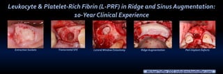
- 1. Leukocyte & Platelet-Rich Fibrin (L-PRF) in Ridge and Sinus Augmentation: 10-Year Clinical Experience Extraction Sockets LateralWindow OsteotomyTranscrestal SFE MichaelToffler DDS (info@michaeltoffler.com) Peri-Implant DefectsRidge Augmentation
- 2. “PRF is easy to handle and modify, providing the defect with not only a matrix permitting cell migration into the surgical site but also with crucial biological cues potentially accelerating the wound healing process.” (Ghanaati et al 2014)
- 3. L-PRF: Simple Preparation • • 24 gauge butterfly needle plastic tube coated internally with silica particles
- 4. 2700 RPM (400g) 12 minutes RBCs Fibrin Clot (PRF) Acellular Plasma (PPP) Intra-Lock.com
- 5. L-PRF:Preparation (cont) • • • WBC and PLT Fibrin clot RBC Li et al 2013
- 6. PRF ‘s naturally polymerized fibrin network sustains a slow release of growth factors and matrix glycoproteins for 7 - 14 days (Ling 2009; Dohan-Ehrenfest et al 2009; He et al 2009; Wu et al 2012; Kawase et al 2015) FRAGMENTSMEMBRANES PLUGS GF-ENRICHED BONE GRAFT MATRIX (PRF-BLOCK®) Standard 12 Minute Protocol Modified 3 Min Protocol PRF BLOCK
- 7. We graft sockets to offset the post-extraction catabolic process and minimize alveolar ridge degradation
- 8. • • •
- 9. The positive effect of growth factors in L-PRF could be particularly relevant when combined with osteoconductive scaffolds with slow healing dynamics (Torres et al 2009; Anitua et al 2010), where bone metabolism is compromised (eg. osteoporosis) as well as in early implantation scenarios (Chen et al 2004).
- 10. My “Socket Cocktail” CaSO4, PRF, D/M/FDB
- 11. PRF has shown promising results in the early phases of extraction socket healing • • • •
- 12. Flapless Extractions • Socket grafted to a level 2 to 3 mm below free gingival margin
- 13. PRF provides an organized matrix at the start of healing, increasing the speed of vascular ingress and wound coverage (Dohan et al 2006) Flapless Extractions 1 wk 4 wks •1 or 2 PRF plugs placed in socket and secured with mattress and interrupted sutures
- 14. In SitesWith Severe Bone Loss, Ridge Augmentation Is Usually Performed 6-8Weeks AfterTooth Removal
- 15. This Delayed Approach Provides Additional Soft-tissue Coverage OverThe Augmented Site 1 WEEK Maintenance of primary closure is essential to maximize bone gain 8 WEEKS after ext’s
- 16. L-PRF over Frenectomy and in Socket 8 wks Ridge augmentation done simultaneously with ext #9 would require a larger flap with extensive release, increasing postop pain and swelling as well as the risk of wound dehiscence.
- 21. PRE-OP
- 22. Toffler M. Guided Bone Regeneration (GBR) Using Cortical Bone Pins in Combination with Leukocyte- and Platelet-Rich Fibrin (L-PRF). Compend Cont Ed Dent 2014;35(3). Salvin Dental
- 23. Cortical Bone Pins: ClinicalAdvantages •No screws to retrieve •Membrane support •Graft retention
- 25. 2 mm twist adjustable stop (hhco-store.com)
- 28. 10-day post-op
- 29. Comparative healing at 6 months • 6 months post-op • Pre-op
- 34. PRF BLOCK®
- 36. PREOP6 MOS
- 39. Consistent Use of L-PRF in Sinus Elevation Surgery for the Last 10 Years • Slow sustained release of key growth factors (Ling 2009; Dohan-Ehrenfest et al 2009; He et al 2009; Wu et al 2012; Lauritano et al 2013; Kawase et al 2015) • Expedited sinus graft healing (Choukroun et al 2006; Inchingolo et al 2010; Tatullo et al 2012; Cruzat et al 2015) • Maintenance of primary wound closure (Khiste & Tare 2013; Baiju et al 2013; Toffler et al 2014) • Membrane protection (Diss et al 2008; Toffler et al 2010) and repair (Choi et al 2006; Shin & Sohn 2005; Baykul & Findik 2014; Toffler & Rosen 2015)
- 40. All My Approaches to Sinus Floor Elevation Incorporate L-PRF Transcrestal/Simultaneous LWOTranscrestal/Staged
- 41. 10 mm long implants “I use a transcrestal approach as often as I can, but prefer using a lateral (visual) approach not only in severely atrophic sites but also in………….
- 42. …….patients with ≤ 4 mm of subantral bone and significant sinus pathology.” Optical and Instrumental Access to Optimize Healing Response
- 43. ……and for extra added safety, platelet-rich fibrin (PRF) lines the sinus membrane prior to grafting
- 44. Single Stage Placement 6 Months Later
- 47. 10 mm
- 49. 4.0 x 11 mm Implant PREOP
- 50. 5 mm core prepped to a depth 3 mm
- 57. PRF inTSFE and Simultaneous Implant Placement
- 58. PRF will be added immediatedly after accessing sinus via direct infracture , drilling or piezosurgery Direct Infracture Drilling/Controlled Erosion Piezosurgery
- 59. Sohn 2010
- 62. 2.8 mm tip to elevate Widen osteotomy to 4.1 mm PRF
- 64. PREOP
- 65. Bone Densification and Placement 5 x 11 mmThommen Implant
- 66. PRF in Membrane Repair
- 68. Perforation Transcrestal Approach 4 mos CBCT 3 years
- 71. At 4 months, full repair and 6.7 mm RSBH 6.73
- 72. 6.73 3-4 mm of TSFE 4 years
- 73. 64 y/ F, heavy smoker Implants placed 10 years ago, referred for tx peri-implant disease Severe sinusitis, failure of implants 13 (floor intact), 14 (OAC) 14 13
- 74. Double Pedicle
- 75. porcine collagen membrane covers plugs PRF
- 76. PRF covers collagen CTG pedicle palate joins facial pedicles CTG
- 77. 1 week 8 weeks Asymptomatic, Successful Closure OAC
- 78. I hope I have demonstrated the potential benefits of incorporating PRF into:
- 79. Extraction Sites • Serves as a matrix to accelerate the healing of wound edges much like a fibrin bandage 7 days
- 80. Ridge Augmentation • Aids in hemostasis • Provides a high quality of gingival maturation (Simonpieri et al 2009) 1 week
- 81. • The strength of PRF membranes enables a biomaterial to be maintained and protected Ridge Augmentation
- 82. Composite Grafts • PRF fragments serve as a biological connector between bone particles facilitating cellular migration, particularly for endothelial cells necessary for neo-angiogenesis (Simonpieri et al 2009; Kobayashi 2011)
- 83. Complex Reconstructive Surgery • Platelet cytokines are gradually released as the fibrin matrix is resorbed, thus creating a perpetual process of healing
- 84. PRF is a welcomed addition for all my SFE procedures, but it is especially helpful in high risk membrane perf/sinus communication cases! FAILED LWO SINGLETOOTH SITESOAF EXT/OAC SINUS SEPTA