
Recommandé
Contenu connexe
Tendances
Tendances (20)
Similaire à Management of Ellis Class IV Fracture
Similaire à Management of Ellis Class IV Fracture (20)
Dernier
Dernier (20)
Management of Ellis Class IV Fracture
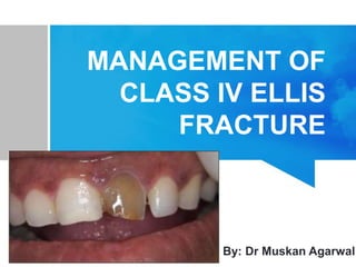
- 1. MANAGEMENT OF CLASS IV ELLIS FRACTURE By: Dr Muskan Agarwal
- 2. CONTENTS 1. CLASS IV ELLIS FRACTURE 2. TREATMENT OF TRAUMATIC INJURIES TO TEETH 3. FRACTURE WITH PULP INVOLVEMENT 4. CROWN FRACTURES 5. CONCLUSION 6. REFERENCE
- 3. CLASS IV ELLIS FRATURE The traumatized teeth that becomes non vital with or without loss of crown structure.
- 4. EMERGENCY: INITIAL CONTACT: • Often these emergencies are communicated by parent to the dentist on the phone . • The dentist should take the following steps: • The clinician should try to alleviate the anxiety.
- 5. If there is an avulsed tooth,following instructions need to be given: i. Insert the sink drain plug to avoid tooth loss. ii. Gently insert the tooth into the socket by holding the crown and not disturbing the root area. iii. If the replacement is nit possible keep it under the tongue and lip and go to dentist fast. iv. Tooth can also be kept in the milk while coming.
- 6. PARENT MANAGEMENT: Dentoalveolar injury can appear alarming to the parent. o A little blood mixed in saliva can give a impression of copious haemorrhage & clotting of the blood on the lips may suggest extensive laceration . o When a child arrives at the clinic the accompanying parent may be in a hysterical or extremely nervous condition & this complicates the situation. o A quick examination will reveal the extent of injury & the results should be communicated to the parents to alleviate their concern.
- 7. INTERMEDIATE TREATMENT: If the tooth becomes non vital, pulp therapy should be instituted.
- 8. In cases of pulpal exposures following can be done: • 1. Pulpectomy • 2. Apexification • 3. Extraction FRACTURE WITH PULP INVOLVEMENT
- 9. • Pulpectomy in apexified permanent teeth is conventional root canal(endodontic) treatment for exposed, infected, and/or necrotic teeth to eliminate pulpal and periradicular infection. PULPECTOMY(CONVENTIONAL ROOT CANAL TREATMENT)
- 10. • In all cases, the entire roof of pulp chamber is removed to gain access to the canals and eliminate all coronal pulp tissue. • Following debridement, disinfection and shaping of root canal system, obturation of the entire root canal is accomplished with a biologically acceptable, non-resorable filling material.
- 11. TECHNIQUE Local anaesthesia Rubber dam isolation
- 12. Cleaning the root canal • It is usually done with files which are available in different files.
- 13. Disinfection of the root canal system with disinfectants
- 14. Final preparation • After thoroughly cleaning and shaping the canals, canals are dried prior to filling the roots.
- 15. Obturating (filling) the root canals • Finally, the canals are sealed with 2 components:- 1. A sealer that sets over time 2. Gutta percha • This serves as the permanent root canal filling
- 16. Root canal treatment completed • Upon treatment of the root canal treatment, a temporary filling is placed over the sealed canals that has two parts :- 1. Cotton pellets soaked in antibacterial solution 2. A solid temporary filling on top
- 17. • A final restoration (usually a crown) is placed and follow-ups are done.
- 18. 2.APEXIFICATION: • Is defined as a method of inducing closure by the formation of osteo-cementum or similar hard tissue or continued apical development of the roots of an incompletely formed tooth in which pulp is no longer vital.
- 19. • Immature permanent tooth which became non-vital have a blunderbuss or divergent root apex that makes canal obturation by a non surgical approach difficult or impossible. 1.Anaesthetise and isolate the tooth. 2.Prepare a conventional access & extirpate the necrotic pulp 3.Instrument the canal, 0.5mm short of the radio graphic apex. 4.Fill the canal with calcium hydroxide or calcium hydroxide &camphorated monochlorophenol.
- 20. 5.Seal the canal with zinc oxide eugenol/IRM. 6.Dressing should be changed in every 3 months till apexification is achieved. 7.Later the canal can be obturated with conventional endodontic treatment.
- 21. MULTIPLE VISIT APEXIFICATION PROCEDURE WITH CALCIUM HYDROXIDE Anaesthesia, rubber dam isolation, and access opening Working length should be at least 2 mm short of radio graphic apex of the tooth.
- 22. Circumferential enlargement is effected by lateral pressure against the walls with a large file. Drying the canal with paper points
- 23. Ca(OH)2 is mixed wit sterile water or anaesthetic solution to a thick consistency. The paste is delivered into the canal with an amalgam carrier and condensed with finger pluggers.
- 24. The entire root canal is filled with Ca(OH)2 paste, ensuring that the material is in contact with the periapical tissues. The access cavity is sealed with RMGIC (Resin modified glass ionomer cement)
- 25. Patient called after 3 months Radiographic evidence of calcific barrier at or near the root apex YES NO OBTURATION USING THERMOPLASTICIZED TECHNIQUE Ca(OH)2 dressing is changed and patient is recalled every 3 months till evidence of calcific barrier is seen
- 26. SINGLE VISIT APEXIFICATION PROCEDURE WITH MTA
- 27. Anaesthesia, rubber dam isolation, and access opening Working length should be at least 2 mm short of radiographic apex of the tooth.
- 28. Circumferential enlargement is effected by lateral pressure against the walls with a large file. Drying the canal with paper points
- 29. MTA is mixed in a 3:1 ratio with sterile distilled water to a wet sand consistency. The paste is delivered into the canal with a MTA carrier and condensed with prefitted pluggers.
- 30. The material is condensed into a 3-4mm apical plug. Moist cotton pellet is placed over the MTA Patient is recalled after 48 hours and the set of MTA is verified and obturation is done using a thermoplasticized technique.
- 32. EXTRACTION • If either of the pulpectomy or apexification is not performed then extraction of the tooth is done.
- 33. CROWN FRACTURES: 1. ENAMEL INVOLVEMENT: Fracture only confined to enamel is rare.A radiograph is necessary to determine the extent of the injury. o Smoothening of fractured enamel & fluoride application to strenghthen the surface layer is usually the treatment of choice. o As an alternative , the acid etch technique may be used effectively to restore the smallest amount of enamel fracture with good esthetic result.
- 35. 2.Crown infarctions: o Very common but often overlooked these fractures appear as lines in the enamel substance , which do not cross the dentio-enamel junction. o Infarctions are caused by direct impact on enamel. o These injuries do not require definitive treatment. o Sealing the infarction line with unfilled resin following an acid etch technique may prevent stains e.g (tobacco, food, wine , tea ,coffee etc)
- 37. o Since no guarantee exists that the infarction line will stop short of dentin or pulp,vitality testing to document the status of pulpal tissue is recommended for all teeth with infarction lines. o The results of vitality testing for tooth or teeth with infarction should be compared to that of adjacent or contralateral control teeth. o Healthy tooth with infarction will respond positively to vitality testing .
- 38. ENAMEL AND DENTIN INVOLVEMENT: o Radiograph and vitality tests are adviced to determine the full extent of injury. Prognosis of the tooth depends on the following factors: • The amount of time the dentin has been exposed(less than 24hrs has good prognosis) • The remaining thickness of the dentin between the fractured tooth surface and pulp. • The stage of development of the pulp.
- 40. • A protective layer of calcium hydroxide or glass ionomer must be applied at the earliest to seal the exposed dentinal tubules. The crown form may then be restored with an acid etch composite resin or a temprorary crown. • An orthodontic band can be used as a temprorary matrix for retention of the dressing covering the exposed dentin.
- 41. 4. Reattachment of fracture segment: • The restorative techniques requires etching the enamel with 35%phosphoric acid gel(30sec). • Rinsing with water(20sec). • Drying with air(5sec). • Conditioning of the dentin and bonding of the dentin(intraoral or extraoral fragments)with any of the adhesive material systems; gluma(gluma bayer AG, FRG) or scotch bond 2. • The strength of the restored crown- fractured tooth using any of the listed systems is 50%of the strength of intact teeth.
- 42. • The restored crown- fractured tooth has a tendency to re-fracture with subsequent traumatic accidents. • This problem may be overcome by placing cast ceramic laminate veneers on crown fractured teeth rather than using dental adhesive material systems. • Laminate veneers provide identical strength to those of intact teeth.
- 44. THANK YOU ;)))