Newborn Care: Jaundice, anaemia and polycythaemia
•
6 j'aime•4,711 vues

Newborn Care was written for healthcare workers providing special care for newborn infants in level 2 hospitals. It covers: resuscitation at birth, assessing infant size and gestational age, routine care and feeding of both normal and high-risk infants, the prevention, diagnosis and management of hypothermia, hypoglycaemia, jaundice, respiratory distress, infection, trauma, bleeding and congenital abnormalities, communication with parents

Recommandé
Contenu connexe
Tendances
Tendances (20)
Similaire à Newborn Care: Jaundice, anaemia and polycythaemia
Similaire à Newborn Care: Jaundice, anaemia and polycythaemia (20)
Plus de Saide OER Africa
Plus de Saide OER Africa (20)
Dernier
Dernier (20)
Newborn Care: Jaundice, anaemia and polycythaemia
- 1. 9 Jaundice, anaemia and polycythaemia JAUNDICE Objectives 9-1 What is jaundice? When you have completed this unit you should be able to: Jaundice is the yellow colour of the skin and sclerae caused by deposits of bilirubin. In • Define jaundice and newborn infants the sclerae (white of the eye) hyperbilirubinaemia. are difficult to see and, therefore, the skin • Describe the excretion of bilirubin. colour is used to detect jaundice. Jaundice is a • List the causes of jaundice. clinical sign and not a laboratory measurement. • List the dangers of hyperbilirubinaemia. • Understand haemolytic disease of the Neonatal jaundice is a yellow colour of the skin newborn. caused by bilirubin • Treat jaundice. • List the common causes and treatment of anaemia. 9-2 What is bilirubin? • List the common causes and treatment Red blood cells contain a red pigment called of polycythaemia. haemoglobin which carries oxygen. Red cells in the fetus and newborn infant live for 3 months only. Therefore, the body is continually forming new red cells in the bone marrow to replace the old red cells which are broken down in the liver and spleen. When red cells die, their haemoglobin is changed into a yellow pigment called bilirubin. This unconjugated bilirubin is carried by
- 2. 164 NEWBORN CARE albumin in the blood stream to the liver to separate the serum from the red cells. The where it is first conjugated (joined to another total serum bilirubin (TSB) is then measured substance) and then excreted in the bile. If with a bilirubinometer and expressed in the concentration of bilirubin in the serum μmol/l. The TSB includes both unconjugated (blood) rises, it becomes visible in the skin and conjugated bilirubin. However, in the causing jaundice. Newborn infants normally newborn infant the TSB usually consists have a high haemoglobin concentration and, mainly of unconjugated bilirubin. therefore, produce a lot of bilirubin. The total serum bilirubin (TSB) cannot be Normal newborn infants produce a lot of bilirubin estimated accurately by clinically assessing the degree of jaundice in the skin 9-3 What is hyperbilirubinaemia? NOTE The degree of jaundice can be measured Hyperbilirubinaemia is defined as concen- with a transcutaneous bilirubinometer. However, tration (level) of total serum bilirubin (TSB) the monitors are expensive, easily damaged and of limited accuracy once phototherapy has been that is higher than the normal range. Normally started. They are useful in screening infants for the bilirubin concentration in the serum phototherapy. is low at birth, less than 35 μmol/l. It then climbs steadily for the first few days before returning again to an adult level of less than 9-5 How is bilirubin excreted in the adult? 35 μmol/l by 2 weeks. The total serum bilirubin Unconjugated bilirubin, formed by the concentration in term infants usually does not breakdown of red cells, is carried by albumin in rise above 200 μmol/l (12 mg/dl). the blood stream to the liver. In the liver cells The upper limit of the total serum bilirubin enzymes combine the unconjugated bilirubin concentration (TSB) in most healthy term with glucuronic acid to form conjugated infants is approximately: bilirubin. This chemical process, called conjugation, makes the bilirubin water soluble. Days after birth 0 0.5 1 Only when the bilirubin is water soluble can TSB (μmol/l) 35 75 125 the liver cells excrete it into the small bile ducts. From here the conjugated bilirubin is Days after birth 2 3 4 5 carried in the bile to the intestine where it is further converted by bacteria into the brown TSB (μmol/l) 150 175 200 200 pigment, stercobilin. In the adult, bilirubin is NOTE The upper limit of the normal total serum not reabsorbed from the intestine. Therefore bilirubin concentration is very controversial. In the final breakdown products of bilirubin are healthy, term, breastfed infants the upper limit passed in the stool. may be as high as 275 μmol/l. Conjugation of bilirubin in the liver is essential 9-4 How is bilirubin measured? before it can be excreted into the bile It is both difficult and inaccurate to assess the concentration of bilirubin in the serum by clinical examination of the degree of 9-6 How is bilirubin excreted in the fetus? jaundice, especially in an infant with a dark The fetus is unable to excrete conjugated skin. Therefore, it is important to measure bilirubin via the stool as it usually does not the bilirubin concentration of the serum if an pass meconium and also lacks bacteria in the infant is jaundiced. Usually a sample of blood is intestines to convert bilirubin into stercobilin. collected into a capillary tube and spun down Instead, fetal bilirubin, is excreted by the
- 3. JAUNDICE , ANAEMIA AND POLYC YTHAEMIA 165 placenta into the mother’s blood. However, Jaundice during the first 24 hours is always the placenta can only remove fat soluble, abnormal unconjugated bilirubin and not water soluble, conjugated bilirubin. To ensure that most of the fetal bilirubin remains unconjugated, NOTE The reabsorption of bilirubin from the intestine back into the blood stream of newborn the enzyme system that controls bilirubin infants (enterohepatic circulation of bilirubin) conjugation in the liver functions very slowly. is due to the enzyme β glucuronidase in the The small amount of conjugated bilirubin bowel wall. This enzyme is also present in that is excreted into the bile is carried to the breast milk resulting in a higher TSB in breastfed fetal intestine. Here a special fetal enzyme infants. As a result, some normal breastfed deconjugates the bilirubin, which is then infants remain jaundiced for more than two reabsorbed back into the fetal blood stream as weeks (breast milk jaundice). unconjugated bilirubin. In this way the fetus ensures that all the bilirubin can be excreted 9-8 What are the causes of jaundice? by the placenta. The main causes of jaundice in the newborn infant are: Bilirubin in the fetus is excreted by the placenta 1. Increased production of bilirubin rather than the liver 2. Slow bilirubin conjugation in the liver 3. Decreased excretion of bile 9-7 How is bilirubin excreted in the 9-9 What are the causes of an increased newborn infant? production of bilirubin? During the first week of life the enzyme system There are many causes of an increased that conjugates bilirubin with glucuronic acid bilirubin production: in the liver functions slowly, as in the fetus. Therefore, unconjugated bilirubin accumulates 1. The normal newborn infants produces a in the serum as the placenta is no longer lot of bilirubin due to a high haemoglobin present to remove it. As a result, newborn concentration. infants commonly become jaundiced due to 2. Cephalhaematoma or bruising. an increased concentration of unconjugated Haemoglobin which has escaped out of bilirubin in the serum. After a few days the damaged blood vessels is rapidly broken rate of liver conjugation increases and the down into bilirubin which is absorbed bilirubin concentration in the serum slowly back into the blood stream. returns to the normal adult range of less than 3. Polycythaemia. Infants with a very high 35 μmol/l. Jaundice at birth or in the first packed cell volume or haemoglobin 24 hours is unusual, however, as the bilirubin concentration have excess haemoglobin is adequately excreted by the placenta up until and, therefore, produce a lot more bilirubin the time of delivery. than normal. 4. Infection. General infections such as Some of the bilirubin that is conjugated and septicaemia and syphilis cause haemolysis excreted by the liver in the first week of life (breakdown of red cells). The released is broken down by the fetal enzyme in the haemoglobin is converted into bilirubin. intestine which continues to function for a few 5. Haemolytic disease of the newborn. weeks after birth. This unconjugated bilirubin Excess haemolysis causes an increased level is reabsorbed by the intestine, adding to the of unconjugated bilirubin. increase of the TSB. NOTE Excessive haemolysis may rarely be due to deficiency of a red cell enzyme (e.g. glucose 6 phosphate dehydrogenase deficiency), an
- 4. 166 NEWBORN CARE abnormal red cell membrane (e.g. spherocytosis) birth as they reabsorb some unconjugated or an abnormal haemoglobin (e.g. alpha bilirubin from the intestine back into the thalassaemia). blood stream. As a result, jaundice with a raised unconjugated bilirubin is common 9-10 What are the causes of slow bilirubin in breastfed infants. conjugation? 2. Hepatitis due to septicaemia, viral 1. Normal, healthy, term infants have a slow infection or syphilis. Swelling of the liver bilirubin conjugation for the first week cells obstructs the flow of bile in the small after delivery. The liver of the newborn bile ducts. infant during the first few days of life, 3. Biliary atresia is destruction of the bile therefore, functions like that of the fetus. ducts caused by a viral infection during the 2. About 10% of clinically healthy, term first weeks of life. infants have a TSB that increases above the Diseases of the liver (hepatitis and biliary normal range. They have a greater than atresia) prevent the excretion of conjugated usual delay in the maturation of their liver bilirubin into the bile. Conjugated bilirubin is, enzymes responsible for conjugation. therefore, reabsorbed into the blood stream, 3. Preterm infants also commonly have a resulting in jaundice. This is called obstructive TSB that rises above the normal range jaundice, and can be diagnosed by finding a due to immaturity of their liver enzymes. high concentration of conjugated bilirubin This is known as jaundice of immaturity. in the serum. If bilirubin cannot be excreted Jaundice is commoner in preterm than in in the bile, the stools become pale. Some of term infants. the excess conjugated bilirubin is excreted by 4. Congenital hypothyroidism due to the the kidneys, giving dark urine. Jaundice due absence of a thyroid gland in the infant to decreased excretion of bilirubin is far less may cause prolonged jaundice due to slow common in newborn infants than jaundice maturation of the liver enzymes. Although due to the excessive production or decreased rare, it is important as these children conjugation of bilirubin. become severely mentally retarded if not diagnosed and treated soon after birth. They need lifelong thyroxine treatment. Pale stools and dark urine suggest that the jaundice is due to liver disease In all these conditions, with an increased production or slow conjugation of bilirubin, the serum bilirubin is unconjugated. 9-12 What is physiological jaundice? All healthy newborn infants have a total serum Prolonged jaundice may be due to hypothyroidism bilirubin concentration (TSB) higher than in adults. This is due to the normal increase in NOTE In some countries all newborn infants are production, slow conjugation and decreased screened for hypothyroidism. Thyroid stimulating excretion of bilirubin. As a result, 50% of hormone (TSH) and thyroxine (T4) are measured normal, term infants have mild jaundice in infant blood collected at birth or within a during the first 2 weeks of life. However, they few days after delivery. A high TSH and low T4 are clinically well, their jaundice disappears suggests hypothyroidism. within 14 days and their TSB does not rise above 200 μmol/l (12 mg/dl). This is known 9-11 What causes a decreased excretion of as physiological jaundice. Mild jaundice is, bilirubin? therefore, very common in normal infants. 1. Healthy breastfed infants have a decreased excretion of bilirubin for a few weeks after
- 5. JAUNDICE , ANAEMIA AND POLYC YTHAEMIA 167 Therefore, a person with both A and D Mild jaundice in healthy infants is very common antigens will have the A positive blood group in the first two weeks of life while another with neither A, B nor D antigens will be blood group O negative. NOTE Some studies suggest that in physiological jaundice the TSB may rise as high as 275 μmol/l. 9-15 What is ABO haemolytic disease? ABO haemolytic disease occurs when the HAEMOLYTIC DISEASE mother is blood group O and her fetus is blood group A or B, the fetus having inherited these blood groups from the father. For reasons 9-13 What is haemolytic disease of the unknown, some group O mothers start newborn? producing anti-A or anti-B antibodies which Haemolytic disease of the newborn is the cross the placenta and cause fetal haemolysis condition where antibodies (immunoglobulins) by attacking the fetal red cells. The haemolysis from the mother cross the placenta into the is not severe enough to damage the fetus but fetal blood stream. Here these antibodies may cause severe jaundice in the newborn destroy the fetal red cells (haemolysis) causing infant. ABO haemolytic disease may occur in a anaemia and an increased production of first pregnancy or any later pregnancy. bilirubin in the fetus and newborn infant. The maternal antibodies, which stick to the The 2 most important causes of haemolytic fetal red cells, give a positive Coomb’s test in the disease of the newborn are: newborn infant, while the haemolysis results in a low packed cell volume and haemoglobin, 1. ABO haemolytic disease and a raised TSB. An infant with ABO 2. Rhesus haemolytic disease haemolytic disease usually appears normal There are also other uncommon forms of at delivery, as the placenta has been able to haemolytic disease in newborn infants. remove the excess bilirubin produced during pregnancy. However, the infant becomes jaundiced within the first 24 hours after birth. 9-14 What are blood groups? The TSB may increase rapidly and reach Red cells have proteins on their surface called dangerous levels. Due to the haemolysis, the antigens, which determine a person’s blood infant becomes anaemic. Unfortunately ABO group. The red cell antigens of a fetus are inheri- haemolytic disease is not preventable nor can ted from both parents and, therefore, may differ it be diagnosed accurately before delivery. from that of the mother. The most important ABO haemolytic disease is the commonest red cell antigens are the ABO antigens and the cause of severe jaundice in term infants. D (Rhesus) antigen. If the A antigen is present on a person’s red cells the blood group will be A. Similarly, the presence of the B antigen ABO haemolytic disease is the commonest cause makes the blood group B. If both antigens are of severe jaundice in term infants present the blood group will be AB while if both antigens are absent the blood group will NOTE In ABO haemolytic disease the mother be O. Most people are blood group O. produces IgG antibodies to A or B. These small antibodies can cross the placenta and, therefore, The D antigen is inherited separately from the differ from the large IgM antibodies to A and B ABO antigens. The presence of the D antigen which are present in the serum of all group O on the red cells makes a person Rhesus positive. adults. It is not understood why some women If the D antigen is missing, then the person is produce IGg antibodies to A and B antigens. Rhesus negative.
- 6. 168 NEWBORN CARE 9-16 How do you diagnose ABO haemolytic as it only occurs if fetal blood crosses the disease at birth? placenta (a fetomaternal bleed) to reach the mother’s blood and, thereby, sensitises her 1. The mother is blood group O. into producing anti-D antibodies. Rhesus 2. The father is blood group A, B or AB. haemolytic disease becomes progressively 3. The infant is blood group A or B. worse with each further pregnancy. Unlike 4. The Coomb’s test is positive in the infant. ABO haemolytic disease, with Rhesus 5. The TSB is often high (above 35 μmol/l) haemolytic disease the degree of fetal while the haemoglobin and packed cell haemolysis is severe and the fetus may go into volume is often low (below 45%) in the heart failure with resulting generalised oedema cord blood. (called hydrops) due to anaemia and die. 6. The infant commonly develops jaundice Fortunately Rhesus haemolytic disease is not within 24 hours of delivery. The jaundice common, because most people are Rh positive. usually increases rapidly and the TSB may Rhesus haemolytic disease can be prevented. reach dangerous levels during the first week. 7. The TSB at 6 hours after delivery is often NOTE The Rhesus blood group is named after the above 80 μmol/l. Rhesus monkey used in early experiments with red cell antigens. Rhesus haemolytic disease can also be caused by other Rhesus antigens (C, c, E Jaundice on day 1 suggests haemolytic disease and e). These forms are less severe than Rhesus haemolytic disease due to the D antigen (Rh D haemolytic disease). 9-17 What is Rhesus haemolytic disease? Fetal red cells may cross the placenta into the Rhesus haemolytic disease is haemolytic mother’s blood: disease of the newborn caused by maternal 1. At delivery (the most common) antibodies to the D antigen. With Rhesus 2. During a miscarriage haemolytic disease, the mother is always 3. With abruptio placentae Rhesus negative (Rh negative or Rh –ve) while 4. During amniocentesis the fetus and infant (and father) are always 5. During external cephalic version Rhesus positive (Rh positive or Rh +ve). Normally the fetal red blood cells do not enter The fetus can die of Rhesus haemolytic disease the maternal circulation during pregnancy or delivery. However, if the fetal capillaries in the placenta are damaged, fetal red blood cells 9-18 How can you prevent Rhesus may cross into the maternal blood. If the fetal haemolytic disease? red cells have the D antigen (Rh positive) but the mother’s do not (Rh negative), then the The Rh positive fetal red cells that cross the fetal cells may be recognised by the mother’s placenta can be destroyed, before they sensitise immune system as foreign. As a result the Rh- the mother, by giving her 100 μg (4 ml) anti-D negative mother will respond by producing immunoglobulin by intramuscular injection antibodies (anti-D) against these foreign red within 72 hours. This is usually given after cells. This process is known as sensitisation. delivery of an Rh-negative mother. However, Rarely an Rh-negative woman may also be anti-D immunoglobulin must also be given sensitised against the D antigen if she receives after any of the above complications of an incompatible blood transfusion with Rh pregnancy. Unfortunately it is useless giving positive red cells. the mother anti-D immunoglobulin if she has already been sensitised and has developed her Rhesus haemolytic disease is more severe own anti-D antibodies. than ABO haemolytic disease. Rhesus haemolytic disease is rare in first pregnancies
- 7. JAUNDICE , ANAEMIA AND POLYC YTHAEMIA 169 bilirubin may then enter the brain of Give all Rhesus-negative mothers anti-D the newborn infant and cause bilirubin immunoglobulin after delivery encephalopathy (kernicterus). Conjugated bilirubin is not toxic to the brain. In clinical practice the TSB is used to assess whether the 9-19 How can you diagnose Rhesus bilirubin is reaching dangerous concentrations haemolytic disease during pregnancy? as the TSB in newborn infants usually consists 1. Mother’s blood group is Rhesus negative. almost entirely of unconjugated bilirubin. 2. Father’s blood group is Rhesus positive. The risk of bilirubin encephalopathy depends 3. Mother has anti-D antibodies in her blood. on: 4. Mother may have had a previous infant with jaundice or a past obstetric history 1. The total serum bilirubin concentration. which suggests a fetomaternal bleed. The higher the TSB, the greater is the Usually the mother would not have chance that unconjugated bilirubin will received anti-D immunoglobulin after her cross the blood brain barrier into the previous deliveries. brain cells. 2. The gestational age. The more preterm The blood group of all pregnant women the infant the higher the risk due to an should be determined at the start of antenatal immature blood brain barrier. care. If you suspect that the pregnancy is 3. The postnatal age. A high TSB in the first complicated by Rhesus haemolytic disease (i.e. few days has a greater chance of increasing a Rh-negative patient with anti-D antibodies), to dangerous levels than the same TSB at a the mother must be referred urgently to a week of age. hospital where specialist care is available. 4. Factors that may make the blood brain barrier more permeable to bilirubin, such All women must have their blood groups as hypoxia, hypothermia, hypoglycaemia identified during pregnancy and infection. NOTE The blood brain barrier is a complex mech- Rhesus haemolytic disease should be anism that prevents toxic substances like bilirubin considered if the infant is jaundiced, pale and crossing from the blood into the brain cells. oedematous in the first 24 hours of life. Severe In well, term infants the TSB becomes Rhesus haemolytic disease affecting the fetus dangerous above 350 μmol/l (20 mg/dl) can be diagnosed during pregnancy if antenatal while in preterm infants the TSB becomes ultrasound examination shows signs of fetal dangerous above 250 μmol/l (15 mg/dl). The oedema and heart failure (hydrops fetalis). dangerous level is lower if factors that make the blood brain barrier more permeable to Rhesus haemolytic disease should be diagnosed bilirubin are also present. during pregnancy A high serum concentration of unconjugated NOTE Antenatal Doppler ultrasound can be used bilirubin can damage the brain to identify anaemic fetuses while new technology can determine the fetal blood group on a sample of maternal blood. 9-21 How do you recognise bilirubin encephalopathy? 9-20 Is jaundice dangerous? 1. The infant is very jaundiced. Jaundice can become dangerous when the 2. The TSB is very high. concentration of unconjugated bilirubin in the blood becomes very high. Unconjugated
- 8. 170 NEWBORN CARE 3. At first the infant is lethargic, hypotonic, from where it can easily be excreted without has a weak cry, poor Moro reflex and feeds first having to be conjugated. Phototherapy poorly with vomiting due to depressed is, therefore, able to rapidly lower the TSB. brain function. Bright light is able to change the shape but 4. Later the infant becomes irritable with a not the chemical composition of the bilirubin high-pitched cry, jitteriness, opisthotonus molecule. Ultraviolet light, which causes and convulsions due to brain irritation. sunburn and severe tissue damage, could kill an infant and is not used for phototherapy. Many infants with bilirubin encephalopathy die while the survivors are usually deaf and NOTE With phototherapy, unconjugated bilirubin mentally retarded with hypotonic cerebral is converted by the process of photoisomerisation palsy. Therefore, every effort must be made to into photobilirubin and lumirubin which are water soluble and, therefore, easily excreted in the stool prevent bilirubin encephalopathy. and urine. Phototherapy does not conjugate bilirubin. Blue light (at a wavelength of ) 455 nm 9-22 How can you prevent bilirubin is the most effective. encephalopathy? By not allowing the TSB to reach dangerous 9-24 What is used to give phototherapy? levels. A number of methods can be used to Phototherapy is usually given with a reduce the TSB: phototherapy unit which consists of a row 1. Give early milk feeds to reduce the amount of fluorescent tubes. Daylight tubes (SABS of bilirubin that is reabsorbed from the No. 5) or white tubes (SABS No. 2) are used. intestine. They should be changed after 1000 hours 2. Prevent preterm delivery. use as, despite still appearing bright, their 3. Give anti-D immunoglobulin to all effectiveness decreases with time and they Rhesus-negative mothers after delivery, produce ultraviolet light which is dangerous. a miscarriage, amniocentesis, abruptio A perspex (clear plastic) sheet must be placed placentae or external cephalic version. below the tubes to reduce heat. A perspex 4. Give phototherapy when the TSB sheet also protects the infant if a fluorescent approaches dangerous levels. tube breaks or comes loose. Sometimes a 5. Do an exchange transfusion when special white halogen spot light or blue LED phototherapy cannot keep the TSB below (light emitting diode) spotlight is also used to dangerous levels or when dangerous levels provide phototherapy. have already been reached. Although exposure to sunlight also lowers the TSB, an infant placed in the sun may rapidly Early milk feeds help to lower the total serum become hyperthermic. Therefore, this form bilirubin concentration of phototherapy must be used with great caution, if at all. NOTE The amount of light given out by the phototherapy unit can be measured with a PHOTOTHERAPY photometer. Only light in the blue part of the spectrum is measured and the energy output is given in μWatts/cm2/nm. An output above 9-23 What is phototherapy? 7μWatts is needed for effective phototherapy. A phototherapy blanket can add to the amount Phototherapy uses white or blue light (i.e. of phototherapy given by phototherapy lights. visible light) to change unconjugated bilirubin However, it can only provide a limited amount in the skin into a water-soluble form of of phototherapy if used alone. An advantage of bilirubin. The water-soluble bilirubin is then LED spot lights is that they emit no ultraviolet carried by albumin in the blood to the liver, light or heat.
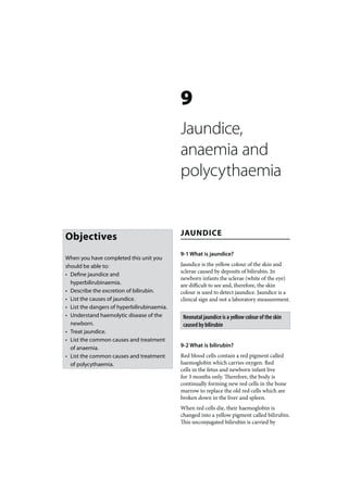
- 9. JAUNDICE , ANAEMIA AND POLYC YTHAEMIA 171 TSB μmol/1 350 300 250 200 150 100 50 0 0 1 2 3 4 5 6 7 Age in days Figure 9-1: Simple phototherapy guide for term infants showing the phototherapy line. 9-25 When should you give phototherapy? encephalopathy. Therefore, prophylactic phototherapy is started immediately after Phototherapy can be used either therapeutically birth if haemolytic disease of the newborn or prophylactically. is suspected or diagnosed. Prophylactic Therapeutic phototherapy should be given phototherapy in preterm infants is usually whenever the TSB is above the normal range started when the TSB reaches 125 μmol/l in and approaches dangerous levels. In practice infants weighing less than 1250 g, at 150 μmol/l a simple chart is used to decide when to give in infants less than 1500 g, and at 200 μmol/l in therapeutic phototherapy. If the TSB for the infants weighing less than 2000 g. infant’s age reaches the phototherapy line, treatment should be started. Phototherapy 9-26 How do you give phototherapy? is usually started earlier in preterm or sick infants. Phototherapy should not be given to 1. Switch on the phototherapy unit and make healthy, term infants who are jaundiced with a sure the tubes are all working. Check TSB below the phototherapy line. the age of the tubes and ensure that the perspex sheet is in position. All infants born to women who are blood 2. Place the infant naked in an incubator or group O should have their TSB measured at 6 bassinet so that the mattress is about 40 cm hours after birth. If the TSB is above 80 μg/dl from the phototherapy tubes. The infant phototherapy should be started. must not wear a nappy. Instead, a nappy With phototherapy it is possible to avoid can be placed under the infant. almost all exchange transfusions. 3. Cover the infant’s eyes with pads as the bright light worries the infant and may Prophylactic phototherapy is given when the possibly damage the retina. Remove the TSB is still below the phototherapy line but eye pads during feeding so that the eyes either the TSB is expected to increase rapidly can be checked for infection and to allow or the infant is at an increased risk of bilirubin the infant and mother to see each other.
- 10. 172 NEWBORN CARE 4. Turning the infant over every hour does and this may interfere with bonding. not make the phototherapy more effective. Conjunctivitis may also be hidden by the 5. Feed the infant milk, at least every 3 to pads. Therefore, it is advisable to remove 4 hours. Breastfeed if possible. Add an extra the eye pads every time the infant is fed. 25 ml/kg/day if the infant is demand fed Replace the pads after the feed. with formula. The lights may be switched off 4. Visible jaundice rapidly disappears under during feeds or the infant may be removed phototherapy even if the TSB remains from the phototherapy unit when fed. high or continues to increase. The TSB 6. Monitor the infant’s temperature hourly, must be monitored in all infants receiving weigh twice a day and measure the TSB phototherapy. daily or more frequently if it approaches 5. Other changes in skin colour may occur. dangerous levels. After a few days the infant may become 7. Allow the mother unrestricted visiting. tanned. Erythema (a pink rash) may If possible, the infant should be given result from excessive heat if a perspex phototherapy next to the mother in the sheet is not placed below the tubes. postnatal ward. Most skin rashes are aggravated by phototherapy, and phototherapy given 9-27 For how long should you give in error to an infant with conjugated phototherapy? hyperbilirubinaemia gives a grey/green colour to the skin known as bronzing. Continue phototherapy until the TSB has been 6. Phototherapy separates the infant from under the phototherapy line for 24 hours. the mother and, therefore, should not be Sometimes the TSB rises above the line again given unless there is a good reason. The after the phototherapy has been stopped and separation caused by phototherapy results treatment has to be restarted. However, once in maternal anxiety and also may prevent the TSB drops it usually stays down. Most the establishment of breastfeeding. phototherapy can be stopped after 3 days. 9-28 What are the dangers of The total serum bilirubin should be measured in phototherapy? all infants receiving phototherapy 1. The infant may become too hot or too cold. It is essential to monitor skin temperature 9-29 Can phenobarbitone be used to treat very carefully during phototherapy. jaundice? 2. The infant may pass loose green stools, due to the large amount of bilirubin There is little evidence that giving oral or excreted into the gut. The infant may also intramuscular phenobarbitone adds to the sweat more than usual. This may lead to effectiveness of phototherapy. It may also cause excessive weight loss due to dehydration. lethargy and poor feeding. Phenobarbitone is, When under phototherapy, breastfed therefore, not recommended to treat neonatal infants should be allowed to feed at least jaundice. It should not be used instead of every 4 hours while bottle-fed infants phototherapy. should receive an extra 25 ml/kg/day as NOTE Phenobarbitone 5 mg/kg intravenously may milk feeds. There is no need to give extra help lower the TSB in infants with severe jaundice. clear feeds. All infants under phototherapy Clearing the meconium from the colon with a should have their weight monitored. glycerine suppository may also help. In infants with 3. The infant’s eyes should be covered with Rh or ABO haemolytic jaundice, immunoglobulin intravenously stops the haemolysis. pads to prevent excessive exposure to bright light. The pads prevent the mother and infant seeing each other, however,
- 11. JAUNDICE , ANAEMIA AND POLYC YTHAEMIA 173 matching. Give phototherapy in the meantime. Routine use of phenobarbitone to reduce Remember that an exchange transfusion has jaundice is not advised its dangers and side-effects. NOTE In an exchange transfusion, twice the 9-30 When is an exchange transfusion infant’s blood volume is exchanged with fresh needed? compatible group O negative donor blood Except for infants with severe Rhesus (160 ml/kg). Usually the exchange is done via a catheter placed in the umbilical vein and haemolytic disease, an exchange transfusion 10–20 ml blood is exchanged at a time. Care is rarely needed today if phototherapy is should be taken to keep the infant warm and to used correctly when indicated. However, an monitor vital signs during the procedure. exchange transfusion may still be needed if there is a delay in starting phototherapy and the progressive increase in TSB cannot be ANAEMIA controlled. The indications for an exchange transfusion are: 9-31 What is anaemia? 1. A TSB above 400 μmol/l (20 mg/dl) in well term infants despite phototherapy. To determine whether an infant has anaemia, 2. A TSB above 250 to 350 μmol/l (15 mg/dl) either of the following can be measured: in well preterm infants depending on the 1. Packed cell volume (PCV), which is weight of the infant. The smaller the infant expressed as a percentage. the lower the TSB at which an exchange 2. Haemoglobin concentration (Hb), which is transfusion is indicated. expressed in g/dl. 3. A lower TSB if the infant is or has been hypoxic, hypothermic, hypoglycaemic or Anaemia is defined as a PCV or Hb that falls infected. below the normal range for the postnatal age 4. A rapidly rising TSB and falling packed cell of the infant. volume on day 1 in an infant with Rhesus The normal PCV at birth is 45–65% and the haemolytic disease. Hb 15–25 g/dl. After delivery the PCV and NOTE The following weight categories are a useful Hb in term infants falls steadily until about guide to the need for an exchange transfusion: 8 weeks of age and then slowly increases. The PCV should not fall below 35% and the Hb Below 1500 g 1500–1999 g below 12 g/dl in a term infant. The life span Well infant 250 μmol/l 300 μmol/l of the red cell in the infant is only 90 days Sick infant or 200 μmol/l 250 μmol/l (3 months) compared to 120 days in the adult. infant with haemolysis It is important that the PCV or Hb is measured on a sample of venous or arterial blood, or 2000–2499 g 2500 g blood collected from a warm heel. Capillary blood sampled from a cold heel will give a Well infant 350 μmol/l 400 μmol/l falsely high reading. Sick infant or 300 μmol/l 350 μmol/l infant with NOTE Anaemia (which is diagnosed technically) haemolysis is not the same as pallor (the clinical sign of pale skin, nail beds and mucous membranes). While All infants who may need an exchange anaemic infants are pale, there are many other transfusion must be transferred urgently to a causes of pallor (e.g. cold or shock). hospital with the staff and equipment needed. Send parental consent and a tube of clotted maternal blood with the infant for cross-
- 12. 174 NEWBORN CARE 9-32 What are the common causes of Packed cells should be given to an infant with anaemia in the infant? anaemia of prematurity if the PCV falls below 25% or the Hb below 8.5 g/dl. An earlier 1. Anaemia of prematurity transfusion is indicated if the PCV is below 2. Repeated removal of small volumes of blood 30% or the Hb below 10 g/dl and the infant for special investigations(e.g. blood gases) develops respiratory distress, signs of heart 3. Haemolytic disease of the newborn failure or patent ductus arteriosus, fails to 4. Haemorrhage before delivery (fetomaternal grow or has severe infection. haemorrhage) 5. Haemorrhage at or after delivery (e.g. The packed cells must be fully cross-matched bleeding from the umbilical cord) with the infant. Usually 10 ml/kg are given 6. Infection (e.g. septicaemia and syphilis). over 4 hours. Very small infants may need a number of transfusions during the first Iron deficiency is a very rare cause of anaemia few months of life before their PCV and Hb in the newborn period. However, iron returns to normal spontaneously. The PCV deficiency anaemia is common after 3 months should be checked a week after the transfusion. of age especially in preterm infants who have their umbilical cord clamped immediately after delivery and do not receive regular iron supplements. POLYCYTHAEMIA 9-33 What is anaemia of prematurity? 9-35 What is polycythaemia? The PCV and Hb of the preterm infant are Polycythaemia (too many red blood cells) is normal at birth but fall faster and to a lower defined as a packed cell volume above 65% or level than those in the term infant. The more a haemoglobin concentration above 25 g/dl. It preterm the infant the faster and lower will is important that the blood is venous, arterial, be the fall in PCV and Hb. In most preterm or capillary blood from a warm foot. infants the PCV falls to 30% and the Hb to 10 g/dl. A Hb or PCV below these levels is Polycythaemic infants appear very red common, especially in very preterm infants, (plethoric). Usually polycythaemia causes and is called ‘anaemia of prematurity’. Anaemia no major problems although the infant may of prematurity is often made worse by repeated become jaundiced. However, if the PCV and blood sampling. Oral iron supplements do not Hb are very high, the blood becomes thick and prevent or correct anaemia of prematurity. sticky causing neurological signs, respiratory distress, hypoglycaemia and heart failure. NOTE The cause of anaemia of prematurity is failure of the immature kidney to secrete erythropoeitin when the PCV and Hb fall below the normal range. 9-36 What are the causes of polycythaemia As a result the bone marrow is not stimulated and in infants? does not release red cells into the blood stream. 1. Chronic fetal hypoxia which is common in: • Underweight for gestational age infants 9-34 When should you treat anaemia in the • Wasted infants newborn infant? 2. Maternal diabetes An infant should be transfused with packed 3. Overtransfusion: cells if the PCV is below 45% or the Hb below • Transfusion of blood from one identical 15 g/dl at birth or during the first 24 hours. twin to the other before delivery via a Thereafter, term infants are usually considered monochorionic placenta (twin-to-twin for transfusion at a PCV below 30% or a Hb transfusion) below 10 g/dl. • Accidental overtransfusion with blood after delivery. This can occur if the
- 13. JAUNDICE , ANAEMIA AND POLYC YTHAEMIA 175 infant is held at a level far below the 3. Does this infant have hyperbili- mother (placenta) before the umbilical rubinaemia? Give reasons for your answer. cord is clamped. No, this infant does not have hyperbili- rubinaemia because the TSB falls within the 9-37 What is the treatment of normal range for a 3 day old infant. polycythaemia? If the polycythaemia causes no clinical problem 4. What is the correct management of this it does not need to be treated. During the first infant? few weeks the PCV and Hb gradually return to The infant should be managed as for a healthy, normal. However, if the infant has neurological normal infant except that the TSB should be signs (very jittery, convulsions or feeds poorly), repeated daily until it starts to fall. respiratory distress, hypoglycaemia or heart failure due to the polycythaemia, then it must be treated by partial plasma exchange 5. Should this infant receive phototherapy? transfusion. The method is the same as an No. There is no reason for phototherapy. exchange transfusion for jaundice except that the infant is given normal saline, fresh 6. Should the mother stop breastfeeding? frozen plasma or stabilised human serum. The exchange is limited to 20 ml/kg. No. she should continue to breastfeed. Although breastfeeding may result in a slightly higher TSB, it is not necessary to stop breast- CASE STUDY 1 feeding. A well, breastfed, term infant develops jaundice on day 3 and the TSB (total serum CASE STUDY 2 bilirubin) is 120 μmol/l. Both the mother and infant are blood group 0+ve. The infant's An infant scores at 32 weeks and weighs packed cell volume is 60% (haemoglobin 20 1600 g at birth. The infant has bilateral g/dl) and the Coomb's test is negative. cephalhaematomas and becomes jaundiced on day 2 with a TSB of 190 μmol/l. No treatment 1. What is the probable cause of this is given. On day 5 the infant becomes lethargic infant’s jaundice? and hypotonic with a weak cry. The TSB is now 370 μmol/l. This infant probably has physiological jaundice caused by the normally high bilirubin 1. Why do you think this infant became production, slow bilirubin conjugation by the jaundiced? liver, and increased bilirubin reabsorption by the intestines. Because the infant was born preterm and has an immature liver with slow conjugation of 2. Why does the infant not have jaundice bilirubin. In addition there is an increased caused by ABO or Rhesus haemolytic production of bilirubin by the breakdown of disease? haemoglobin in the cephalhaematomas. Because both the mother and infant have the 2. How should this infant have been same ABO blood groups and are both Rhesus treated on day 2? positive, the infant’s Coomb’s test is negative, and the PCV (Hb) is normal. With haemolytic Phototherapy should have been started as soon disease, the TSB would probably be much as the TSB was above the phototherapy line. higher and the PCV low.
- 14. 176 NEWBORN CARE 3. Why should you be worried if a jaundiced 4. When should the phototherapy be infant becomes lethargic and hypotonic stopped? with a weak cry? When the TSB has been below the photo- Because these are early signs of bilirubin therapy line for 24 hours. Thereafter, the TSB encephalopathy. Remember that they may should still be monitored for a few days as the also be early signs of other problems such as TSB may increase again due to continuing septicaemia. haemolysis. 4. How would you treat this infant’s hyperbilirubinaemia? CASE STUDY 4 The TSB is so high that the infant must be given an exchange transfusion as soon as A preterm infant who weighed 1200 g at birth possible. In the meantime, phototherapy is now a month old. For the past week the must be started. Do not drain the cephal- infant has not gained weight but otherwise haematomas. appears to be well. The PCV is 22%. The infant is being treated with iron supplements. As the infant now weights 1800 g, the mother wants CASE STUDY 3 to take him home. 1. Does this infant have anaemia? A term infant becomes jaundiced at 12 hours. The mother is blood group O+ve and the Yes, because the PCV is below 30%. infant is blood group B+. Phototherapy is started and after 24 hours the infant no longer 2. What is your diagnosis? appears jaundiced. Anaemia of prematurity. Many preterm infants fail to produce red cells for a few weeks after 1. What is the likely cause of this infant’s birth. jaundice? Haemolytic disease as the jaundice was 3. How should this infant be treated? noticed within the first 24 hours. The blood groups suggest ABO haemolytic disease (i.e. The infant needs a transfusion with packed mother group O and infant blood group B). cells. Normally, 10 ml/kg of blood is given over A positive Coomb’s test would confirm the 4 hours. diagnosis of haemolytic disease. 4. Why should this infant not be taken 2. What investigation is needed? home yet? The infant’s TSB must be measured. Because the PCV (and Hb) may still continue to fall. Once the infant has been transfused he can be discharged. He should be brought 3. Do you think that the phototherapy can back to the clinic or hospital to have his PCV be stopped safely on day 2 as the jaundice checked after a week. had cleared? Explain your answer. No. Phototherapy may clear the jaundice 5. Should this infant receive iron although the TSB remains high. supplements? Yes. Iron supplements will help to prevent iron deficiency in a few months time. However,
- 15. JAUNDICE , ANAEMIA AND POLYC YTHAEMIA 177 340 320 300 280 Micro mol/L TSB (total serum bilirubin) 260 240 220 200 180 160 140 120 100 38+ wks or 3000+ g 35–37w 6d or 2500–2999 g 80 34–34w 6d or 2000–2499 g 60 32–33w 6d or 1500–1999 g 30–31w 6d or 1250–1499 g 40 28–29w 6d or 1000–1249 g 20 < 28w or < 1000 g 0 6h 12h 24h 36h 48h 60h 72h 84h 96h 108h120h Time (age of baby in hours) Figure 9-2: Detailed phototherapy chart for infants in different birth weight and gestational age categories (e.g. 34 to 34 weeks and 6 days or 34 to less than 35 weeks). (From: A R Horn: Neonatal Medicine, University of Cape Town.) Start intensive phototherapy when the TSB is above the line according to gestation or weight. iron supplements will not prevent or correct 2. What is a normal PCV at birth? the anaemia of prematurity. At birth the normal PCV is 45 to 65%. During the first few weeks the PCV gradually drops. CASE STUDY 5 3. What precautions should be taken if a sample of capillary blood is collected for a A term infants is born at home and then taken PCV? to the nearest hospital. On examination the infant appears well but is noted to be very The heel must be warm and should not be plethoric. A capillary sample of blood is taken. squeezed. Otherwise the PCV result will be The PCV is 70%. falsely high. 1. What is the diagnosis? 4. How should this infant be managed? The infant has polycythaemia. This is As the infant appears well there is no special suggested clinically by the red colour and treatment needed. However, the infant should confirmed by the high PVC. be observed for jaundice.