2. Neonatal Resuscitation
The NRP has been carefully designed in a modular fashion, with each
module emphasizing a particular skill vital for a successful
resuscitation. The modules represent the progressive steps that would
occur during a full resuscitative effort of a severely depressed neonate.
The modules are as follows:
1. Preparation for delivery
2. Initial stabilization
3. Ventilation - bag and mask
4. Chest compressions
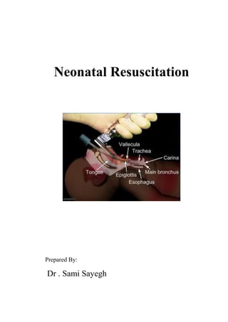
5. Endotracheal intubation
6. Medications
A nurse might be trained up to and including chest compressions and
how to assist with endotracheal intubation, while physicians would be
trained to provide all resuscitative steps. For either nurse or physician,
the instructional material is the same for each module, thus ensuring a
common ground for good communication and teamwork during an
actual resuscitation.
Each resuscitation should go through A,B,C, D
A : means airway.
B: means brething.
C : means circulation .
D : means drugs.
3. Asphyxia - The Basics
Apnea
1. The asphyxiated infant passes through a series of events:
o rapid breathing and fall in heart rate
o primary apnea
o irregular gasping, further fall in heart rate and drop in
blood pressure
o secondary apnea
2. Most infants in primary apnea will resume breathing when
stimulated. Once in secondary apnea, infants are unresponsive
to stimulation.
3. Apnea at birth should be treated as secondary apnea of
unknown duration (i.e. began in utero) and resuscitation should
begin at once.
Clearing Fetal Lung Fluid
1. The first few breaths of a normal infant are usually adequate to
expand the lungs and clear the alveolar lung fluid.
2. The pressure required to open the alveoli for the first time may
be two to three times that for normal breaths.
3. Expect problems in lung fluid clearance with:
o apnea at birth
o weak initial respiratory effort caused by:
prematurity
depression by asphyxia, maternal drugs, or
anaesthesia
Pulmonary Circulation
1. At birth, pulmonary blood flow increases rapidly as the lung
arterioles open up and blood is no longer diverted through the
ductus arteriosus.
2. With asphyxia, hypoxemia and acidosis perpetuate pulmonary
vasoconstriction and maintain the fetal pattern of circulation.
4. Systemic Circulation and Cardiac Function
1. Early in asphyxia, vasoconstriction in the gut, kidneys, muscles
and skin redistributes blood flow to the heart and brain as an
attempt to preserve function.
2. With progressive hypoxemia and acidosis, myocardial function
deteriorates and cardiac output declines.
Preparation for Delivery
Anticipate Need for Resuscitation
1. Antepartum and intrapartum history may help to alert delivery-
room staff about the possibility of a depressed or asphyxiated
newborn.
Antepartum Factors Intrapartum Factors
Age > 35 years
Maternal diabetes
Pregnancy-induced
hypertension
Chronic hypertension
Other maternal illness
(e.g. CVS, thyroid,
neuro)
Previous Rh
sensitization
Drug therapy
(e.g. magnesium,
lithium
adrenergic-blockers)
Maternal substance
abuse
Abnormal presentation
Operative delivery
Premature labour
Premature rupture of
membranes
Precipitous labour
Prolonged labour
Indices of fetal distress
(FHR abnormalities,
biophysical profile)
Maternal narcotics
(within 4 hrs of delivery)
General anaesthesia
Meconium-stained fluid
Prolapsed cord
Placental abruption
5. No prenatal care
Previous stillbirth
Bleeding - 2nd/3rd
trimester
Hydramnios
Oligohydramnios
Multiple gestation
Post-term gestation
Small-for-dates fetus
Fetal malformations
Placenta previa
Uterine tetany
Personnel
1. At every delivery, at least one individual should be capable of
performing a complete resuscitation (i.e. including endotracheal
intubation and the use of medications). In many cases, this is
the person delivering the infant.
2. A second person who will be primarily responsible for the
infant, must be present in the delivery room as well, even for
cases when a normal infant is expected. This person must be
able to initiate a resuscitation and if a complete resuscitation
becomes necessary, assist the fully-trained person.
3. When neonatal asphyxia is anticipated, two individuals whose
sole responsibility is to the infant, should be present in the
delivery room and be prepared to work as a team to perform a
complete resuscitation. The person delivering the mother must
not be considered as one of the two resuscitators.
4. With multiple births, a team is needed for each infant.
5. There should be no delay in initiating resuscitation; waiting a
few minutes for someone "on-call" to arrive is an unacceptable
practice and invites disaster.
6. Equipment
1. Equipment and medications should be checked as a daily
routine and then prior to anticipated need. Used items should
be replenished as soon as possible after a resuscitation.
2. The delivery room should be kept relatively warm and the
radiant heater should be preheated when possible. Prewarming
of towels and blankets can also be helpful in preventing
excessive heat loss from the neonate.
Resuscitation Equipment in the Delivery Room
Radiant Heater
Stethoscope
ECG monitor
Wall oxygen with flowmeter and
tubing
Neonatal resuscitation bag
(with manometer)
Face masks, Oral airways:
- newborn and premature
Medications:
- Epinephrine (1:10,000)
- Naloxone (0.4 or 1 mg · ml-1)
- Volume expander
- Sodium bicarb (0.5mEq · ml-1)
Suction with manometer
Bulb syringe
Suction catheters:
- 5F or 6F, 8F and 10F
Endotracheal tubes:
- 2.5, 3.0, 3.5, and 4.0 mm
ET tube stylet
Laryngoscope with straight
blades:
- No. 0 & 1
Umbilical vessel catheterization
tray
Umbilical catheters:
- 3.5 & 5F
Needles, syringes
Feeding tube 8F + syringe
7. Initial Stabilization
Prevent Heat Loss
1. Place the infant under an overhead radiant heater to minimize
radiant and convective heat loss.
2. Dry the body and head to remove amniotic fluid and prevent
evaporative heat loss. This will also provide gentle stimulation
to initiate or help maintain breathing.
Open the Airway
1. Position the infant supine or on his or her side with the neck
either in a neutral position or slightly extended. Avoid
overextension or flexion which may produce airway
obstruction. A slight Trendelenburg position may also be
helpful.
2. A folded towel (approximately 2.5 cm thick) placed under the
infant's shoulders may be useful if the infant has a large occiput.
3. If the infant has absent, slow or difficult respirations, apply
suction first to the mouth and then nose. If the nose were
cleared first the infant may gasp and aspirate secretions in the
pharynx. If mechanical suction with an 8F or 10F catheter is
used, make sure the vacuum does not exceed -13.3 kPa (-100
mmHg). Limit suctioning to 5 seconds at a time and monitor
heart rate for bradycardia which may be associated with deep
oropharyngeal stimulation.
4. If meconium is present in the amniotic fluid, special suctioning
may be required in the depressed infant. (See Endotracheal
Intubation below.)
Tactile Stimulation
1. If drying and suctioning do not induce effective breathing,
additional safe methods include:
o slapping or flicking the soles of the feet
o rubbing the back gently
2. Do not waste time continuing tactile stimulation if there is no
response after 10 - 15 seconds.
8. Evaluate the Infant
1. Respirations: Infants who are apneic or gasping despite brief
stimulation attempts should receive positive-pressure
ventilation. If there is adequate spontaneous breathing, go to
next step.
2. Heart Rate: Monitor either by auscultating the apical beat or by
palpating the base of the umbilical cord. If the heart rate is
below 100 bpm, begin positive-pressure ventilation, even if the
infant is making some respiratory efforts. If the heart rate is
above 100 bpm, go to the next step.
3. Colour: The presence of central cyanosis indicates that although
there is enough oxygen passing through the lungs to maintain
the heart rate, the infant is still not well oxygenated. Free-flow
100% oxygen at 5 l·min-1 using a mask held closely to the
infant's face should be administered until the infant becomes
pink, when the oxygen should be gradually withdrawn.