SlideShare une entreprise Scribd logo
1  sur  25
Télécharger pour lire hors ligne
1

ELECTROPHORESIS
Notes on electrophoresis.. By Dr. Ashish V. Jawarkar
Contact: pathologybasics@gmail.com Facebook: facebook.com/pathologybasics
2

OVERVIEW
1. History
2. Zonal electrophoresis

Principle

Method
3. Buffer
4. Support media
5. Ancillary techniques before performing electrophoresis
 Precipitation
 Column separation
- Gel filteration
- Ion exchange chromatography (anion exchange, cation exchange)
- Hydrophobic chromatography
- Affinity chromatography
- Capillary electrophoresis
6. Summary of workflow
7. Applications
a.
Serum protein electrophoresis
b.
CSF electrophoresis
c.
Urine electrophoresis
d.
Hemoglobin electrophoresis

Notes on electrophoresis.. By Dr. Ashish V. Jawarkar
Contact: pathologybasics@gmail.com Facebook: facebook.com/pathologybasics
3

* HISTORY
1. The technique of electrophoresis was introduced by Tiselius.
2. His technique is known as MOVING BOUNDARY ELECTROPHORESIS / FRONTAL
ELECTROPHORESIS
3. He separated proteins dissolved in electrolyte solution by application of electric current
through U shaped quartz tube that held the solution.
4. At ph 7.6, the solution got separated into four distinct protein fractions, albumin, α, β
and γ. These separated proteins were identified by checking the change in refractive
index across the solution.
5. As there was no solid support to the medium, these separated proteins did not form
distinct zones.

Notes on electrophoresis.. By Dr. Ashish V. Jawarkar
Contact: pathologybasics@gmail.com Facebook: facebook.com/pathologybasics
4

* ZONAL ELECTROPHORESIS
1. In Teselius’ technique, the proteins cound not form distinct bands
2. This problem was solved by introducing an anticonvection solid support medium – like
filter paper.
3. Now on applying electric current, the proteins separated into discrete bands at ph 8.6
4. The α band got further split into α1 and α2.
5. Other solid media that are frequently used nowadays include cellulose acetate
membrane, agarose gel, starch gel and polyacrideamide gel.

Anode

Cathode

Principle:
1. The proteins to be separated are applied at anode.
2. The support medium has negative charge – the electromotive force (determined by
charge of proteins and pH) tends to move the support medium towards anode (positive
pole) – but support medium cannot move.
3. The buffer ions have positive charge – they tend to move towards cathode.
4. This net result is flow of buffer towards cathode – this force called electro osmotic force
(endosmosis).
5. The buffer ions carry with them water molecules and protein molecules. Proteins here
move not only on the basis of their charge, but also determined by their molecular
weight.
6. This explains the movement of slightly negatively charged γ globulins towards the
cathode.
7. The actual distance traveled by the protein is dependent on these two forces described
above.

Notes on electrophoresis.. By Dr. Ashish V. Jawarkar
Contact: pathologybasics@gmail.com Facebook: facebook.com/pathologybasics
5

Method:
1. The sample to be separated is soaked into a gel.
2. Each end of the gel is then inserted into separate buffer chambers in which electrodes
are mounted.
3. An electric current is then passed through the gel for a period of, about 30 minutes
usually.
4. After this the gel is treated with a mild fixative such as acetic acid.
5. This fixative precipitates the proteins at the positions on the gel to which they have
migrated to.
6. They are then stained and the gel is dried.
7. The protein patterns can now be visually inspected for qualitiative identification of
abnormal proteins.
8. To find out the quantity of each type of protein present, densitometric scanners are
used, they generate tracings and quantitate relative percentages of proteins in each
fraction.
9. These percentages when multiplied by total proteins can give an idea about the
concentration of protein in each fraction.
Eg. If total protein is 9 gm/dl and albumin fraction obtained is 50% - = 4.5 gm/dl
(9 x 50/100) is the albumin concentration
10. On varying the buffer media / solid media – different applications of electrophoresis can
be developed.

Notes on electrophoresis.. By Dr. Ashish V. Jawarkar
Contact: pathologybasics@gmail.com Facebook: facebook.com/pathologybasics
6

* Varying buffer concentration and its application
1. If endosmosis is set at a high, this maximizes cathodal movement.
2. Immunoglobulins can be separated optimally and this helps in detecting oligoclonal
bands in CSF

Notes on electrophoresis.. By Dr. Ashish V. Jawarkar
Contact: pathologybasics@gmail.com Facebook: facebook.com/pathologybasics
7

* Varying solid media and its applications
PAGE – Polyacrylamide Gel Electrophoresis
1. Polyacrylamide is an inert support medium
2. It is useful for standard separation of native proteins based on charge

Notes on electrophoresis.. By Dr. Ashish V. Jawarkar
Contact: pathologybasics@gmail.com Facebook: facebook.com/pathologybasics
8

SDS-PAGE – Sodium dodecyl sulphate Polyacrylamide gel electrophoresis
1. SDS denatures proteins according to their molecular weight
2. SDS-PAGE is very powerful for resolving proteins and separating them into subunits
according to their molecular weight
3. Very useful for research purposes

Notes on electrophoresis.. By Dr. Ashish V. Jawarkar
Contact: pathologybasics@gmail.com Facebook: facebook.com/pathologybasics
9

TWO DIMENSIONAL ELECTROPHORESIS (2D ELECTROPHORESIS)
2. Standard separation of proteins is done in one direction
3. This is followed by SDS-PAGE in perpendicular direction
4. This results in hundreds of identifiable protein peaks

Notes on electrophoresis.. By Dr. Ashish V. Jawarkar
Contact: pathologybasics@gmail.com Facebook: facebook.com/pathologybasics
10

ISOELECTRIC FOCUSSING
1. Gives superior resolution to closely migrating proteins
2. Here the gel used contains a gradient of pH established by using ampholytes
3. As each protein reaches the gel location where the pH equals its own, the net charge
becomes zero
4. It cannot move further
5. Thus proteins move here strictly on the basis of their pH

Notes on electrophoresis.. By Dr. Ashish V. Jawarkar
Contact: pathologybasics@gmail.com Facebook: facebook.com/pathologybasics
11

* ANCILLARY TECHNIQUES
PRECIPITATION
1. The sample to be tested usually contains a lot of undesired proteins.
2. These undesired proteins can be removed by precipitation so that a single minor protein
can be isolated by electrophoresis.
3. First the value of total proteins is found out
4. Then Sodium sulfate, sodium sulfite, ammonium sulfate or methanol is used to
precipitate globulins.
5. Protein estimation from the remaining solution after precipitation can give an idea
about the albumin concentration – electrophoresis can give an idea about the types of
proteins apart from globulins.
6. globulin = total protein – albumin
7. estimation of albumin : globulin ratio is useful in various disorders
Normal :
A:G ratio

>1:5 (1-3)

Decreased A:G ratio
Decreased albumin
Malnutrition
Malabsorption
Liver failure
Proteinuria
Accumulation of ascetic fluid
Enteropathy

Increased A:G ratio
************

Increased globulins
Acute or chronic infections
Myelomas

Notes on electrophoresis.. By Dr. Ashish V. Jawarkar
Contact: pathologybasics@gmail.com Facebook: facebook.com/pathologybasics
12

COLUMN SEPARATION
(1) Gel filteration:
1. Media such as agarose beads/sephadex is used
2. They have beads in them with pores of various sizes
3. Largest proteins cannot pass through pores in the beads – they pass through spaces in
beads and are eluted first
4. Medium sized proteins pass through medium sized pores and appear second.
5. Small sized proteins pass through small sized pores and are eluted last.

Notes on electrophoresis.. By Dr. Ashish V. Jawarkar
Contact: pathologybasics@gmail.com Facebook: facebook.com/pathologybasics
13

(2) Ion Exchange Chromatgraphy
1. This is of two types – Anion exchange and Cation exchange chromatography
2. This technique uses charged particles which are placed in the column.
3. When proteins having the opposite charge as the column pass through it, they are
retained for a longer time, neutral elute the second while same charged particles elute
first.

ANION EXCHANGE CHROMATOGRAPHY
1. Media such as DEAE or QAE are used with have net positive charge.
2. Albumin, α and some β globulins which are anions stick to positively charged matrix.
3. γ globulins that have no charge are eluted faster.
CATION EXCHANGE CHROMATOGRAPHY
1. Media such as carboxymethylcellulose are used which have a negative charge.
2. Positively charged cations stick to the matrix
3. Albumin is eluted first here

Notes on electrophoresis.. By Dr. Ashish V. Jawarkar
Contact: pathologybasics@gmail.com Facebook: facebook.com/pathologybasics
14

(3) Hydrophobic chromatography
1. In this technique, support medium interacts with proteins according to their
hydrophobic nature.
2. Here ‘high salts’ and ‘low salts’ are used
3. high salts structure water around themselves and expose the hydrophobic ends of
proteins
4. these hydrophobic ends get attracted toward the medium and stick to it
5. according to hydrophobic nature of proteins, they stick or are eluted
6. ‘low salts’ are used to elute as they donot / expose less number of hydrophobic ends.

Notes on electrophoresis.. By Dr. Ashish V. Jawarkar
Contact: pathologybasics@gmail.com Facebook: facebook.com/pathologybasics
15

(4) Affinity chromatography
1. This is based on specific binding between a protein of interest and another protein (can
be its antibody/antigen) bound to the solid medium.
2. Bound protein can then be eluted in its purified form.
3. examples
a. to separate FVII-vWF – column is incorporated with monoclonal antibody to
vWF
b. Igs can be separated by using Staphylococcal protein A in gel matrix
c. Attaching dihydroxyboronate to medium helps separate glycosylated
hemoglobin
d. To separate digoxin – digibind is used
CroFab – crotalid snake venom

Notes on electrophoresis.. By Dr. Ashish V. Jawarkar
Contact: pathologybasics@gmail.com Facebook: facebook.com/pathologybasics
16

(5) Capillary electrophoresis
1. This procedure is similar to HPLC in that a column is employed where separation of
molecules is done on the basis of size/hydrophobicity or strereospecificity.
2. Applicable to large molecules such as DNA.

Notes on electrophoresis.. By Dr. Ashish V. Jawarkar
Contact: pathologybasics@gmail.com Facebook: facebook.com/pathologybasics
17

* SUMMARY OF WORK FLOW

Notes on electrophoresis.. By Dr. Ashish V. Jawarkar
Contact: pathologybasics@gmail.com Facebook: facebook.com/pathologybasics
18

* APPLICATIONS
(1) SERUM PROTEIN ELECTROPHORESIS
Following patterns are observed in normal conditions and various disorders on
electrophoresis.
Normal:

Cirrhosis of liver:

When liver function is sufficiently diminished, protein synthesizing capacity is
compromised and concentrations of albumin and proteins in the alpha and beta bands are
decreased. An additional common finding is beta-gamma bridging due to increased IgA.

Beta-gamma bridging

Alb

α1

α2

β

γ

Notes on electrophoresis.. By Dr. Ashish V. Jawarkar
Contact: pathologybasics@gmail.com Facebook: facebook.com/pathologybasics
19

The Nephrotic Syndrome -1. Renal disease involving the glomeruli is always associated with increased urinary protein
loss. When protein loss is greater than 3-4 g/day, the protein synthesizing capacity of
the liver is exceeded and hypoproteinemia, accompanied by anasarca, develops to
cause the nephrotic syndrome.
2. The massive urine protein loss is due to increased permeability of glomeruli to protein.
The permeability increase may be minimal so that only albumin and other smaller
molecular weight proteins are selectively filtered (selective nephrosis, as in Minimal
Change Disease) or may be greater so that larger proteins are also filtered (nonselective
nephrosis, as in membranous golmerulonephritis) as is the case in the example shown.
3. Alpha-2-macroglobulin is sufficiently large so that it is not filtered and increased
synthesis (from the general hepatic protein synthesis) causes its accumulation.
4. Lipoproteins are also sufficiently large to accumulate and hyperlipidemia is a
characteristic of the nephrotic syndrome, although lipoproteins are not stained with the
protein stain used in visualizing proteins.

Normal
Abnormal

Alb

α1

α2

β

γ

Alpha-1-Antitrypsin Deficiency:
A genetic defect causes a deficiency of alpha-1-antitrypsin. The antiprotease deficiency
results in a propensity to develop emphysema. Since alpha-1-antitrypsin is the major
component of the alpha-1 band, deficiency is suggested by a reduced alpha-1 band. Deficiency is
confirmed by specific immunochemical quantification.

Normal
Abnormal

Alb α1

α2

β

γ

Notes on electrophoresis.. By Dr. Ashish V. Jawarkar
Contact: pathologybasics@gmail.com Facebook: facebook.com/pathologybasics
20

Acute Inflammation
The alpha-1 and alpha-2 bands are increased during the inflammatory response from
increased hepatic synthesis of acute phase reactant proteins.

Normal
Abnormal

Alb α1

α2 β

γ

Chronic Inflammation -Immunoglobulin synthesis by antigen activated B lymphocytes transformed to plasma
cells is demonstrated by the increased polyclonal gamma band.

Normal
Abnormal

Alb

α1

α2

β

γ

Notes on electrophoresis.. By Dr. Ashish V. Jawarkar
Contact: pathologybasics@gmail.com Facebook: facebook.com/pathologybasics
21

Immunoglobulin Deficiency -Deficient immunoglobulin synthesis is revealed by a markedly diminished gamma band.
Effected individuals are prone to recurrent infection

Monoclonal Gammopathy -1. An unusually sharp band in the gamma region strongly suggests the presence of a
homogeneous immunoglobulin and, thus, the malignant proliferation of plasma cells
from a single cell (multiple myeloma) in contrast to the broad, heterogeneous, or
polyclonal, gamma band as exhibited above in chronic inflammation from
immunoglobulin synthesis by many different clones of plasma cells.
2. Homogeneous immunoglobulins are also found in Waldenstrom's macroglobulinemia
(where the sharp gamma band is always IgM). Specimens which exhibit a narrow
gamma band are further examined by immunofixation electrophoresis as described
below

Alb

α1

α2

β

γ

(2) CSF ELECTROPHORESIS

Notes on electrophoresis.. By Dr. Ashish V. Jawarkar
Contact: pathologybasics@gmail.com Facebook: facebook.com/pathologybasics
22

1. The diagnosis of Multiple sclerosis is ultimately a clinical one based on neurologic
history and physical examination.
2. Nevertheless, advanced laboratory results such as elevated IgG indices and presence of
oligoclonal bands of IgG or Kappa /lambda light chains on CSF electrophoresis, as well as
neuroimaging techniques have proven to be invaluable in diagnosis of MS.
Agarose gel electrophoresis
1. Concentrated CSF is used as sample
2. Shows discrete bands of IgG – the oligoclonal bands
3. Presence of two or more bands is necessary for diagnosis
4. False positive results are obtained in SSPE, CNS infections like neurosyphilis,
Crypotococcal meningitis, GBS, transverse myelitis, burkitt’s lymphoma etc.
5. Sensitivity is less than other methods of electrophoresis
Staining
Commassie brilliant blue/paragon violet/silver staining
1. silver staining is more sensitive then CBB
2. however these techniques must be simultaneously carried out on patients serum also
because polyclonal gammopathy can lead to diffusion of immunoglobulin into CSF and
lead to false positive results. This is seen in case of liver diseases, SLE, RA and chronic
granulomatous diseases.
ImmunoFixation Electrophoresis
1. Immunofixation electrophoresis (IFE) is used to demonstrate that a narrow gamma band
is due to a homogeneous immunoglobulin.
2. In the illustration below, 6 replicates of the specimen are loaded on to separate lanes of
an IFE gel.
3. Following electrophoresis, the protein in each lane is stained differently.
4. The first lane (SP) is stained for total protein.
5. The protein in each of the other lanes is "stained" with specific antisera for
immunoglobulin heavy and light chains, respectively, as illustrated in the figure.
6. The finding of the preponderance of only one light chain associated with a
predominantly staining heavy chain confirms the molecular homogeneity of the
immunoglobulin and also provides identification.
7. IFE identifies the narrow band as monoclonal IgG, lambda.
8. Sometimes malignant plasma cells synthesize excess light chains and less frequently
only light chains are synthesized. Almost never is excess heavy chain or only heavy chain
synthesized. Excess free lambda light chain is exhibited in the illustration.

Notes on electrophoresis.. By Dr. Ashish V. Jawarkar
Contact: pathologybasics@gmail.com Facebook: facebook.com/pathologybasics
23

a. Lymphocytic pleocytosis and 2 plasma cells in the CSF cytospin preparation.
b. 4b. Isoelectric focusing/immunofixation revealed CSF-restricted oligoclonal IgG
bands as well as identical paraprotein bands in CSF and serum. Anode is on the
left.
c. 4c. Serum immunofixation electrophoresis revealed the presence of IgGκ
monoclonal band (arrow). Anode is at the top. ELP, electrophoresis (general
protein stain); G, A, M, K, L: immunofixation for γ, α, μ, κ and λ chain,
respectively.
d. 4 d. Isoelectric focusing/affinity-mediated immunoblotting analysis: Paraprotein
bands did not react with Borrelia antigen but partly obscured CSF-restricted
oligoclonal IgG pattern. Paired CSF and serum samples tested for (from left to
right) "total" IgG, IgGκ, IgGλ; Anti-Borrelia IgG, IgGκ, IgGλ. Anode is at the top.

Notes on electrophoresis.. By Dr. Ashish V. Jawarkar
Contact: pathologybasics@gmail.com Facebook: facebook.com/pathologybasics
24

(3) URINE ELECTROPHORESIS
1.

2.
3.

4.

5.
6.

7.

The main reason for performing urine protein electrophoresis is to find a light chain
myeloma producing an excess of free light chains (Bence Jones protein), an important
part of a myeloma screen.
A band in the urine protein electropherogram may also result from an intact monoclonal
immunoglobulin, especially if the patient has poor renal function.
Immunofixation is important in defining the nature of the band and in distinguishing
between Bence Jones protein and an intact monoclonal protein originating from the
serum.
From the urine electropherogram we can also tell if the proteinuria is of glomerular
origin with a predominance of albumin, or if it has tubular components with excretion of
smaller molecular weight proteins such as retinol binding protein and alpha-1
microglobulin.
Fragmented albumin in urine is occasionally seen but is of unknown significance.
Historically, urine has been concentrated by either removal of water from the specimen
leaving the proteins in higher concentration, or by centrifugation whereby the proteins
are spun away from the majority of the water.
Demonstration of the protein components of urine from concentrated specimens was
originally performed on cellulose acetate and later on agarose and high resolution
agarose gel.

Notes on electrophoresis.. By Dr. Ashish V. Jawarkar
Contact: pathologybasics@gmail.com Facebook: facebook.com/pathologybasics
25

(4) HEMOGLOBIN ELECTROPHORESIS
Rationale:
1. Haemoglobin (Hb) abnormalities are the most frequent genetic disease, affecting
approximately 7 per cent of the world population.
2. Today, abnormal Hbs are generally discovered during a systematic study performed
within programmes for prevention of thalassaemias or sickle cell disease.
3. In several regions (India, Turkey, Irak, Iran, Gaza strip, Saudi Arabia, Cyprus, etc.), these
are found during a premarital screening. In other regions, like west Europeans countries,
the research for the main Hb disorders is often limited to populations at risk. It is done
either as preconceptional or neonatal screening programmes.
Methods and patterns:
1. Hb S [β6 Glu>Val], the first abnormal Hb described, was discovered in 1949 by Pauling et
al using moving boundary electrophoresis.
2. Zone electrophoresis performed on cellulose acetate strips (CAE) is still used in many
clinical laboratories.
3. This technique has a resolution lower than that of IEF, but because of its simplicity
remains among the more popular methods used in Hb screening. In this technique, the
Hb molecules are separated at alkaline pH. Under these conditions all Hbs have a
negative charge and migrate towards the cathode. Hb S, which has an additional
positive charge compared to Hb A migrates more slowly.
4. In IEF, a pH gradient is established by carrier ampholytes submitted to an electric
current and the Hb molecules migrate across this gradient until they reach the position
where their net charge is zero (isoelectric point). The molecules will then concentrate in
a sharp band as illustrated in Fig.
5. This technique allows separating molecules with isoelectric points differing only by 0.02
pH unit. Hb D-Punjab [β121 Glu>Gln], not detectable from Hb S by CAE, could easily be
recognized by this technique. The disadvantage of this method is its relatively high cost
and the requirement for a well-experienced laboratory staff.
6. This technique can help in diagnosing various hemoglobinopathies.

Notes on electrophoresis.. By Dr. Ashish V. Jawarkar
Contact: pathologybasics@gmail.com Facebook: facebook.com/pathologybasics

Contenu connexe

Tendances

VANILLYLMANDELIC ACID ESTIMATION
VANILLYLMANDELIC ACID ESTIMATIONVANILLYLMANDELIC ACID ESTIMATION
VANILLYLMANDELIC ACID ESTIMATIONRaghwendra sah
 
Interpreting serum protein electrophoresis
Interpreting serum protein electrophoresisInterpreting serum protein electrophoresis
Interpreting serum protein electrophoresisDr. Rajesh Bendre
 
CYTOLOGY OF CSF
CYTOLOGY OF CSFCYTOLOGY OF CSF
CYTOLOGY OF CSFMusa Khan
 
Cytochemical staining checked
Cytochemical staining checkedCytochemical staining checked
Cytochemical staining checkedBALRAM KRISHAN
 
Serum Protein and Albumin-Globulin Ratio
Serum Protein and Albumin-Globulin RatioSerum Protein and Albumin-Globulin Ratio
Serum Protein and Albumin-Globulin RatioASHIKH SEETHY
 
Gel card technology ppt nc
Gel card technology ppt ncGel card technology ppt nc
Gel card technology ppt ncNainshi Bhatt
 
SERUM ELECTROPHORESIS
SERUM ELECTROPHORESIS SERUM ELECTROPHORESIS
SERUM ELECTROPHORESIS naren
 
Amylase estimation practical
Amylase estimation  practicalAmylase estimation  practical
Amylase estimation practicalbinaya tamang
 
Periodic Acid Schiff and Alcian Blue Periodic Acid Schiff
Periodic Acid Schiff and Alcian Blue Periodic Acid SchiffPeriodic Acid Schiff and Alcian Blue Periodic Acid Schiff
Periodic Acid Schiff and Alcian Blue Periodic Acid SchiffNuhaJuma
 
estimation of blood glucose.pptx
estimation of blood glucose.pptxestimation of blood glucose.pptx
estimation of blood glucose.pptxKiranKhatwate
 
Overview to Diagnosis of Acute leukemia
Overview to Diagnosis of Acute leukemiaOverview to Diagnosis of Acute leukemia
Overview to Diagnosis of Acute leukemiaAhmed Makboul
 
Hb electrophoresis (principle materials and procedure)
Hb electrophoresis (principle materials and procedure)Hb electrophoresis (principle materials and procedure)
Hb electrophoresis (principle materials and procedure)hussainshahid55
 
Osmotic fragility test
Osmotic fragility testOsmotic fragility test
Osmotic fragility testSivaranjini N
 
Serum proteins
Serum proteinsSerum proteins
Serum proteinsCsalam
 

Tendances (20)

VANILLYLMANDELIC ACID ESTIMATION
VANILLYLMANDELIC ACID ESTIMATIONVANILLYLMANDELIC ACID ESTIMATION
VANILLYLMANDELIC ACID ESTIMATION
 
Interpreting serum protein electrophoresis
Interpreting serum protein electrophoresisInterpreting serum protein electrophoresis
Interpreting serum protein electrophoresis
 
CYTOLOGY OF CSF
CYTOLOGY OF CSFCYTOLOGY OF CSF
CYTOLOGY OF CSF
 
Creatinine estimation
Creatinine estimationCreatinine estimation
Creatinine estimation
 
Cytochemical staining checked
Cytochemical staining checkedCytochemical staining checked
Cytochemical staining checked
 
Serum Protein and Albumin-Globulin Ratio
Serum Protein and Albumin-Globulin RatioSerum Protein and Albumin-Globulin Ratio
Serum Protein and Albumin-Globulin Ratio
 
Gel card technology ppt nc
Gel card technology ppt ncGel card technology ppt nc
Gel card technology ppt nc
 
SERUM ELECTROPHORESIS
SERUM ELECTROPHORESIS SERUM ELECTROPHORESIS
SERUM ELECTROPHORESIS
 
Amylase estimation practical
Amylase estimation  practicalAmylase estimation  practical
Amylase estimation practical
 
morphology of red blood cells
morphology of red blood cellsmorphology of red blood cells
morphology of red blood cells
 
Periodic Acid Schiff and Alcian Blue Periodic Acid Schiff
Periodic Acid Schiff and Alcian Blue Periodic Acid SchiffPeriodic Acid Schiff and Alcian Blue Periodic Acid Schiff
Periodic Acid Schiff and Alcian Blue Periodic Acid Schiff
 
estimation of blood glucose.pptx
estimation of blood glucose.pptxestimation of blood glucose.pptx
estimation of blood glucose.pptx
 
Overview to Diagnosis of Acute leukemia
Overview to Diagnosis of Acute leukemiaOverview to Diagnosis of Acute leukemia
Overview to Diagnosis of Acute leukemia
 
Lap score
Lap scoreLap score
Lap score
 
Blood groups
Blood groupsBlood groups
Blood groups
 
Reticulocyte
ReticulocyteReticulocyte
Reticulocyte
 
Special stains
Special stainsSpecial stains
Special stains
 
Hb electrophoresis (principle materials and procedure)
Hb electrophoresis (principle materials and procedure)Hb electrophoresis (principle materials and procedure)
Hb electrophoresis (principle materials and procedure)
 
Osmotic fragility test
Osmotic fragility testOsmotic fragility test
Osmotic fragility test
 
Serum proteins
Serum proteinsSerum proteins
Serum proteins
 

En vedette

Electrophoresis ppt.
Electrophoresis ppt.Electrophoresis ppt.
Electrophoresis ppt.gulamrafey
 
Electrophoresis, the types of electrophoresis and samples used
Electrophoresis, the types of electrophoresis and samples usedElectrophoresis, the types of electrophoresis and samples used
Electrophoresis, the types of electrophoresis and samples usedMohit Adhikary
 
Techniques of electrophoresis
Techniques of electrophoresisTechniques of electrophoresis
Techniques of electrophoresisSayanti Sau
 
Electrophoresis (2)
Electrophoresis (2)Electrophoresis (2)
Electrophoresis (2)ceutics1315
 
Electrophoresis principle and types by Dr. Anurag Yadav
Electrophoresis principle and types by Dr. Anurag YadavElectrophoresis principle and types by Dr. Anurag Yadav
Electrophoresis principle and types by Dr. Anurag YadavDr Anurag Yadav
 
Haemeglobin Electrophoresis
Haemeglobin ElectrophoresisHaemeglobin Electrophoresis
Haemeglobin Electrophoresisrudy184
 
Electrophoresis by Dr. Anurag Yadav
Electrophoresis by Dr. Anurag YadavElectrophoresis by Dr. Anurag Yadav
Electrophoresis by Dr. Anurag YadavDr Anurag Yadav
 
Lab diag nephrotic synd 2003
Lab diag nephrotic synd 2003Lab diag nephrotic synd 2003
Lab diag nephrotic synd 2003Ravi Jain
 
Xray Production
Xray ProductionXray Production
Xray Productionlidgor
 
HEMOGLOBIN DERIVATIVES
HEMOGLOBIN DERIVATIVESHEMOGLOBIN DERIVATIVES
HEMOGLOBIN DERIVATIVESYESANNA
 
Xray Beam
Xray BeamXray Beam
Xray Beamlidgor
 

En vedette (20)

Electrophoresis ppt.
Electrophoresis ppt.Electrophoresis ppt.
Electrophoresis ppt.
 
Electrophoresis, the types of electrophoresis and samples used
Electrophoresis, the types of electrophoresis and samples usedElectrophoresis, the types of electrophoresis and samples used
Electrophoresis, the types of electrophoresis and samples used
 
Electrophoresis
ElectrophoresisElectrophoresis
Electrophoresis
 
Techniques of electrophoresis
Techniques of electrophoresisTechniques of electrophoresis
Techniques of electrophoresis
 
Electrophoresis (2)
Electrophoresis (2)Electrophoresis (2)
Electrophoresis (2)
 
Electrophoresis principle and types by Dr. Anurag Yadav
Electrophoresis principle and types by Dr. Anurag YadavElectrophoresis principle and types by Dr. Anurag Yadav
Electrophoresis principle and types by Dr. Anurag Yadav
 
Anti hiv
Anti hivAnti hiv
Anti hiv
 
Hyperlipidimea
HyperlipidimeaHyperlipidimea
Hyperlipidimea
 
Electophorosis
ElectophorosisElectophorosis
Electophorosis
 
Antiviral
AntiviralAntiviral
Antiviral
 
Haemeglobin Electrophoresis
Haemeglobin ElectrophoresisHaemeglobin Electrophoresis
Haemeglobin Electrophoresis
 
Gel electrophoresis
Gel electrophoresisGel electrophoresis
Gel electrophoresis
 
Electrophoresis
ElectrophoresisElectrophoresis
Electrophoresis
 
Antiviral Lecture
Antiviral LectureAntiviral Lecture
Antiviral Lecture
 
Electrophoresis by Dr. Anurag Yadav
Electrophoresis by Dr. Anurag YadavElectrophoresis by Dr. Anurag Yadav
Electrophoresis by Dr. Anurag Yadav
 
Lab diag nephrotic synd 2003
Lab diag nephrotic synd 2003Lab diag nephrotic synd 2003
Lab diag nephrotic synd 2003
 
Xray Production
Xray ProductionXray Production
Xray Production
 
Presentation gel electrophoresis
Presentation gel electrophoresisPresentation gel electrophoresis
Presentation gel electrophoresis
 
HEMOGLOBIN DERIVATIVES
HEMOGLOBIN DERIVATIVESHEMOGLOBIN DERIVATIVES
HEMOGLOBIN DERIVATIVES
 
Xray Beam
Xray BeamXray Beam
Xray Beam
 

Similaire à Electrophoresis

Similaire à Electrophoresis (20)

Protein Electrophoresis 2 nd year student
Protein Electrophoresis 2 nd year student Protein Electrophoresis 2 nd year student
Protein Electrophoresis 2 nd year student
 
Electrophoresis
ElectrophoresisElectrophoresis
Electrophoresis
 
ALLOZYME ELECTROPHORESIS.pdf
ALLOZYME ELECTROPHORESIS.pdfALLOZYME ELECTROPHORESIS.pdf
ALLOZYME ELECTROPHORESIS.pdf
 
Slab gel electrophoresis
Slab gel electrophoresisSlab gel electrophoresis
Slab gel electrophoresis
 
Elecrophoresis
ElecrophoresisElecrophoresis
Elecrophoresis
 
Electrophoresis
Electrophoresis  Electrophoresis
Electrophoresis
 
Protein Electrophoresis
Protein ElectrophoresisProtein Electrophoresis
Protein Electrophoresis
 
Iso electric focusing(ranjith)
Iso electric focusing(ranjith)Iso electric focusing(ranjith)
Iso electric focusing(ranjith)
 
Electrophoresis 2015
Electrophoresis 2015Electrophoresis 2015
Electrophoresis 2015
 
Moving Boundary & Isoelctric focusing(JEEV).pptx
Moving Boundary & Isoelctric focusing(JEEV).pptxMoving Boundary & Isoelctric focusing(JEEV).pptx
Moving Boundary & Isoelctric focusing(JEEV).pptx
 
Electrophoresis.pptx
Electrophoresis.pptxElectrophoresis.pptx
Electrophoresis.pptx
 
Electrophoresis
ElectrophoresisElectrophoresis
Electrophoresis
 
(5) Electrophoresis.pptx
(5) Electrophoresis.pptx(5) Electrophoresis.pptx
(5) Electrophoresis.pptx
 
Electrophoresis
ElectrophoresisElectrophoresis
Electrophoresis
 
Gel electrophoresis
Gel electrophoresisGel electrophoresis
Gel electrophoresis
 
Other electrophoresis
Other electrophoresisOther electrophoresis
Other electrophoresis
 
immunoblotting techniques
immunoblotting techniquesimmunoblotting techniques
immunoblotting techniques
 
68. Electrophoresis
68. Electrophoresis68. Electrophoresis
68. Electrophoresis
 
Electrophoresis sem4 paper1 MITHIBAI COLLEGE
Electrophoresis sem4 paper1 MITHIBAI COLLEGEElectrophoresis sem4 paper1 MITHIBAI COLLEGE
Electrophoresis sem4 paper1 MITHIBAI COLLEGE
 
Characterization of proteins
Characterization of proteinsCharacterization of proteins
Characterization of proteins
 

Plus de Ashish Jawarkar

Ch 6 diseases of the immune system part 1
Ch 6 diseases of the immune system part 1Ch 6 diseases of the immune system part 1
Ch 6 diseases of the immune system part 1Ashish Jawarkar
 
MALE AND FEMALE GENITAL TRACT
MALE AND FEMALE GENITAL TRACTMALE AND FEMALE GENITAL TRACT
MALE AND FEMALE GENITAL TRACTAshish Jawarkar
 
Ch 2 adaptations, cell injury, cell death
Ch 2 adaptations, cell injury, cell deathCh 2 adaptations, cell injury, cell death
Ch 2 adaptations, cell injury, cell deathAshish Jawarkar
 
Ch 4 hemorragic disorders,thromboembolic diseases, shock
Ch 4 hemorragic disorders,thromboembolic diseases, shockCh 4 hemorragic disorders,thromboembolic diseases, shock
Ch 4 hemorragic disorders,thromboembolic diseases, shockAshish Jawarkar
 
Ch 3 inflammation and repair
Ch 3 inflammation and repairCh 3 inflammation and repair
Ch 3 inflammation and repairAshish Jawarkar
 
CYTOLOGY OF BREAST LESIONS??!
CYTOLOGY OF BREAST LESIONS??! CYTOLOGY OF BREAST LESIONS??!
CYTOLOGY OF BREAST LESIONS??! Ashish Jawarkar
 
Immunohistochemistry in breast lesions
Immunohistochemistry in breast lesionsImmunohistochemistry in breast lesions
Immunohistochemistry in breast lesionsAshish Jawarkar
 
Immunohistochemistry of Prostatic lesions
Immunohistochemistry of Prostatic lesionsImmunohistochemistry of Prostatic lesions
Immunohistochemistry of Prostatic lesionsAshish Jawarkar
 
Robbins Chapter 1.. Cell as a unit of health and disease
Robbins Chapter 1.. Cell as a unit of health and diseaseRobbins Chapter 1.. Cell as a unit of health and disease
Robbins Chapter 1.. Cell as a unit of health and diseaseAshish Jawarkar
 
Pathology of the digestive system
Pathology of the digestive systemPathology of the digestive system
Pathology of the digestive systemAshish Jawarkar
 
Cellular adaptations, injury and death.. Lecture 1
Cellular adaptations, injury and death.. Lecture 1Cellular adaptations, injury and death.. Lecture 1
Cellular adaptations, injury and death.. Lecture 1Ashish Jawarkar
 
Quality control in clinical laboratories
Quality control in clinical laboratoriesQuality control in clinical laboratories
Quality control in clinical laboratoriesAshish Jawarkar
 
CSF - Cerebrospinal fluid examination - from tapping to pathological diagnosis
CSF - Cerebrospinal fluid examination - from tapping to pathological diagnosisCSF - Cerebrospinal fluid examination - from tapping to pathological diagnosis
CSF - Cerebrospinal fluid examination - from tapping to pathological diagnosisAshish Jawarkar
 
LABORATORY DIAGNOSIS OF HIV - AIDS
LABORATORY DIAGNOSIS OF HIV - AIDSLABORATORY DIAGNOSIS OF HIV - AIDS
LABORATORY DIAGNOSIS OF HIV - AIDSAshish Jawarkar
 
Intracellular accumulations
Intracellular accumulationsIntracellular accumulations
Intracellular accumulationsAshish Jawarkar
 

Plus de Ashish Jawarkar (20)

Ch 6 diseases of the immune system part 1
Ch 6 diseases of the immune system part 1Ch 6 diseases of the immune system part 1
Ch 6 diseases of the immune system part 1
 
MALE AND FEMALE GENITAL TRACT
MALE AND FEMALE GENITAL TRACTMALE AND FEMALE GENITAL TRACT
MALE AND FEMALE GENITAL TRACT
 
Ch 2 adaptations, cell injury, cell death
Ch 2 adaptations, cell injury, cell deathCh 2 adaptations, cell injury, cell death
Ch 2 adaptations, cell injury, cell death
 
Ch 4 hemorragic disorders,thromboembolic diseases, shock
Ch 4 hemorragic disorders,thromboembolic diseases, shockCh 4 hemorragic disorders,thromboembolic diseases, shock
Ch 4 hemorragic disorders,thromboembolic diseases, shock
 
Ch 3 inflammation and repair
Ch 3 inflammation and repairCh 3 inflammation and repair
Ch 3 inflammation and repair
 
CYTOLOGY OF BREAST LESIONS??!
CYTOLOGY OF BREAST LESIONS??! CYTOLOGY OF BREAST LESIONS??!
CYTOLOGY OF BREAST LESIONS??!
 
Histotechniques
HistotechniquesHistotechniques
Histotechniques
 
Immunohistochemistry in breast lesions
Immunohistochemistry in breast lesionsImmunohistochemistry in breast lesions
Immunohistochemistry in breast lesions
 
Immunohistochemistry of Prostatic lesions
Immunohistochemistry of Prostatic lesionsImmunohistochemistry of Prostatic lesions
Immunohistochemistry of Prostatic lesions
 
Cardiovascular system
Cardiovascular systemCardiovascular system
Cardiovascular system
 
Ch7 Neoplasia
Ch7 NeoplasiaCh7 Neoplasia
Ch7 Neoplasia
 
Amyloidosis
AmyloidosisAmyloidosis
Amyloidosis
 
Ch12 Heart Part 1
Ch12 Heart Part 1Ch12 Heart Part 1
Ch12 Heart Part 1
 
Robbins Chapter 1.. Cell as a unit of health and disease
Robbins Chapter 1.. Cell as a unit of health and diseaseRobbins Chapter 1.. Cell as a unit of health and disease
Robbins Chapter 1.. Cell as a unit of health and disease
 
Pathology of the digestive system
Pathology of the digestive systemPathology of the digestive system
Pathology of the digestive system
 
Cellular adaptations, injury and death.. Lecture 1
Cellular adaptations, injury and death.. Lecture 1Cellular adaptations, injury and death.. Lecture 1
Cellular adaptations, injury and death.. Lecture 1
 
Quality control in clinical laboratories
Quality control in clinical laboratoriesQuality control in clinical laboratories
Quality control in clinical laboratories
 
CSF - Cerebrospinal fluid examination - from tapping to pathological diagnosis
CSF - Cerebrospinal fluid examination - from tapping to pathological diagnosisCSF - Cerebrospinal fluid examination - from tapping to pathological diagnosis
CSF - Cerebrospinal fluid examination - from tapping to pathological diagnosis
 
LABORATORY DIAGNOSIS OF HIV - AIDS
LABORATORY DIAGNOSIS OF HIV - AIDSLABORATORY DIAGNOSIS OF HIV - AIDS
LABORATORY DIAGNOSIS OF HIV - AIDS
 
Intracellular accumulations
Intracellular accumulationsIntracellular accumulations
Intracellular accumulations
 

Dernier

Call Girls Service in Bommanahalli - 7001305949 with real photos and phone nu...
Call Girls Service in Bommanahalli - 7001305949 with real photos and phone nu...Call Girls Service in Bommanahalli - 7001305949 with real photos and phone nu...
Call Girls Service in Bommanahalli - 7001305949 with real photos and phone nu...narwatsonia7
 
Call Girl Lucknow Mallika 7001305949 Independent Escort Service Lucknow
Call Girl Lucknow Mallika 7001305949 Independent Escort Service LucknowCall Girl Lucknow Mallika 7001305949 Independent Escort Service Lucknow
Call Girl Lucknow Mallika 7001305949 Independent Escort Service Lucknownarwatsonia7
 
Call Girls Jp Nagar Just Call 7001305949 Top Class Call Girl Service Available
Call Girls Jp Nagar Just Call 7001305949 Top Class Call Girl Service AvailableCall Girls Jp Nagar Just Call 7001305949 Top Class Call Girl Service Available
Call Girls Jp Nagar Just Call 7001305949 Top Class Call Girl Service Availablenarwatsonia7
 
Call Girls ITPL Just Call 7001305949 Top Class Call Girl Service Available
Call Girls ITPL Just Call 7001305949 Top Class Call Girl Service AvailableCall Girls ITPL Just Call 7001305949 Top Class Call Girl Service Available
Call Girls ITPL Just Call 7001305949 Top Class Call Girl Service Availablenarwatsonia7
 
Call Girls Service In Shyam Nagar Whatsapp 8445551418 Independent Escort Service
Call Girls Service In Shyam Nagar Whatsapp 8445551418 Independent Escort ServiceCall Girls Service In Shyam Nagar Whatsapp 8445551418 Independent Escort Service
Call Girls Service In Shyam Nagar Whatsapp 8445551418 Independent Escort Serviceparulsinha
 
VIP Call Girls Lucknow Nandini 7001305949 Independent Escort Service Lucknow
VIP Call Girls Lucknow Nandini 7001305949 Independent Escort Service LucknowVIP Call Girls Lucknow Nandini 7001305949 Independent Escort Service Lucknow
VIP Call Girls Lucknow Nandini 7001305949 Independent Escort Service Lucknownarwatsonia7
 
Call Girls Jayanagar Just Call 7001305949 Top Class Call Girl Service Available
Call Girls Jayanagar Just Call 7001305949 Top Class Call Girl Service AvailableCall Girls Jayanagar Just Call 7001305949 Top Class Call Girl Service Available
Call Girls Jayanagar Just Call 7001305949 Top Class Call Girl Service Availablenarwatsonia7
 
Housewife Call Girls Bangalore - Call 7001305949 Rs-3500 with A/C Room Cash o...
Housewife Call Girls Bangalore - Call 7001305949 Rs-3500 with A/C Room Cash o...Housewife Call Girls Bangalore - Call 7001305949 Rs-3500 with A/C Room Cash o...
Housewife Call Girls Bangalore - Call 7001305949 Rs-3500 with A/C Room Cash o...narwatsonia7
 
97111 47426 Call Girls In Delhi MUNIRKAA
97111 47426 Call Girls In Delhi MUNIRKAA97111 47426 Call Girls In Delhi MUNIRKAA
97111 47426 Call Girls In Delhi MUNIRKAAjennyeacort
 
Bangalore Call Girls Marathahalli 📞 9907093804 High Profile Service 100% Safe
Bangalore Call Girls Marathahalli 📞 9907093804 High Profile Service 100% SafeBangalore Call Girls Marathahalli 📞 9907093804 High Profile Service 100% Safe
Bangalore Call Girls Marathahalli 📞 9907093804 High Profile Service 100% Safenarwatsonia7
 
call girls in Connaught Place DELHI 🔝 >༒9540349809 🔝 genuine Escort Service ...
call girls in Connaught Place  DELHI 🔝 >༒9540349809 🔝 genuine Escort Service ...call girls in Connaught Place  DELHI 🔝 >༒9540349809 🔝 genuine Escort Service ...
call girls in Connaught Place DELHI 🔝 >༒9540349809 🔝 genuine Escort Service ...saminamagar
 
Kolkata Call Girls Services 9907093804 @24x7 High Class Babes Here Call Now
Kolkata Call Girls Services 9907093804 @24x7 High Class Babes Here Call NowKolkata Call Girls Services 9907093804 @24x7 High Class Babes Here Call Now
Kolkata Call Girls Services 9907093804 @24x7 High Class Babes Here Call NowNehru place Escorts
 
Call Girls Hebbal Just Call 7001305949 Top Class Call Girl Service Available
Call Girls Hebbal Just Call 7001305949 Top Class Call Girl Service AvailableCall Girls Hebbal Just Call 7001305949 Top Class Call Girl Service Available
Call Girls Hebbal Just Call 7001305949 Top Class Call Girl Service Availablenarwatsonia7
 
Book Call Girls in Yelahanka - For 7001305949 Cheap & Best with original Photos
Book Call Girls in Yelahanka - For 7001305949 Cheap & Best with original PhotosBook Call Girls in Yelahanka - For 7001305949 Cheap & Best with original Photos
Book Call Girls in Yelahanka - For 7001305949 Cheap & Best with original Photosnarwatsonia7
 
Glomerular Filtration rate and its determinants.pptx
Glomerular Filtration rate and its determinants.pptxGlomerular Filtration rate and its determinants.pptx
Glomerular Filtration rate and its determinants.pptxDr.Nusrat Tariq
 
Book Call Girls in Kasavanahalli - 7001305949 with real photos and phone numbers
Book Call Girls in Kasavanahalli - 7001305949 with real photos and phone numbersBook Call Girls in Kasavanahalli - 7001305949 with real photos and phone numbers
Book Call Girls in Kasavanahalli - 7001305949 with real photos and phone numbersnarwatsonia7
 
Call Girl Service Bidadi - For 7001305949 Cheap & Best with original Photos
Call Girl Service Bidadi - For 7001305949 Cheap & Best with original PhotosCall Girl Service Bidadi - For 7001305949 Cheap & Best with original Photos
Call Girl Service Bidadi - For 7001305949 Cheap & Best with original Photosnarwatsonia7
 
Russian Call Girl Brookfield - 7001305949 Escorts Service 50% Off with Cash O...
Russian Call Girl Brookfield - 7001305949 Escorts Service 50% Off with Cash O...Russian Call Girl Brookfield - 7001305949 Escorts Service 50% Off with Cash O...
Russian Call Girl Brookfield - 7001305949 Escorts Service 50% Off with Cash O...narwatsonia7
 
Call Girl Koramangala | 7001305949 At Low Cost Cash Payment Booking
Call Girl Koramangala | 7001305949 At Low Cost Cash Payment BookingCall Girl Koramangala | 7001305949 At Low Cost Cash Payment Booking
Call Girl Koramangala | 7001305949 At Low Cost Cash Payment Bookingnarwatsonia7
 
Hemostasis Physiology and Clinical correlations by Dr Faiza.pdf
Hemostasis Physiology and Clinical correlations by Dr Faiza.pdfHemostasis Physiology and Clinical correlations by Dr Faiza.pdf
Hemostasis Physiology and Clinical correlations by Dr Faiza.pdfMedicoseAcademics
 

Dernier (20)

Call Girls Service in Bommanahalli - 7001305949 with real photos and phone nu...
Call Girls Service in Bommanahalli - 7001305949 with real photos and phone nu...Call Girls Service in Bommanahalli - 7001305949 with real photos and phone nu...
Call Girls Service in Bommanahalli - 7001305949 with real photos and phone nu...
 
Call Girl Lucknow Mallika 7001305949 Independent Escort Service Lucknow
Call Girl Lucknow Mallika 7001305949 Independent Escort Service LucknowCall Girl Lucknow Mallika 7001305949 Independent Escort Service Lucknow
Call Girl Lucknow Mallika 7001305949 Independent Escort Service Lucknow
 
Call Girls Jp Nagar Just Call 7001305949 Top Class Call Girl Service Available
Call Girls Jp Nagar Just Call 7001305949 Top Class Call Girl Service AvailableCall Girls Jp Nagar Just Call 7001305949 Top Class Call Girl Service Available
Call Girls Jp Nagar Just Call 7001305949 Top Class Call Girl Service Available
 
Call Girls ITPL Just Call 7001305949 Top Class Call Girl Service Available
Call Girls ITPL Just Call 7001305949 Top Class Call Girl Service AvailableCall Girls ITPL Just Call 7001305949 Top Class Call Girl Service Available
Call Girls ITPL Just Call 7001305949 Top Class Call Girl Service Available
 
Call Girls Service In Shyam Nagar Whatsapp 8445551418 Independent Escort Service
Call Girls Service In Shyam Nagar Whatsapp 8445551418 Independent Escort ServiceCall Girls Service In Shyam Nagar Whatsapp 8445551418 Independent Escort Service
Call Girls Service In Shyam Nagar Whatsapp 8445551418 Independent Escort Service
 
VIP Call Girls Lucknow Nandini 7001305949 Independent Escort Service Lucknow
VIP Call Girls Lucknow Nandini 7001305949 Independent Escort Service LucknowVIP Call Girls Lucknow Nandini 7001305949 Independent Escort Service Lucknow
VIP Call Girls Lucknow Nandini 7001305949 Independent Escort Service Lucknow
 
Call Girls Jayanagar Just Call 7001305949 Top Class Call Girl Service Available
Call Girls Jayanagar Just Call 7001305949 Top Class Call Girl Service AvailableCall Girls Jayanagar Just Call 7001305949 Top Class Call Girl Service Available
Call Girls Jayanagar Just Call 7001305949 Top Class Call Girl Service Available
 
Housewife Call Girls Bangalore - Call 7001305949 Rs-3500 with A/C Room Cash o...
Housewife Call Girls Bangalore - Call 7001305949 Rs-3500 with A/C Room Cash o...Housewife Call Girls Bangalore - Call 7001305949 Rs-3500 with A/C Room Cash o...
Housewife Call Girls Bangalore - Call 7001305949 Rs-3500 with A/C Room Cash o...
 
97111 47426 Call Girls In Delhi MUNIRKAA
97111 47426 Call Girls In Delhi MUNIRKAA97111 47426 Call Girls In Delhi MUNIRKAA
97111 47426 Call Girls In Delhi MUNIRKAA
 
Bangalore Call Girls Marathahalli 📞 9907093804 High Profile Service 100% Safe
Bangalore Call Girls Marathahalli 📞 9907093804 High Profile Service 100% SafeBangalore Call Girls Marathahalli 📞 9907093804 High Profile Service 100% Safe
Bangalore Call Girls Marathahalli 📞 9907093804 High Profile Service 100% Safe
 
call girls in Connaught Place DELHI 🔝 >༒9540349809 🔝 genuine Escort Service ...
call girls in Connaught Place  DELHI 🔝 >༒9540349809 🔝 genuine Escort Service ...call girls in Connaught Place  DELHI 🔝 >༒9540349809 🔝 genuine Escort Service ...
call girls in Connaught Place DELHI 🔝 >༒9540349809 🔝 genuine Escort Service ...
 
Kolkata Call Girls Services 9907093804 @24x7 High Class Babes Here Call Now
Kolkata Call Girls Services 9907093804 @24x7 High Class Babes Here Call NowKolkata Call Girls Services 9907093804 @24x7 High Class Babes Here Call Now
Kolkata Call Girls Services 9907093804 @24x7 High Class Babes Here Call Now
 
Call Girls Hebbal Just Call 7001305949 Top Class Call Girl Service Available
Call Girls Hebbal Just Call 7001305949 Top Class Call Girl Service AvailableCall Girls Hebbal Just Call 7001305949 Top Class Call Girl Service Available
Call Girls Hebbal Just Call 7001305949 Top Class Call Girl Service Available
 
Book Call Girls in Yelahanka - For 7001305949 Cheap & Best with original Photos
Book Call Girls in Yelahanka - For 7001305949 Cheap & Best with original PhotosBook Call Girls in Yelahanka - For 7001305949 Cheap & Best with original Photos
Book Call Girls in Yelahanka - For 7001305949 Cheap & Best with original Photos
 
Glomerular Filtration rate and its determinants.pptx
Glomerular Filtration rate and its determinants.pptxGlomerular Filtration rate and its determinants.pptx
Glomerular Filtration rate and its determinants.pptx
 
Book Call Girls in Kasavanahalli - 7001305949 with real photos and phone numbers
Book Call Girls in Kasavanahalli - 7001305949 with real photos and phone numbersBook Call Girls in Kasavanahalli - 7001305949 with real photos and phone numbers
Book Call Girls in Kasavanahalli - 7001305949 with real photos and phone numbers
 
Call Girl Service Bidadi - For 7001305949 Cheap & Best with original Photos
Call Girl Service Bidadi - For 7001305949 Cheap & Best with original PhotosCall Girl Service Bidadi - For 7001305949 Cheap & Best with original Photos
Call Girl Service Bidadi - For 7001305949 Cheap & Best with original Photos
 
Russian Call Girl Brookfield - 7001305949 Escorts Service 50% Off with Cash O...
Russian Call Girl Brookfield - 7001305949 Escorts Service 50% Off with Cash O...Russian Call Girl Brookfield - 7001305949 Escorts Service 50% Off with Cash O...
Russian Call Girl Brookfield - 7001305949 Escorts Service 50% Off with Cash O...
 
Call Girl Koramangala | 7001305949 At Low Cost Cash Payment Booking
Call Girl Koramangala | 7001305949 At Low Cost Cash Payment BookingCall Girl Koramangala | 7001305949 At Low Cost Cash Payment Booking
Call Girl Koramangala | 7001305949 At Low Cost Cash Payment Booking
 
Hemostasis Physiology and Clinical correlations by Dr Faiza.pdf
Hemostasis Physiology and Clinical correlations by Dr Faiza.pdfHemostasis Physiology and Clinical correlations by Dr Faiza.pdf
Hemostasis Physiology and Clinical correlations by Dr Faiza.pdf
 

Electrophoresis

  • 1. 1 ELECTROPHORESIS Notes on electrophoresis.. By Dr. Ashish V. Jawarkar Contact: pathologybasics@gmail.com Facebook: facebook.com/pathologybasics
  • 2. 2 OVERVIEW 1. History 2. Zonal electrophoresis  Principle  Method 3. Buffer 4. Support media 5. Ancillary techniques before performing electrophoresis  Precipitation  Column separation - Gel filteration - Ion exchange chromatography (anion exchange, cation exchange) - Hydrophobic chromatography - Affinity chromatography - Capillary electrophoresis 6. Summary of workflow 7. Applications a. Serum protein electrophoresis b. CSF electrophoresis c. Urine electrophoresis d. Hemoglobin electrophoresis Notes on electrophoresis.. By Dr. Ashish V. Jawarkar Contact: pathologybasics@gmail.com Facebook: facebook.com/pathologybasics
  • 3. 3 * HISTORY 1. The technique of electrophoresis was introduced by Tiselius. 2. His technique is known as MOVING BOUNDARY ELECTROPHORESIS / FRONTAL ELECTROPHORESIS 3. He separated proteins dissolved in electrolyte solution by application of electric current through U shaped quartz tube that held the solution. 4. At ph 7.6, the solution got separated into four distinct protein fractions, albumin, α, β and γ. These separated proteins were identified by checking the change in refractive index across the solution. 5. As there was no solid support to the medium, these separated proteins did not form distinct zones. Notes on electrophoresis.. By Dr. Ashish V. Jawarkar Contact: pathologybasics@gmail.com Facebook: facebook.com/pathologybasics
  • 4. 4 * ZONAL ELECTROPHORESIS 1. In Teselius’ technique, the proteins cound not form distinct bands 2. This problem was solved by introducing an anticonvection solid support medium – like filter paper. 3. Now on applying electric current, the proteins separated into discrete bands at ph 8.6 4. The α band got further split into α1 and α2. 5. Other solid media that are frequently used nowadays include cellulose acetate membrane, agarose gel, starch gel and polyacrideamide gel. Anode Cathode Principle: 1. The proteins to be separated are applied at anode. 2. The support medium has negative charge – the electromotive force (determined by charge of proteins and pH) tends to move the support medium towards anode (positive pole) – but support medium cannot move. 3. The buffer ions have positive charge – they tend to move towards cathode. 4. This net result is flow of buffer towards cathode – this force called electro osmotic force (endosmosis). 5. The buffer ions carry with them water molecules and protein molecules. Proteins here move not only on the basis of their charge, but also determined by their molecular weight. 6. This explains the movement of slightly negatively charged γ globulins towards the cathode. 7. The actual distance traveled by the protein is dependent on these two forces described above. Notes on electrophoresis.. By Dr. Ashish V. Jawarkar Contact: pathologybasics@gmail.com Facebook: facebook.com/pathologybasics
  • 5. 5 Method: 1. The sample to be separated is soaked into a gel. 2. Each end of the gel is then inserted into separate buffer chambers in which electrodes are mounted. 3. An electric current is then passed through the gel for a period of, about 30 minutes usually. 4. After this the gel is treated with a mild fixative such as acetic acid. 5. This fixative precipitates the proteins at the positions on the gel to which they have migrated to. 6. They are then stained and the gel is dried. 7. The protein patterns can now be visually inspected for qualitiative identification of abnormal proteins. 8. To find out the quantity of each type of protein present, densitometric scanners are used, they generate tracings and quantitate relative percentages of proteins in each fraction. 9. These percentages when multiplied by total proteins can give an idea about the concentration of protein in each fraction. Eg. If total protein is 9 gm/dl and albumin fraction obtained is 50% - = 4.5 gm/dl (9 x 50/100) is the albumin concentration 10. On varying the buffer media / solid media – different applications of electrophoresis can be developed. Notes on electrophoresis.. By Dr. Ashish V. Jawarkar Contact: pathologybasics@gmail.com Facebook: facebook.com/pathologybasics
  • 6. 6 * Varying buffer concentration and its application 1. If endosmosis is set at a high, this maximizes cathodal movement. 2. Immunoglobulins can be separated optimally and this helps in detecting oligoclonal bands in CSF Notes on electrophoresis.. By Dr. Ashish V. Jawarkar Contact: pathologybasics@gmail.com Facebook: facebook.com/pathologybasics
  • 7. 7 * Varying solid media and its applications PAGE – Polyacrylamide Gel Electrophoresis 1. Polyacrylamide is an inert support medium 2. It is useful for standard separation of native proteins based on charge Notes on electrophoresis.. By Dr. Ashish V. Jawarkar Contact: pathologybasics@gmail.com Facebook: facebook.com/pathologybasics
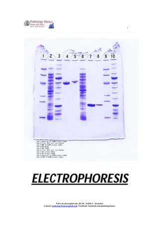
  • 8. 8 SDS-PAGE – Sodium dodecyl sulphate Polyacrylamide gel electrophoresis 1. SDS denatures proteins according to their molecular weight 2. SDS-PAGE is very powerful for resolving proteins and separating them into subunits according to their molecular weight 3. Very useful for research purposes Notes on electrophoresis.. By Dr. Ashish V. Jawarkar Contact: pathologybasics@gmail.com Facebook: facebook.com/pathologybasics
  • 9. 9 TWO DIMENSIONAL ELECTROPHORESIS (2D ELECTROPHORESIS) 2. Standard separation of proteins is done in one direction 3. This is followed by SDS-PAGE in perpendicular direction 4. This results in hundreds of identifiable protein peaks Notes on electrophoresis.. By Dr. Ashish V. Jawarkar Contact: pathologybasics@gmail.com Facebook: facebook.com/pathologybasics
  • 10. 10 ISOELECTRIC FOCUSSING 1. Gives superior resolution to closely migrating proteins 2. Here the gel used contains a gradient of pH established by using ampholytes 3. As each protein reaches the gel location where the pH equals its own, the net charge becomes zero 4. It cannot move further 5. Thus proteins move here strictly on the basis of their pH Notes on electrophoresis.. By Dr. Ashish V. Jawarkar Contact: pathologybasics@gmail.com Facebook: facebook.com/pathologybasics
  • 11. 11 * ANCILLARY TECHNIQUES PRECIPITATION 1. The sample to be tested usually contains a lot of undesired proteins. 2. These undesired proteins can be removed by precipitation so that a single minor protein can be isolated by electrophoresis. 3. First the value of total proteins is found out 4. Then Sodium sulfate, sodium sulfite, ammonium sulfate or methanol is used to precipitate globulins. 5. Protein estimation from the remaining solution after precipitation can give an idea about the albumin concentration – electrophoresis can give an idea about the types of proteins apart from globulins. 6. globulin = total protein – albumin 7. estimation of albumin : globulin ratio is useful in various disorders Normal : A:G ratio >1:5 (1-3) Decreased A:G ratio Decreased albumin Malnutrition Malabsorption Liver failure Proteinuria Accumulation of ascetic fluid Enteropathy Increased A:G ratio ************ Increased globulins Acute or chronic infections Myelomas Notes on electrophoresis.. By Dr. Ashish V. Jawarkar Contact: pathologybasics@gmail.com Facebook: facebook.com/pathologybasics
  • 12. 12 COLUMN SEPARATION (1) Gel filteration: 1. Media such as agarose beads/sephadex is used 2. They have beads in them with pores of various sizes 3. Largest proteins cannot pass through pores in the beads – they pass through spaces in beads and are eluted first 4. Medium sized proteins pass through medium sized pores and appear second. 5. Small sized proteins pass through small sized pores and are eluted last. Notes on electrophoresis.. By Dr. Ashish V. Jawarkar Contact: pathologybasics@gmail.com Facebook: facebook.com/pathologybasics
  • 13. 13 (2) Ion Exchange Chromatgraphy 1. This is of two types – Anion exchange and Cation exchange chromatography 2. This technique uses charged particles which are placed in the column. 3. When proteins having the opposite charge as the column pass through it, they are retained for a longer time, neutral elute the second while same charged particles elute first. ANION EXCHANGE CHROMATOGRAPHY 1. Media such as DEAE or QAE are used with have net positive charge. 2. Albumin, α and some β globulins which are anions stick to positively charged matrix. 3. γ globulins that have no charge are eluted faster. CATION EXCHANGE CHROMATOGRAPHY 1. Media such as carboxymethylcellulose are used which have a negative charge. 2. Positively charged cations stick to the matrix 3. Albumin is eluted first here Notes on electrophoresis.. By Dr. Ashish V. Jawarkar Contact: pathologybasics@gmail.com Facebook: facebook.com/pathologybasics
  • 14. 14 (3) Hydrophobic chromatography 1. In this technique, support medium interacts with proteins according to their hydrophobic nature. 2. Here ‘high salts’ and ‘low salts’ are used 3. high salts structure water around themselves and expose the hydrophobic ends of proteins 4. these hydrophobic ends get attracted toward the medium and stick to it 5. according to hydrophobic nature of proteins, they stick or are eluted 6. ‘low salts’ are used to elute as they donot / expose less number of hydrophobic ends. Notes on electrophoresis.. By Dr. Ashish V. Jawarkar Contact: pathologybasics@gmail.com Facebook: facebook.com/pathologybasics
  • 15. 15 (4) Affinity chromatography 1. This is based on specific binding between a protein of interest and another protein (can be its antibody/antigen) bound to the solid medium. 2. Bound protein can then be eluted in its purified form. 3. examples a. to separate FVII-vWF – column is incorporated with monoclonal antibody to vWF b. Igs can be separated by using Staphylococcal protein A in gel matrix c. Attaching dihydroxyboronate to medium helps separate glycosylated hemoglobin d. To separate digoxin – digibind is used CroFab – crotalid snake venom Notes on electrophoresis.. By Dr. Ashish V. Jawarkar Contact: pathologybasics@gmail.com Facebook: facebook.com/pathologybasics
  • 16. 16 (5) Capillary electrophoresis 1. This procedure is similar to HPLC in that a column is employed where separation of molecules is done on the basis of size/hydrophobicity or strereospecificity. 2. Applicable to large molecules such as DNA. Notes on electrophoresis.. By Dr. Ashish V. Jawarkar Contact: pathologybasics@gmail.com Facebook: facebook.com/pathologybasics
  • 17. 17 * SUMMARY OF WORK FLOW Notes on electrophoresis.. By Dr. Ashish V. Jawarkar Contact: pathologybasics@gmail.com Facebook: facebook.com/pathologybasics
  • 18. 18 * APPLICATIONS (1) SERUM PROTEIN ELECTROPHORESIS Following patterns are observed in normal conditions and various disorders on electrophoresis. Normal: Cirrhosis of liver: When liver function is sufficiently diminished, protein synthesizing capacity is compromised and concentrations of albumin and proteins in the alpha and beta bands are decreased. An additional common finding is beta-gamma bridging due to increased IgA. Beta-gamma bridging Alb α1 α2 β γ Notes on electrophoresis.. By Dr. Ashish V. Jawarkar Contact: pathologybasics@gmail.com Facebook: facebook.com/pathologybasics
  • 19. 19 The Nephrotic Syndrome -1. Renal disease involving the glomeruli is always associated with increased urinary protein loss. When protein loss is greater than 3-4 g/day, the protein synthesizing capacity of the liver is exceeded and hypoproteinemia, accompanied by anasarca, develops to cause the nephrotic syndrome. 2. The massive urine protein loss is due to increased permeability of glomeruli to protein. The permeability increase may be minimal so that only albumin and other smaller molecular weight proteins are selectively filtered (selective nephrosis, as in Minimal Change Disease) or may be greater so that larger proteins are also filtered (nonselective nephrosis, as in membranous golmerulonephritis) as is the case in the example shown. 3. Alpha-2-macroglobulin is sufficiently large so that it is not filtered and increased synthesis (from the general hepatic protein synthesis) causes its accumulation. 4. Lipoproteins are also sufficiently large to accumulate and hyperlipidemia is a characteristic of the nephrotic syndrome, although lipoproteins are not stained with the protein stain used in visualizing proteins. Normal Abnormal Alb α1 α2 β γ Alpha-1-Antitrypsin Deficiency: A genetic defect causes a deficiency of alpha-1-antitrypsin. The antiprotease deficiency results in a propensity to develop emphysema. Since alpha-1-antitrypsin is the major component of the alpha-1 band, deficiency is suggested by a reduced alpha-1 band. Deficiency is confirmed by specific immunochemical quantification. Normal Abnormal Alb α1 α2 β γ Notes on electrophoresis.. By Dr. Ashish V. Jawarkar Contact: pathologybasics@gmail.com Facebook: facebook.com/pathologybasics
  • 20. 20 Acute Inflammation The alpha-1 and alpha-2 bands are increased during the inflammatory response from increased hepatic synthesis of acute phase reactant proteins. Normal Abnormal Alb α1 α2 β γ Chronic Inflammation -Immunoglobulin synthesis by antigen activated B lymphocytes transformed to plasma cells is demonstrated by the increased polyclonal gamma band. Normal Abnormal Alb α1 α2 β γ Notes on electrophoresis.. By Dr. Ashish V. Jawarkar Contact: pathologybasics@gmail.com Facebook: facebook.com/pathologybasics
  • 21. 21 Immunoglobulin Deficiency -Deficient immunoglobulin synthesis is revealed by a markedly diminished gamma band. Effected individuals are prone to recurrent infection Monoclonal Gammopathy -1. An unusually sharp band in the gamma region strongly suggests the presence of a homogeneous immunoglobulin and, thus, the malignant proliferation of plasma cells from a single cell (multiple myeloma) in contrast to the broad, heterogeneous, or polyclonal, gamma band as exhibited above in chronic inflammation from immunoglobulin synthesis by many different clones of plasma cells. 2. Homogeneous immunoglobulins are also found in Waldenstrom's macroglobulinemia (where the sharp gamma band is always IgM). Specimens which exhibit a narrow gamma band are further examined by immunofixation electrophoresis as described below Alb α1 α2 β γ (2) CSF ELECTROPHORESIS Notes on electrophoresis.. By Dr. Ashish V. Jawarkar Contact: pathologybasics@gmail.com Facebook: facebook.com/pathologybasics
  • 22. 22 1. The diagnosis of Multiple sclerosis is ultimately a clinical one based on neurologic history and physical examination. 2. Nevertheless, advanced laboratory results such as elevated IgG indices and presence of oligoclonal bands of IgG or Kappa /lambda light chains on CSF electrophoresis, as well as neuroimaging techniques have proven to be invaluable in diagnosis of MS. Agarose gel electrophoresis 1. Concentrated CSF is used as sample 2. Shows discrete bands of IgG – the oligoclonal bands 3. Presence of two or more bands is necessary for diagnosis 4. False positive results are obtained in SSPE, CNS infections like neurosyphilis, Crypotococcal meningitis, GBS, transverse myelitis, burkitt’s lymphoma etc. 5. Sensitivity is less than other methods of electrophoresis Staining Commassie brilliant blue/paragon violet/silver staining 1. silver staining is more sensitive then CBB 2. however these techniques must be simultaneously carried out on patients serum also because polyclonal gammopathy can lead to diffusion of immunoglobulin into CSF and lead to false positive results. This is seen in case of liver diseases, SLE, RA and chronic granulomatous diseases. ImmunoFixation Electrophoresis 1. Immunofixation electrophoresis (IFE) is used to demonstrate that a narrow gamma band is due to a homogeneous immunoglobulin. 2. In the illustration below, 6 replicates of the specimen are loaded on to separate lanes of an IFE gel. 3. Following electrophoresis, the protein in each lane is stained differently. 4. The first lane (SP) is stained for total protein. 5. The protein in each of the other lanes is "stained" with specific antisera for immunoglobulin heavy and light chains, respectively, as illustrated in the figure. 6. The finding of the preponderance of only one light chain associated with a predominantly staining heavy chain confirms the molecular homogeneity of the immunoglobulin and also provides identification. 7. IFE identifies the narrow band as monoclonal IgG, lambda. 8. Sometimes malignant plasma cells synthesize excess light chains and less frequently only light chains are synthesized. Almost never is excess heavy chain or only heavy chain synthesized. Excess free lambda light chain is exhibited in the illustration. Notes on electrophoresis.. By Dr. Ashish V. Jawarkar Contact: pathologybasics@gmail.com Facebook: facebook.com/pathologybasics
  • 23. 23 a. Lymphocytic pleocytosis and 2 plasma cells in the CSF cytospin preparation. b. 4b. Isoelectric focusing/immunofixation revealed CSF-restricted oligoclonal IgG bands as well as identical paraprotein bands in CSF and serum. Anode is on the left. c. 4c. Serum immunofixation electrophoresis revealed the presence of IgGκ monoclonal band (arrow). Anode is at the top. ELP, electrophoresis (general protein stain); G, A, M, K, L: immunofixation for γ, α, μ, κ and λ chain, respectively. d. 4 d. Isoelectric focusing/affinity-mediated immunoblotting analysis: Paraprotein bands did not react with Borrelia antigen but partly obscured CSF-restricted oligoclonal IgG pattern. Paired CSF and serum samples tested for (from left to right) "total" IgG, IgGκ, IgGλ; Anti-Borrelia IgG, IgGκ, IgGλ. Anode is at the top. Notes on electrophoresis.. By Dr. Ashish V. Jawarkar Contact: pathologybasics@gmail.com Facebook: facebook.com/pathologybasics
  • 24. 24 (3) URINE ELECTROPHORESIS 1. 2. 3. 4. 5. 6. 7. The main reason for performing urine protein electrophoresis is to find a light chain myeloma producing an excess of free light chains (Bence Jones protein), an important part of a myeloma screen. A band in the urine protein electropherogram may also result from an intact monoclonal immunoglobulin, especially if the patient has poor renal function. Immunofixation is important in defining the nature of the band and in distinguishing between Bence Jones protein and an intact monoclonal protein originating from the serum. From the urine electropherogram we can also tell if the proteinuria is of glomerular origin with a predominance of albumin, or if it has tubular components with excretion of smaller molecular weight proteins such as retinol binding protein and alpha-1 microglobulin. Fragmented albumin in urine is occasionally seen but is of unknown significance. Historically, urine has been concentrated by either removal of water from the specimen leaving the proteins in higher concentration, or by centrifugation whereby the proteins are spun away from the majority of the water. Demonstration of the protein components of urine from concentrated specimens was originally performed on cellulose acetate and later on agarose and high resolution agarose gel. Notes on electrophoresis.. By Dr. Ashish V. Jawarkar Contact: pathologybasics@gmail.com Facebook: facebook.com/pathologybasics
  • 25. 25 (4) HEMOGLOBIN ELECTROPHORESIS Rationale: 1. Haemoglobin (Hb) abnormalities are the most frequent genetic disease, affecting approximately 7 per cent of the world population. 2. Today, abnormal Hbs are generally discovered during a systematic study performed within programmes for prevention of thalassaemias or sickle cell disease. 3. In several regions (India, Turkey, Irak, Iran, Gaza strip, Saudi Arabia, Cyprus, etc.), these are found during a premarital screening. In other regions, like west Europeans countries, the research for the main Hb disorders is often limited to populations at risk. It is done either as preconceptional or neonatal screening programmes. Methods and patterns: 1. Hb S [β6 Glu>Val], the first abnormal Hb described, was discovered in 1949 by Pauling et al using moving boundary electrophoresis. 2. Zone electrophoresis performed on cellulose acetate strips (CAE) is still used in many clinical laboratories. 3. This technique has a resolution lower than that of IEF, but because of its simplicity remains among the more popular methods used in Hb screening. In this technique, the Hb molecules are separated at alkaline pH. Under these conditions all Hbs have a negative charge and migrate towards the cathode. Hb S, which has an additional positive charge compared to Hb A migrates more slowly. 4. In IEF, a pH gradient is established by carrier ampholytes submitted to an electric current and the Hb molecules migrate across this gradient until they reach the position where their net charge is zero (isoelectric point). The molecules will then concentrate in a sharp band as illustrated in Fig. 5. This technique allows separating molecules with isoelectric points differing only by 0.02 pH unit. Hb D-Punjab [β121 Glu>Gln], not detectable from Hb S by CAE, could easily be recognized by this technique. The disadvantage of this method is its relatively high cost and the requirement for a well-experienced laboratory staff. 6. This technique can help in diagnosing various hemoglobinopathies. Notes on electrophoresis.. By Dr. Ashish V. Jawarkar Contact: pathologybasics@gmail.com Facebook: facebook.com/pathologybasics