Module 2
•Télécharger en tant que PPTX, PDF•
0 j'aime•195 vues

This document discusses the morphology and types of chromosomes. It begins by defining what a chromosome is and where they are located in prokaryotic and eukaryotic cells. It then describes the different structural features of chromosomes visible under a light microscope, including chromatids, centromeres, secondary constrictions, telomeres, and satellites. It explains the different types of chromosomes based on centromere position, number of centromeres, size, and composition. The key differences between heterochromatin and euchromatin are also summarized.

Recommandé
Contenu connexe
Tendances
Tendances (20)
Similaire à Module 2
Similaire à Module 2 (20)
Plus de PILLAI ASWATHY VISWANATH
Plus de PILLAI ASWATHY VISWANATH (20)
Dernier
Dernier (20)
Module 2
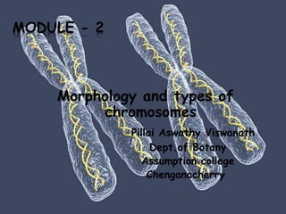
- 1. MODULE - 2 Morphology and types of chromosomes Pillai Aswathy Viswanath Dept.of Botany Assumption college Chenganacherry
- 2. Introduction A chromosome is an organised structure of DNA and protein that is found in the nucleus of eukaryotic cells. In prokaryots they localised in the cytoplasam Chromosome is a single dsDNA in coiled and condensed form Chromosome other wise it called as chromatin
- 3. The difference is that chromatin is less condensed extented DNA While chromosomes are highly condensed DNA DNA is condensed with the help of histones protein in eukaryotes and polyamines in prokarotes
- 5. There are two kinds of eukarotic chromosomes,namely autosomes and sex chromosomes Autosomes carry the genes which control somatic or non sexual characters and sex chromosomes contain the genes which control sexual characteristics The somatic chromosome number is the number of chromosomes found in somatic cell and is represented by 2n (Diploid) The gametic chromosome number is half of the somatic chromosome numbers and represented by n (Haploid).
- 6. Haploid cells have only one copy of each chromosome. In animals, gametes (sperm and eggs) are haploid. Diploid cells have two homologous copies of each chromosome. Both the copies are ordinarily identical in morphology, gene content and gene order and hence known as homologous chromosomes. Each pair of chromosomes made up of two homologs. Homologous chromosome is inherited from separate parents; one homolog comes from the mother and the other comes from the father.
- 8. Chromosome number Normally all individual of a species have the same chromosome number. The number of chromosomes varies from species to species. Sometimes,changes may occur in the somatic number.Such changes are called ploidy changes Ploidy refers to the number of basic chromosome sets for eg. A diploid has 2 sets where as a hexaploid has 6 sets.
- 9. The basic chromosome number x,also called the monoploid number is the number of different chromosomes that make up a single complete set. Generally somatic cells contain two copies of each chromosome except the sex chromosomes.
- 10. Chromosome size The size of the chromosome shows a remarkable variation depending upon the stage of cell division. longest and thinnest during interphase and hence not visible under light microscope. smallest and thickest during mitotic metaphase. Chromosome size is not proportional to the number of genes present on the chromosome.
- 11. Chromosome morphology Mitotic metaphase is the most suitable stage for studies on chromosome morphology. The outer covering or sheath of a chromosome is known as pellicle, which encloses the matrix. Within the matrix lies the chromatin. The chromosome morphology changes during cell division. During Interphase: the chromosomes remain in form of chromatin .They are thin, coiled, elastic, thread-like structures
- 12. As cells enter mitosis, their chromosomes become highly condensed Prophase: distinct thread like structures called chromatid Metaphase and anaphase: they become fully condensed and take the shapes eukaryotic nuclear chromosomes. This cyclic change in shape and size of chromosomes during cell cycle is called chromosomal cycle
- 14. In mitotic metaphase chromosomes, the following structural features can be seen under the light microscope. 1. Chromatid 2. Centromere 3. Secondary constriction 4. Telomere 7. Matrix 8. Satellite
- 15. Chromatid Each metaphase chromosome appears to be longitudinally divided into two identical parts each of which is called chromatid. Chromatids of a chromosome appear to be joined together at a point known as centromere. Two chromatids making up a chromosome are referred to as sister chromatids. The chromatids of homologous chromosomes are known as nonsister chromatids.
- 16. A chromosome consists of two chromatids and each chromatid consists of thread like coiled structures called chromonema (plural chromonemata). Matrix The mass of acromatic material which surrounds the chromonemata is called matrix. The matrix is enclosed in a sheath which is known as pellicle.
- 17. Centromere (primary constriction) Chromosome has a constriction point called the centromere which divides the chromosome into two arms. The short arm of the chromosome is labeled the "p" arm. The long arm of the chromosome is labeled the "q" arm. Centromere usually not located exactly in the center of the chromosomes and in some cases,is located almost at the chromosome’s end
- 19. Based on the position of centromere, chromosomes are called: i. Metacentric (centromere median). ii. Sub-metacentric (centromere is submedian), iii. Acrocentric (centromere subterminal and capped by telomere), iv. Telocentric (centromere terminal),
- 20. Metacentric chromosome The centromere is located in the centre of chromosomes, i.e. the centromere is median. The centromere is localized approximately midway between each end and thereby two arms are roughly equal in length. Metacentric chromosome take V shape during anaphase.
- 21. Submetacentric chromosome Centromere is located on one side of the central point of a chromosome. Centromere is submedian giving one longer and one shorter arms. Submetacentric chromosome may be J or L shaped during anaphase.
- 22. Acrocentric chromosome The centromere located close to one end of chromosomes. The centromere is more terminally placed and forms very unequal arm length (The "acro-" in acrocentric refers to the Greek word for "peak"). The p (short) arm is so short that is hard to observe, but still present. Acrocentric chromosome may be rod shape during anaphase.
- 23. Telocentric chromosome Centromere located at one end of chromosome (at terminal part of chromosome) lies at one end. Telocentic chromosome may be rod shape during anaphase.
- 25. Based on the no. Of centromere Acentric ( without cetromere) monocentric (one centromere); dicentric (e.g. in wheat, maize etc.) polycentric (with few distributed centromer e.g. Luzula. Ascaris
- 26. Kinetochore A complex of proteins associated with the centromere of a chromosome during cell division, to which the microtubules of the spindle attach. The structure of kinetochore is complex and is seen during late prophase. Kinetochore proteins serve as motor proteins for the poleward anaphasic movement of chromosome
- 27. Kinetochore serve as a site for the attachment of spindle fibers during cell division.
- 28. Fuction of centromer Plays a role in the differentiation of chromosomal arms Plays a vital role in the correct distribution of daughter chromosomes to the corresponding daughter cells in mitosis Serves as an attachment site for chromosomal spindle so that the daughter chromosomes are pulled apart to opposite poles
- 29. Secondary constriction In some chromosome addition to centromere / primary constriction, one or more constrictions in the chromosome are present termed secondary constrictions. They are always constant in their positions ,number and extent among the members of species. Secondary constrictions are of two types : NOR (nucleolus organizer region) Joint
- 30. The NOR (nucleolus organizer region) are specialized for the organization of nucleolus It is best known as the site of ribosome biogenesis ( contains the genes which code for 18S and 28S rRNA The joints sometimes develop due to breaking and fusion of chromosome segments. Represents the site of fusion and fission of chromosome segments
- 32. Satellite A Satellite chromosome or SAT Chromosome is a chromosome segment that is separated from the main body of the chromosome by secondary construction
- 33. Telomeres The terminal ends of chromosomes are called telomeres. A telomere is a short repeated DNA sequence (GC rich) complexed with proteins. They are synthesized separately and later add to the chromosomal tips They play critical roles in chromosome replication and maintenance of chromosomal length.
- 34. They are highly stable and do not fuse or unite with telomeres of other chromosomes due to polarity effect. Any broken end of a chromosome is unstable and can join with a piece of any other chromosome. But the telomeres impart stability to the chromosome, which retains its identity and individuality through cell cycle and for many cell generations.
- 35. Composition of chromosomes The material of which chromosomes are composed is called chromatin. N.Fleming introduced the term chromatin in 1879. Chromatin was classified into two groups by cytologists on the basis of its affinity to basic dyes like acetocarmine or feulgen reagent at prophase. The darkly stained regions were called heterochromatin, while lightly stained regions were called euchromatin. This differential staining capacity of different parts of a chromosomes is known as ‘heteropycnosis’
- 37. Heterochromatin? Heterochromatin is the tightly packed form of chromatin present in the cells of eukaryotes. It is usually present at the periphery of the nucleus. Due to its highly packed nature, it is visible during the staining of DNA of a cell. Also, this intensely stained DNA has two types; they are the constitutive and facultative heterochromatin.
- 38. a) Constitutive :- It is present in all cells at identical positions on both homologous chromosomes of a pair. Constitutive heterochromatin is basically responsible for forming the centromere or the telomere while attracting signals for both gene expression and repression.
- 39. b) Facultative:- It varies in state in different cell types, at different stages or sometimes, from one homologous chromosome to another. Facultative heterochromatin becomes repetitive under special signals or environments; otherwise, it stays quiet with a highly condensed structure.
- 40. Euchromatin? Euchromatin is the loosely-packed DNA structures in the cells. Usually, they are present towards the inner core of the nucleus. Euchromatin is present in both prokaryotes and eukaryotes. In fact, euchromatin is the only type of chromatin present in the prokaryotic genetic material. Moreover, its loosely packed structure causes less visibility during the DNA staining, unlike heterochromatin.
- 41. The uncondensed nature of euchromatin is mainly due to the loose wrapping of histone proteins around the DNA strand. Therefore, the access of DNA is easy to initiate the DNA transcription. Moreover, euchromatin contains the most active genes of an organism. It is because euchromatin participates actively in the transcription of DNA into mRNA.
- 43. What is the difference between heterochromatin and euchromatin? Heterochromatin and euchromatin are two varieties of chromatin present in living organisms. The key difference between heterochromatin and euchromatin is that the heterochromatin is the highly packed form of chromatin in the nucleus while euchromatin is the loosely packed form of chromatin in the nucleus. Heterochromatin is inactive while euchromatin is active. Consequently, heterochromatin contains more dna, while euchromatin contains less dna.
- 44. Heterochromatin is less abundant. But, around 90% of the total human genome is euchromatin. A further difference between heterochromatin and euchromatin is that heterochromatin is only present in eukaryotes, but, euchromatin is present in both prokaryotes and eukaryotes.
- 45. Questions Morphology of chromosomes? Types of chromosomes? Hetrochromatin? Euchromatin? Difference? Terms? Like Kinetochore,Chromatid,etc.