Section ii a biochemistry carbohydrate
•Télécharger en tant que PPT, PDF•
15 j'aime•1,069 vues

carbohydrate

Recommandé
Contenu connexe
Tendances
Tendances (20)
Similaire à Section ii a biochemistry carbohydrate
Similaire à Section ii a biochemistry carbohydrate (20)
Plus de PNK SINGH
Plus de PNK SINGH (20)
Dernier
Dernier (20)
Section ii a biochemistry carbohydrate
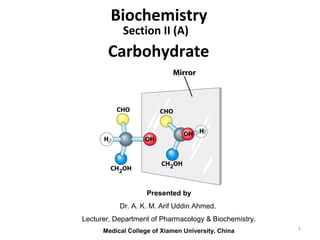
- 1. Presented by Dr. A. K. M. Arif Uddin Ahmed. Lecturer, Department of Pharmacology & Biochemistry. Medical College of Xiamen University, China Section II (A) Biochemistry Carbohydrate 1
- 3. CARBOHYDRATES ARE ALDEHYDE OR KETONE DERIVATIVES OF POLYHYDRIC ALCOHOLS. They are the end products of photosynthesis. Contain three elements - C, H, O, According to the formula (CH2O)n where n ≥ 3. Example:- 3
- 4. Plants: photosynthesis chlorophyll 6 CO2 + 6 H2O C6H12O6 + 6 O2 sunlight (+)-glucose (+)-glucose starch or cellulose respiration C6H12O6 + 6 O2 6 CO2 + 6 H2O + energy Animals plant starch (+)-glucose (+)-glucose glycogen glycogen (+)-glucose (+)-glucose fats or aminoacids respiration (+)-glucose + 6 O2 6 CO2 + 6 H2O + energy 4
- 5. Functions of carbohydrate •Carbohydrates are essential to all living organisms and are the most abundant class of biological molecules. •The metabolic breakdown of monosaccharides provides most of the energy used to power biological processes. •In structural material (cell walls, connective tissue) •Important for cell signalling, cell-cell interactions 5
- 6. Classification of carbohydrates • Monosaccharides- glucose, fructose • Oligosaccharides • Disaccharides, trisaccharides, tetrasaccharides, pentasaccharides, hexasaccharides, (up to 10 monosaccharides ) • Most important are the disaccharides- maltose, lactose, sucrose . • Polysaccharides • Homopolysaccharides- starch, glycogen, cellulose, chitin, inulin. • Heteropolysaccharides • Complex carbohydrates 6
- 7. Monosaccharides The monosaccharide commonly found in humans are classified according to the number of carbons they contain in their backbone structures. Classifications of monosaccharide Trioses (C3 H6 O3 ) Glyceraldehyde Dihydroxyacetone Tetroses (C4 H8 O4 ) Erythrose Erythrulose Pentoses (C5 H10 O5 ) Ribose Ribulose Hexoses (C6 H12 O6 ) Glucose Fructose Heptoses (C7 H14 O7 ) — Sedoheptulose ALDOSES KETOSES 7
- 8. Aldoses Aldoses are monosaccharides • with an aldehyde group • with many hydroxyl (-OH) groups. triose (3C atoms) tetrose (4C atoms) pentose (5 C atoms) hexose (6 C atoms) O ║ C─H aldose │ H─ C─OH │ H─ C─OH │ CH2OH Erythose, an aldotetrose 8
- 9. Ketoses Ketoses are monosaccharides • with a ketone group • with many hydroxyl (-OH) groups. CH2OH │ C=O ketose │ H─ C─OH │ H─ C─OH │ H─C─OH │ CH2OH Fructose, a ketohexose 9
- 10. Identify each as aldo- or keto- and as tetrose, pentose, or hexose: H CH2OH OHC H H H OH OH OH C C C HC O CH2OH HHO CH2OH O H OHC C C aldohexose ketopentose 10
- 13. Epimers (two sugars that differ only in the configuration around one carbon atom) 13
- 14. Monosaccharides have asymmetric centers Three ways to represent the two stereoisomers of glyceraldehyde 14
- 15. D & L sugars are mirror images of one another. They have the same name, e.g., D-glucose & L-glucose. Other stereoisomers have unique names, e.g., glucose, mannose, galactose, etc. The number of stereoisomers is 2n , where n is the number of asymmetric centers. The 6-C aldoses have 4 asymmetric centers. Thus there are 16 stereoisomers (8 D-sugars and 8 L-sugars). O H O H C C H – C – OH HO – C – H HO – C – H H – C – OH H – C – OH HO – C – H H – C – OH HO – C – H CH2OH CH2OH D-glucose L-glucose 15
- 16. Formation of hemiacetals and hemiketals 16
- 17. Cyclic Structures Cyclic structures • are the prevalent form of monosaccharides with 5 or 6 carbon atoms. • form when the hydroxyl group on C-5 reacts with the aldehyde group or ketone group. O O 17
- 18. The Formation of Cyclic Glucose 18
- 19. Alpha, Beta anomers The α and β anomes of D-glucose interconvert in aqueous solution by a process called mutarotation. Isomeric forms of monosaccharides that differ only in the configuration of the number-1 carbon atom are called anomers. 19
- 20. Cyclic Structure of Fructose Fructose • is a ketohexose. • forms a cyclic structure. • reacts the —OH on C-5 with the C=O on C-2. D-fructose β-D-fructoseα-D-fructose O CH2OH OH OH OH CH2OH O OH CH2OH OH OH CH2OH H OH H OH HHO O CH2OH C C C C CH2OH 20
- 23. Some hexose derivatives important in biology 23
- 24. Reducing Sugars • Sugars that contain aldehyde groups that are oxidized to carboxylic acids are classified as reducing sugars. • Common test reagents are : • Benedicts reagent (CuSO4 / citrate) • Fehlings reagent (CuSO4 / tartrate) • They are classified as reducing sugars since they reduce the Cu2+ to Cu+ which forms as a red precipitate, copper (I) oxide. 24
- 25. Glucose and other sugars capable of reducing Cu2+ are called reducing sugars. Sucrose is non reducing sugar. 25 Reducing Sugars
- 26. Physiologic Importance of Pentoses. Sugar Source Biochemical and Clinical Importance D-Ribose Nucleic acids and metabolic intermediate Structural component of nucleic acids and coenzymes, including ATP, NAD(P), and flavin coenzymes D-Ribulose Metabolic intermediate Intermediate in the pentose phosphate pathway D-Arabinose Plant gums Constituent of glycoproteins D-Xylose Plant gums, proteoglycans, glycosaminoglycans Constituent of glycoproteins L-Xylulose Metabolic intermediate Excreted in the urine in essential pentosuria 26
- 27. Physiologic Importance of Hexoses. Sugar Source Biochemical Importance Clinical Significance D-Glucose Fruit juices, hydrolysis of starch, cane or beet sugar, maltose and lactose The main metabolic fuel for tissues; "blood sugar" Excreted in the urine (glucosuria) in poorly controlled diabetes mellitus as a result of hyperglycemia D-Fructose Fruit juices, honey, hydrolysis of cane or beet sugar and inulin, enzymic isomerization of glucosesyrups for food manufacture Readily metabolized either via glucoseor directly Hereditary fructose intolerance leads to fructose accumulation and hypoglycemia D-Galactose Hydrolysis of lactose Readily metabolized to glucose; synthesized in the mammary gland for synthesis of lactose in milk. A constituent of glycolipids and glycoproteins Hereditary galactosemia as a result of failure to metabolize galactose leads to cataracts D-Mannose Hydrolysis of plant mannan gums Constituent of glycoproteins 27
- 28. Oligosaccharide Some common Disaccharides: Maltose, a cleavage product of starch (e.g., amylose), is a disaccharide with an α(1→ 4) glycosidic link between C1 - C4 OH of 2 glucoses. It is the α anomer (C1 O points down). Lactose, milk sugar, is composed of galactose & glucose, with β(1→4) linkage from the anomeric OH of galactose. Its full name is β-D-galactopyranosyl-(1→ 4)-α-D-glucopyranose Sucrose, common table sugar, has a glycosidic bond linking the anomeric hydroxyls of glucose & fructose.Because the configuration at the anomeric C of glucose is α (O points down from ring), the linkage is α(1→2). The full name of sucrose is α-D-glucopyranosyl-(1→2)-β-D-fructopyranose.) 28
- 29. Important Disaccharides A disaccharide consists of two monosaccharides. Monosaccharides Disaccharide glucose + glucose maltose + H2O glucose + galactose lactose + H2O glucose + fructose sucrose + H2O 29
- 30. Disaccharides contain a glycosidic bond Formation of maltose anomeric carbon Glu(α1→4)Glu The glycosidic bond protects the anomeric carbon from oxidation. 30
- 31. Some common disaccharides reducing sugar non reducing sugar reducing sugar 31
- 32. Maltose Maltose is • a disaccharide also known as malt sugar. • composed of two D-glucose molecules. • obtained from the hydrolysis of starch. • used in cereals, candies, and brewing. • found in both the α- and β - forms. 32
- 33. Formation of Maltose Free α-OH 33
- 34. Lactose Lactose • is a disaccharide of β-D- galactose and α- or β-D- glucose. • contains a β -1,4- glycosidic bond. • is found in milk and milk products. α- form α- form 34
- 35. Sucrose Sucrose or table sugar • is obtained from sugar cane and sugar beets. • consists of α-D-glucose and β-D-fructose.. • has an α,β-1,2-glycosidic bond. α-D-glucose β -D-fructose 35
- 36. Sweetness of Sweeteners Sugars and artificial sweeteners • differ in sweetness. • are compared to sucrose (table sugar), which is assigned a value of 100. 60 000 36
- 37. Learning Check Identify the monosaccharides in each of the following: A. lactose (1) α-D-glucose (2) β-D-fructose (3) β-D-galactose B. maltose (1) α-D-glucose (2) β-D-fructose (3) β-D-galactose C. sucrose (1) α-D-glucose (2) β-D-fructose (3) β-D-galactose 37
- 38. Sugar Source Clinical Significance Isomaltose Enzymic hydrolysis of starch (the branch points in amylopectin) Maltose Enzymic hydrolysis of starch (amylase); germinating cereals and malt Lactose Milk (and many pharmaceutical preparations as a filler) Lack of lactase (alactasia) leads to lactose intolerance—diarrhea and flatulence; may be excreted in the urine in pregnancy Lactulose Heated milk (small amounts), mainly synthetic Not hydrolyzed by intestinal enzymes, but fermented by intestinal bacteria; used as a mild osmotic laxative Sucrose Cane and beet sugar, sorghum and some fruits and vegetables Rare genetic lack of sucrase leads to sucrose intolerance—diarrhea and flatulence Trehalose Yeasts and fungi; the main sugar of insect hemolymph Physiologic Importance of Disaccharides. 38
- 39. Polysaccharides • Homopolysaccharides:- (starch, glycogen, cellulose, Chitin, inulin) • Heteropolysaccharides:- (peptidoglycan, agarose, hyaluronate, chondroitin sulfate, keratan sulfate, heparin) • Characteristics of polysaccharides: • polymers (MW from 200,000) • White and amorphous products (glassy) • not sweet • not reducing; do not give the typical aldose or ketose reactions) • form colloidal solutions or suspensions 39
- 40. Polysaccharides (also called Glycans) Most carbohydrates found in nature occur as polysaccharides. 40
- 41. Starch • most common storage polysaccharide in plants • composed of 10 – 30% α−amylose and 70- 90% amylopectin depending on the source • Common sources are grains , potatoes, peas, beans, wheat 41
- 42. Electron micrographs of starch granules 42
- 43. Amylose and amylopectin, the polysaccharides of starch amylopectin occurs every 24 to 30 residues Strands of amylopectin form double helical structures with each other or with amylose strands 43
- 44. Structures of Amylose and Amylopectin 44
- 45. Amylose Amylose is • a polymer of α-D- glucose molecules. • linked by α-1,4 glycosidic bonds. • a continuous (unbranched) chain. 45
- 46. Amylopectin Amylopectin • is a polymer of α-D- glucose molecules. • is a branched-chain polysaccharide. • has α-1,4-glycosidic bonds between the glucose units. • has α-1,6 bonds to branches. 46
- 47. Dextrins • Starches like amylose and amylopectin hydrolyze to dextrins (smaller polysaccharides) • Contain 3-8 glucose units 47
- 48. Glycogen • also known as animal starch • stored in muscle and liver • present in cells as granules (high MW) • contains both α(1,4) links and α(1,6) branches at every 8 to 12 glucose unit • complete hydrolysis yields glucose 48
- 49. Electron micrographs of glycogen 49
- 50. Glycogen, the glucose storage polymer in animals, is similar in structure to amylopectin. But glycogen has more α(1→6) branches. The highly branched structure permits rapid glucose release from glycogen stores, e.g., in muscle during exercise. The ability to rapidly mobilize glucose is more essential to animals than to plants. H O OH H OHH OH CH2OH H O H H OHH OH CH2OH H O HH H O O H OHH OH CH2 H H H O H OHH OH CH2OH H OH HH O O H OHH OH CH2OH H O H O 1 4 6 H O H OHH OH CH2OH H H H O H OHH OH CH2OH H H O 1 OH 3 4 5 2 glycogen Glycogen 50
- 51. Glycogen Molecule B branched G A AA A A A A A B C B BB B B B B A linear 50 000 glucose units 2 000 non-reducing ends All chains of same length (11~15 Glucose/chain) 2 branching points/B chain 51
- 52. Cellulose, a major constituent of plant cell walls, consists of long linear chains of glucose with β(1→4) linkages. Every other glucose is flipped over, due to β linkages. This promotes intra-chain and inter-chain H-bonds and van der Waals interactions, that cause cellulose chains to be straight & rigid. cannot be digested by humans because humans cannot break down - β(1→4) glycosidic bonds. cellulose H O OH H OHH OH CH2OH H O H OHH OH CH2OH H O H H O O H OHH OH CH2OH H H O H OHH OH CH2OH H H OHH O O H OHH OH CH2OH H O H H H H 1 6 5 4 3 1 2 Cellulose 52
- 53. Chitin – exoskeleton of anthropods. Linear, unbranched homopolymer. N-acetylglucosamine units in (β1→4) linkage. Only difference from cellulose is the acetylated amino group instead of – OH at C-2. Chitin 53
- 54. Functions of homopolysaccharides Starch homopolymer, called a glucosan or glucan. most important dietary source of carbohydrate. constituents are amylose (13–20%), nonbranching helical structure, and amylopectin (80–85%), Glycogen storage polysaccharide. D-glucopyranose residues (in 1 4 glucosidic linkage) with branching by means of 1 6 glucosidic bonds . Dextrins are intermediates in the hydrolysis of starch. Cellulose insoluble -D-glucopyranose 1 4 bonds cross-linking hydrogen bonds. Chitin exoskeleton of crustaceans and insects. Inulin polysaccharide of fructose ,used to determine the glomerular filtration rate, 54
- 55. Peptidoglycan of Bacterial cell walls Alternating (β1→4) linked GlcNAc and Mur2Ac residues. Lysozyme in tears and saliva cleaves the linkage. Penicillin and β-lactamase Heteropolysaccharides 55
- 56. Used in Agarose gel electrophoresis & nucleic acids separation Agarose in seaweeds (Algae) Heteropolysaccharides 56
- 57. Repeating units of some common glycosaminoglycans of extracellular matrix linear polymers composed of repeating disaccharide units Glucoronic acid N-Acetylglucosamine Heteropolysaccharides 57
- 58. Glycosaminoglycans (mucopolysaccharides) are linear polymers of repeating disaccharides. The constituent monosaccharides tend to be modified, with acidic groups, amino groups, sulfated hydroxyl and amino groups, etc. Glycosaminoglycans tend to be negatively charged, because of the prevalence of acidic groups. H O H H OHH OH COO− H H O OH H H NHCOCH3H CH2OH H OO D-glucuronate O 1 23 4 5 6 1 23 4 5 6 N-acetyl-D-glucosamine hyaluronate 58
- 59. Hyaluronate (hyaluronan) is a glycosaminoglycan with a repeating disaccharide consisting of 2 glucose derivatives, glucuronate (glucuronic acid) & N-acetyl-glucosamine. The glycosidic linkages are β(1→3) & β(1→4). H O H H OHH OH COO− H H O OH H H NHCOCH3H CH2OH H OO D-glucuronate O 1 23 4 5 6 1 23 4 5 6 N-acetyl-D-glucosamine hyaluronate 59
- 60. Heparin or Heparan sulfate is initially synthesized on a membrane-embedded core protein as a polymer of alternating N-acetylglucosamine and glucuronate residues. Later, in segments of the polymer, glucuronate residues may be converted to the sulfated sugar iduronic acid, while N-acetylglucosamine residues may be deacetylated and/or sulfated. H O H OSO3 − H OH H COO− O H H NHSO3 − H OH CH2OSO3 − H H H O O heparin or heparan sulfate - examples of residues iduronate-2-sulfate N-sulfo-glucosamine-6-sulfate 60
- 61. Heparin, a soluble glycosaminoglycan found in granules of mast cells, has a structure similar to that of heparan sulfates, but is more highly sulfated. When released into the blood, it inhibits clot formation by interacting with the protein antithrombin. Heparin has an extended helical conformation. heparin: (IDS-SGN)5 PDB 1RID C O N S Charge repulsion by the many negatively charged groups may contribute to this conformation. Heparin shown has 10 residues, alternating IDS (iduronate-2-sulfate) & SGN (N-sulfo-glucosamine-6-sulfate). 61
- 62. 62
- 63. Glycoconjugates (Proteoglycans, Glycoproteins, Glycolipids) Glycoconjugate: Biologically active molecule made from a carbohydrate covalently linked to a protein or lipid (glycoprotein or glycolipid) -- found at cell surfaces Both glycoproteins and glycolipids are important in: Cell-cell recognition and adhesion, Cell migration during development Blood clotting, The immune response, Wound healing, etc. In all these cases, the carbohydrate parts serve as the information carrier by providing specific, high affinity recognition sites. Complex carbohydrates 63
- 64. Some proteoglycans of the extracellular matrix bind non- covalently to hyaluronate via protein domains called link modules. E.g.: • Multiple copies of the aggrecan proteoglycan associate with hyaluronate in cartilage to form large complexes. • Versican, another proteoglycan, binds hyaluronate in the extracellular matrix of loose connective tissues. H O H H OHH OH COO− H H O OH H H NHCOCH3H CH2OH H OO D-glucuronate O 1 23 4 5 6 1 23 4 5 6 N-acetyl-D-glucosamine hyaluronate Proteoglycans 64
- 65. The core protein of a syndecan heparan sulfate proteoglycan includes a single transmembrane α-helix, as in the simplified diagram above. The core protein of a glypican heparan sulfate proteoglycan is attached to the outer surface of the plasma membrane via covalent linkage to a modified phosphatidylinositol lipid. heparan sulfate glycosaminoglycan cytosol core protein transmembrane α-helix Some cell surface heparan sulfate glycosaminoglycans remain covalently linked to core proteins embedded in the plasma membrane. Proteoglycans 65
- 66. - (Smaller and diverse) carbohydrate (1~70% by mass)-protein conjugates. - Glycoproteins are found on the outer surface of plasma membrane, in the extracellular matrix, in the blood, and in specific organelles, Golgi complexes, lysosomes, and secretory granules. • Why glycoproteins? – The biological advantages of adding oligosaccharides to proteins: - Increase polarity and solubility of the proteins. - May influence the folding process. - Protect from proteolytic enzymes. - Responsible for specific biological activities: Intracellular targeting of proteins Cell-cell interactions, Tissue development Extracellular signaling -Carbohydrate forms a glycosidic linkage with the – OH of Ser or Thr through its anomeric end (O-linked), or an N-glycosyl link through the amide of Asn (N-linked). Glycoproteins 66
- 67. Glycoproteins 67
- 68. O-linked oligosaccharide chains of glycoproteins vary in complexity. They link to a protein via a glycosidic bond between a sugar residue & a serine or threonine OH. O-linked oligosaccharides have roles in recognition, interaction, and enzyme regulation. Glycoproteins 68
- 69. N-acetylglucosamine (GlcNAc) is a common O-linked glycosylation of protein serine or threonine residues. Many cellular proteins, including enzymes & transcription factors, are regulated by reversible GlcNAc attachment. Often attachment of GlcNAc to a protein OH alternates with phosphorylation, with these 2 modifications having opposite regulatory effects (stimulation or inhibition). Glycoproteins 69
- 70. N-linked oligosaccharides of glycoproteins tend to be complex and branched. First N-acetylglucosamine is linked to a protein via the side-chain N of an asparagine residue in a particular 3-amino acid sequence. H O OH HN H H HNH OH CH2OH H C CH3 O C CH2 CH O HN C HN O HC C HN HC R O C R O Asn X Ser or Thr N-acetylglucosamine Initial sugar in N-linked glycoprotein oligosaccharide Glycoproteins 70
- 71. Additional monosaccharides are added, and the N-linked oligosaccharide chain is modified by removal and addition of residues, to yield a characteristic branched structure. Glycoproteins 71
- 72. Many proteins secreted by cells have attached N-linked oligosaccharide chains. Genetic diseases have been attributed to deficiency of particular enzymes involved in synthesizing or modifying oligosaccharide chains of these glycoproteins. Such diseases, and gene knockout studies in mice, have been used to define pathways of modification of oligosaccharide chains of glycoproteins and glycolipids. Carbohydrate chains of plasma membrane glycoproteins and glycolipids usually face the outside of the cell. They have roles in cell-cell interaction and signaling, and in forming a protective layer on the surface of some cells. Glycoproteins 72
- 73. Lectinsare glycoproteins that recognize and bind to specific oligosaccharides. Concanavalin A & wheat germ agglutinin are plant lectins that have been useful research tools. Examples of animal lectins: Mannan-binding lectin (MBL) is a glycoprotein found in blood plasma. It binds cell surface carbohydrates of disease-causing microorganisms & promotes phagocytosis of these organisms as part of the immune response. Recognition/binding of CHO moieties of glycoproteins, glycolipids & proteoglycans by animal lectins is a factor in: • cell-cell recognition • adhesion of cells to the extracellular matrix • interaction of cells with chemokines and growth factors • recognition of disease-causing microorganisms • initiation and control of inflammation. Glycoproteins 73
- 74. Glycolipids and Blood Group - Blood group antigens are immunochemical markers made of glycolipids on the surface of red blood cells. - Those with type A cells have type A antigens on their cell surfaces, B have B antigens, AB have both, O carry the O antigen - The only difference appears at the terminal sugar Glycolipids 74
- 75. Thank You 75