1. From the
Archives of the AFIP
This article meets the Bronchogemc Carcinoma:
criteriafor 1.0 credit
hour in Category 1 of Radiologic-Pathologic Correlation1
theAMA Physician’s Melissa L. Rosado-de-Christenson, Lt Col, USAF, MC Philip
#{149} A. Templeton, MD
Recognition Award.
CesarA. Moran, Maf, USAF, MC
To obtain credit, see
the questionnaire at
the end oftbe article.
Bronchogenic carcinoma is the leading cause of death from cancer in men
and women in the United States. Although the cause ofthis malignancy is
probably multifactorial, approximately 85% of lung cancer deaths are attrib-
utable to cigarette smoking. Patients may present with symptoms of airway
obstruction caused by central tumors, symptoms related to direct tumor in-
vasion of surrounding structures, or symptoms produced by distant metasta-
ses. There are four major cell types: adenocarcinoma, squamous cell carci-
noma, undifferentiated large cell carcinoma, and small cell carcinoma.
Adenocarcinoma and undifferentiated large cell carcinoma are generally pe-
ripheral lesions manifesting as solitary nodules or masses, whereas squa-
mous cell carcinoma and small cell carcinoma are typically central and may
manifest as hilar masses, atelectasis, or pneumonia. The prognosis for pa-
tients with bronchogenic carcinoma is poor, with an overall 5-year survival
of 10%-15%. In general, patients with squamous cell carcinoma have the
best prognosis, those with adenocarcinoma and undifferentiated large cell
carcinoma have an intermediate prognosis, and those with small cell carci-
noma have the worst prognosis.
U INTRODUCTION
The term “bronchogenic carcinoma” is synonymous with the terms “lung cancer”
and “lung carcinoma. “ Its use has been criticized, since not all of these tumors onigi-
Abbreviations: H-E hematoxylin, PA = posteroanterior
Index terms: Adenocarcinoma, 60.3212 Lung
#{149} neoplasms, 60.31 1, 60.320, 60.3214, 60.32 16
RadioGraphlcs 1994; 14:429-446
From the Departments of Radiologic Pathology (M.L.R.) and Pulmonary and Mediastinal Pathology (CAM.), Armed
Forces Institute of Pathology, Bldg 54, Rm M.121, Alaska and Fern Sts, Washington, DC 20306-6000; the Department of
Radiology and Nuclear Medicine, Uniformed Services University ofthe Health Sciences, Bethesda, Md (M.L.R.); and the
Department of Radiology, University of Maryland Medical System, Baltimore (PAT.). Received October 25, 1993; revi.
sion requested November 12 and received December 15; accepted December 16. Address reprint requestso M.L.R.
t
The opinions and assertions contained herein are the private views of the authors and are not to be construed as official
or as reflecting the views of the Department of the Air Force or the Department of Defense.
EdUors note-This material was previously presented at the American College of Radiology Categorical Course on Im.
agingofCancers on September 12, 1992. Figures lb. 3, 4a. 8c, 11, 14, 16b, 18, and 19b are reprinted, with permission,
from a chapter in an ACR syllabus (Rosado.de-Christenson ML, Moran CA. Primary lung cancer: pathology and present.
ing features. In: Bragg DG, Thompson WM, eds. Categorical course on imaging of cancers: diagnosis, staging, and fol.
low-up challenges. ACR 1992; 1-8).
C RSNA, 1994
429
2. nate in the bronchial epithelium. However, produced by the primary tumor. Central tu-
the term is used by some pathologists to refer mons may cause coughing, wheezing, hemop-
to those primary malignant neoplasms of the tysis, and pneumonia. Although a rare clinical
lung related to exposure to inhaled carcino- manifestation, diffuse lung involvement by
gens, mainly cigarette smoke (1). We use this bronchioloalveolar carcinoma may produce
term throughout this article. bronchorrhca, the expectoration of large
Bronchogenic carcinoma is a disease of amounts ofmucus (5). With direct invasion of
great importance in both the United States local extnapulmonary structures such as the
and the rest ofthe industrialized world. In the panietal pleura, chest wall, and mediastinal
United States, the number of deaths from this structures, patients may present with pleuritic
malignancy increased by 440% between 1957- on local chest pain, dyspnea or cough, the
1959 and 1987-1989 (2). Bronchogenic canci- Pancoast syndrome, the superior vena cava
noma has become the leading cause of mor- syndrome, on hoarseness (5, 1 1) . Patients may
tality resulting from cancer in the United also present with symptoms produced by dis-
States, the most common malignancy of men tant metastases, typically to the central ncr-
in the world, and the leading cause of mortal- vous system, bone, liver, or adrenal glands. In
ity from cancer in male patients in 35 differ- addition, patients may present with pananeo-
ent countries (3). Bronchogenic carcinoma is plastic syndromes, that is, systemic manifesta-
the sixth leading cancer in women worldwide, tions of the primary tumor unrelated to dis-
and in 1987 it surpassed breast cancer as tant metastases (5, 1 1, 12). These may include
the most common fatal malignancy of U.S. cachexia of malignancy, clubbing and hyper-
women, accounting for 2 1 % of cancer-related trophic osteoarthropathy, nonbacterial throm-
deaths in female patients (3-6). According to boric endocanditis, migratory thrombophlebi-
American Cancer Society estimates, there tis, and various neunologic and cutaneous
were 161,000 new cases and 143,000 deaths syndromes. Paraneoplastic syndromes may
from lung cancer in 1991 in the United States also be secondary to secretion of ectopic hon.
(7,8). It is estimated that there will be mones by tumor cells and may result in hypcr-
170,000 new cases ofbnonchogenic canci- calcemia, the syndrome of inappropriate se-
noma in 1993, with a projected male-female cretion of antidiunctic hormone, Cushing
ratioofl.4:1 (4). syndrome from corticotropin secretion, gync-
Cigarette smoking is the most important comastia, and acromegaly (5, 1 1 , 12). Approxi-
causative factor in the development of bron- mately 10% of patients, usually those with
chogenic carcinoma, with approximately peripheral tumors, have no symptoms (1 1).
80%-90% of deaths directly attributable to
tobacco use. The risk is related to the number U HISTOLOGIC CLASSIFICATION
of cigarettes smoked, depth of inhalation, and The World Health Organization histologic
age at which smoking began (6,8). Passive classification of lung tumors is based on mor-
smoking by indirect exposure is also thought phologic features identified with light micros-
to play a role and may account for 25% of copy. Primary tumors of the lung are classified
bronchogenic carcinomas in nonsmokers on the basis of their best differentiated areas
(3,6). Cessation of smoking can reduce the and are graded on the basis of their least dif-
risk to approach that of the nonsmoking ferentiated areas (13). Four cell types account
population after a period of 10-20 years (6). for oven 95% ofall primary lung neoplasms:
Radon gas may be the second leading con- adenocarcinoma, squamous cell carcinoma,
tnibuton to lung cancer and may be respon- undifferentiated large cell carcinoma, and
sible for up to 20,000 deaths per year (9). small cell carcinoma (14). Mixtures of these
Other important epidemiologic factors in- cell types may occur within the same primary
elude occupational exposure to asbestos, ra- neoplasm. Adenosquamous carcinoma (com-
diation exposure for uranium miners, expo- bined adenocarcinoma and squamous cell
sure to other carcinogens, and concomitant carcinoma) is the most common of these mul-
lung disease including chronic pulmonary tidiffcrcntiatcd tumors. Combinations of
scans and pulmonary fibrosis (1,6,10). small cell and squamous cell carcinoma as
well as small cell and adenocarcinoma have
U CLINICAL PRESENTATION also been described (1,14-17).
Patients with bronchogenic carcinoma are A practical classification based on the treat-
typically men in the 6th or 7th decade of life ment options for bronchogenic carcinoma
( 1 1). They commonly present with symptoms divides the histologic types into non-small
cell and small cell carcinomas. In fact, the
rapid growth and early metastatic spread of
430 U Continuing Education Volume 14 Number 2
3. , . . . .5.; -
,:( , .. ., - . . ; .
I . ..__.4 I.,
. L .. . ‘. . - -.. ..
‘: .- , ‘-‘ ,
t’ : . ‘ .
. ,
,
:‘‘ -
,
f S #{149}
‘*
:: , #{149}#{149} - ,l-
?#{149},,i ,‘ ‘ f
y - ,.: --i;
* ,4 ,. 4J#{248}’S
:
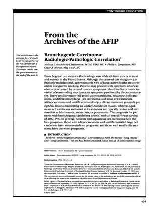
Figure 1. Adenocarcinoma.
(a) High-power photomicrograph
(original magnification, x 1 50; he-
matoxylin-eosin [ H-E I stain) shows
a well-differentiated adenocarci-
noma characterized by the forma-
tion ofglands and papillary struc-
tunes (*). Note the desmoplastic
reaction and fibrosis that surrounds
the glandular elements (f). (b) Cut
surface of an adenocarcinoma of the
night lower lobe shows a well-mar-
ginated, lobulated, subpleural, pe-
nipheral lung mass.
small cell carcinoma, as well as its responsive- lung diseases that produce focal or diffuse
ness to chemotherapy and radiation therapy fibrosis, including tuberculosis, pulmonary
are unique features that distinguish it from infarction, chronic interstitial pneumonitis
the non-small cell carcinomas (4, 18). Never- and fibrosis, sclenoderma, bnonchiectasis,
theless, to describe the varied nadiologic chronic pneumonia, and honeycomb lung
manifestations and pathologic features of (1,10,13,14,17,19).
bronchogenic carcinomas, we discuss each of Microscopically, adcnocarcinomas arc char-
the four cell types separately. actenized by the formation of glands and pap-
illary structures (Fig la). The neoplastic cells
. Adenocarcinoma have round to oval nuclei, prominent flu-
Adenocarcinoma is the most frequently diag- cleoli, and moderate amounts of cytoplasm.
nosed cell type and accounts for approxi- Histochemical stains (mucicarmine) are useful
mately 50% of all bronchogenic carcinomas for demonstration of the characteristic intra-
(4) . The increasing frequency of adenocarci- and extracellulan mucosubstance (14,17).
noma and the decrease in the diagnosis of These tumors have been associated with lung
squamous cell carcinoma have been observed scars. The degree of scarring can be extensive,
since the middle 1960s and are believed to suggesting a preexistent scar and giving rise to
represent an actual change in the biologic the concept of ‘ ‘scar carcinoma. ‘ ‘ Although a
features of these cell types rather than a re- small number of adenocarcinomas probably
flection of modern changes in diagnostic cni- arise in scar tissue, there is evidence that in
tenia. Adenocancinoma is also the most corn- the majority of cases the fibrosis on scar ne-
mon cell type seen in women and nonsmokers. suits from a desmoplastic host reaction in-
Although it is weakly associated with cigarette cited by the tumor (14,15,17,20,21) (Fig la).
smoking, most patients with adenocarcinoma
have a history of tobacco use. Adenocarcinoma
has also been associated with concomitant
March 1994 Rosado-de-Christenson et al U RadioGraphics U 431
4. Figure 2. Adenocancinoma in an asymptomatic
58-year-old male smoker with a radiographic ab-
normality found incidentally on a preoperative
radiograph obtained before cataract surgery.
(a) Posteroantenior (PA) chest radiograph shows a
lobulated 1.5-cm solitary nodule (arrow) in the
right upper lobe overlying the first anterior rib.
(b) Chest computed tomographic (CT) scan (lung
window) shows large bullae surrounding a well-
marginated, lobulated soft-tissue nodule. (c) Cut
surface of the tumor demonstrates the nodule (ar-
rowhead) within the collapsed bullae. Histologic
evaluation revealed a poorly differentiated adeno-
carcinoma with central fibrosis.
b. C.
On gross examination, adenocarcinoma lated, irregular, or poorly defined borders
typically manifests as a peripheral, subpleural (Figs 2, 3). Peripheral adenocancinomas may
nodule or mass that usually results in retrac- directly invade the pleura and grow cincum-
tion of the overlying pleura. Like most lung fenentially around the lung, thus mimicking
cancers, adenocarcinoma typically affects the diffuse malignant mesothelioma on initial cx-
upper lobes and exhibits an expansile (so- amination (19). Thin-section CT ofsmall (<2
called hilic) growth pattern that destroys and cm) peripheral carcinomas manifesting as soli-
displaces the adjacent lung panenchyma. The tary pulmonary nodules may demonstrate air
borders of the tumor may be rounded, lobu- bronchograms or air bnonchiolograms in 65%
lated, or poorly defined. Lobulation reflects of cases. This finding may help differentiate
the histologic heterogeneity oflung cancer these bronchogenic carcinomas from benign
and results from differential growth rates in lung tumors (25).
different areas within the tumor (Fig ib). Ill- CT can also demonstrate chest wall invasion
defined bonders may relate to invasion of the by peripheral pulmonary lesions (Fig 3). How-
adjacent lung, fibrosis, or interstitial edema even, absence of direct evidence of extrapul-
(22-24). monary involvement does not necessarily cx-
The typical radiologic manifestation of ad- dude it (26). CT is less accurate than the
enocancinoma is a solitary pulmonary nodule clinical presence of local chest pain in the cx-
on mass that may have well-manginated, lobu- clusion of chest wall invasion. Magnetic reso-
nance (MR) imaging may allow the distinction
of tumor from adjacent chest wall muscula-
ture and may improve the accuracy of CT in
the demonstration of chest wall invasion.
432 U Continuing Education Volume 14 Number 2
5. a. b.
Figure 3. Adenocarcinoma in a 41-year-old man with right shoulder pain for several months. (a) Apical br-
dotic chest radiograph demonstrates a right apical mass with poorly marginated borders. (b) Chest CT scan
(lung window) shows a homogeneous peripheral right upper lobe mass with irregular borders. There is tu-
mon involvement of a posterior rib (arrow). An en bloc resection of the right upper lobe and the involved
chest wall was performed.
a.
Figure 4. Bronchioboalveolar carcinoma. (a) High-power photomicrognaph (original magnification, x 150;
H-E stain) demonstrates the lepidic growth pattern. Columnar peglike cells line the alveolar walls. The pul-
monary interstitium (arrows) remains intact. (b) Cut surface demonstrates a heterogeneous parenchymal
lesion that resembles a consolidation.
Chest wall involvement is best seen as in- approximately 2%-6% of all lung neoplasms,
creased signal intensity on T2-weighted MR although its frequency may be increasing
images (27). (14). The tumor is characterized by wdll-dif-
Lung cancer has been reported to occur in fenentiated histologic features and is typically
close relation to preexisting bullac and may located peripherally beyond a recognizable
manifest as a nodular opacity within the bulla bronchus. Microscopically, these tumors cx-
(Fig 2), thickening ofthe bulla wall, change in hibit the so-called lepidic pattern of growth,
the size of the bulla, or spontaneous pneumo- which is characterized by cuboidal or colum-
thorax (28). In the study by Tsutsui et al (28), nan peglike cells that line the walls ofdistal air
12 of 25 lung cancers associated with bullac spaces. The pulmonary interstitium is re-
were adenocarcinomas. spected and serves as a ‘scaffolding’
‘ for tu- ‘
Bronchioloalveolar carcinoma represents a mon growth (24) (Fig 4). Bronchioloalveolar
subtype of adenocancinoma that accounts for
March 1994 Rosado-de-Christenson et al U RadioGraphics U 433
6. 5a. Sb.
6a. 6b.
Figures 5, 6. (5) Bronchioloalveolar carcinoma in a 56-year old man with no symptoms. (a) PA chest ra-
diograph demonstrates an ill-defined peripheral nodule in the left lower lung zone. (b) Chest CT scan dem-
onstrates a subpleural lobulated solitary pulmonary nodule in the left lower lobe. A 2-cm bronchioboalveolar
carcinoma was found at surgery. (6) Bronchioloalveolar carcinoma in a 39-year-old man with blood-tinged
sputum and pleuritic chest pain. (a) PA chest radiograph demonstrates a cavitary consolidation of the lingu-
bar segment of the left upper lobe. (b) Chest CT scan (lung window) demonstrates a cavity within the paren-
chymal consolidation. Air bronchograms are seen near the cavity. At surgery, an 8.4 x 6.4 x 3.5-cm cavitary
bronchioloalveolar carcinoma with direct extension to the visceral pleura was found. Although radiologic
studies may show a pneumonic pattern, the most common manifestation of bnonchioboabveolar carcinoma is
that ofa solitary pulmonary nodule.
carcinomas may exhibit tracheobronchial dis- The most common nadiologic manifestation
semination as neoplastic cells detach from the of the bronchioloalvcolan subtype of adeno-
primary tumor and attach to alveolar septa carcinoma is that of a well-circumscribed pe-
elsewhere in the lung, commencing growth in niphenal solitary pulmonary nodule or mass
a new location. The cells commonly produce (31) (Fig 5). Cavitation, an infrequent finding
abundant mucus (29-31). in adenocarcinomas, may be seen in bron-
chioloalvcolan carcinoma (Fig 6). In the study
by Theros (24), which reviewed 1,267 periph-
crab primary neoplasms of the lung, bnonchio-
434 U Continuing Education Volume 14 Number 2
7. “ ‘V .
a. b.
Figure 7. Bronchioboalveolar carcinoma in a 35-year-old woman with a chronic pulmonary consolidation.
(a) PA chest radiograph shows a right middle lobe consolidation. (b) PA chest radiograph obtained years
1 #{189}
later shows bibasilar multinodular consolidations with air bronchograms, which represented tracheobron-
chial tumor dissemination. The surgical clips over the right lower lung were placed during the initial biopsy.
loalveolar carcinoma was the second most ated with cigarette smoking. Its histogenesis is
common cell type (after squamous cell carci- thought to relate to chronic inflammation and
noma) to radiographically demonstrate cavita- injury of the bronchial epithelium, which can
tion. The lepidic pattern of tumor growth may result in squamous metaplasia. This may sub-
result in lesions of heterogeneous radiologic sequently progress to dysplasia, carcinoma
opacity, with air bronchograms and poorly in situ, and ultimately invasive carcinoma
manginated borders mimicking pneumonia (1,5, 10). Squamous cell carcinoma is the only
(29-31) (Figs 6, 7a). Less commonly, patterns cell type in which in situ changes are necog-
of multiple nodules (Fig 7) or extensive con- nized, and thus it may be diagnosed with cyto-
solidation involving one on more lobes may be logic examination of the sputum of affected
seen (1 1,29-32). High-resolution CT may patients. Therefore, it is the most common
demonstrate air attenuation and pseudocavi- cell type diagnosed when it is radiologically
tation within the nodules corresponding to occult (1,14,17). Unfortunately, less than 1%
small bronchi and cystic spaces (33). Patients of bronchogenic carcinomas are detected at
with extensive consolidation on multifocal this stage (4). Squamous cell carcinoma is also
disease have a poor prognosis (29-31). the most common cell type associated with
hypercalccmia. The hypercalcemia is thought
. Squamous Cell Carcinoma to be caused by a parathyroid hormone-like
Squamous cell carcinoma accounts for ap- substance produced by the tumor (17).
proximately one-third of all bronchogenic can-
cinomas (4). This cell type is strongly associ-
March 1994 Rosado-de-Christenson et al U RadioGraphics U 435
8. Figure 8. Squamous cell carcinoma. (a) High-power photomicrograph (original magnification, X 150; H-E
stain) shows neoplastic cells with moderate amounts of eosinophilic cytoplasm. Well-differentiated keratiniz-
ing areas (arrowheads) are seen intermixed with the malignant cells. (b) Gross specimen shows an irregular,
exophytic, endobronchial mucosal tumor that partially obstructs the lumen of the main stem bronchus. The
tumor invaded the bronchial wall and the adjacent lung parenchyma. Scale is in centimeters. Linear chest
tomogram (C) and bronchogram (d) show the characteristic growth pattern of these tumors in a patient with
a squamous cell carcinoma of the night main stem bronchus. Note the irregular narrowing (arrow) of the
bronchial lumen, which may result in postobstructive pneumonia or atelcsis.
., .
,
‘ #J%
*,
:‘
q’S,#{149}
‘ .
., ,U-..’
), :,,.
:.
.- ..
;. -..
.
4 #{149}
,
d?t.pj.
c’.::.c:
*. :‘
1i
l
. ‘ ‘.
,,..
, ..
‘
.v
.-. . --,..
,-,
:
. .4-..,..’ -. ‘,
L#{149} , ,
#{149} . ,
‘ I ,,,, , . b
. ‘I
. ..
..-
:‘ ‘
‘ - - .
‘A”
a. b.
C. d.
Microscopically, squamous cell carcinoma is dens ofcontiguous cells), individual cell kera-
characterized by the presence of intercellular tinization (characterized by intense eosino-
bridges (fine parallel lines between the bor- philia ofindividual cells), and the formation
of keratin pearls (laminated whorls of cosino-
philic cells) in well-differentiated tumors
(13,14,16,17,19) (Fig 8a). The term “squa-
436 U Continuing Education Volume 14 Number 2
9. Figure 9. Squamous cell carcinoma in a 57-year-old man. PA (a) and lateral (b) chest radiographs demon-
strate a complete consolidation of the right upper lobe. At bronchoscopy, an endobronchial tumor of the
r t main stem bronchus was identified.
a. b.
mous’ ‘ means flat and refers to the flattened Therefore, all cases of pneumonia occurring
appearance of the tumor cells. Because squa- in adults should be followed to complete na-
mous cell carcinoma mimics the differentia- diologic resolution to exclude the presence
tion of the epidermis by producing keratin, it of a causative endobronchial lesion such as
is also called epidermoid carcinoma (1). Squa- bronchogenic carcinoma.
mous cell carcinoma incites a strong inflam- Lobar or complete lung atelectasis may also
matory host response with resultant adhe- result from these central endobronchial Ic-
sions, across which the tumor can invade sions (Figs 10, 11). Because ofthe presence
adjacent structures (15). of a central mass, the lobe is unable to corn-
Squamous cell carcinomas are centrally lo- pletely collapse and a bulging contour of the
cated within the main, lobar, and segmental atelectatic lung may be produced by the pni-
bronchi in approximately two-thirds of cases. mary tumor, giving rise to the radiographic “S
On gross examination, these endobronchial sign of Golden” (32). Early lesions may mani-
tumors range from a focal irregular growth in fest with lobular thickening of the bronchial
the bronchial mucosa to a polypoid mass that wall (32,35). Larger tumors may produce a
obstructs on narrows the bronchial lumen hilar on penihilan mass (1 1). Approximately
(1,14,34) (Fig 8b). Virtually all central squa- one-third of squamous cell carcinomas are
mous cell carcinomas can be identified on peripheral and appear as solitary pulmonary
endoscopic examination (13, 15). These tu- nodules or masses (14).
mors commonly grow through the bronchial Squamous cell carcinoma is the most corn-
wall, subsequently invading adjacent lymph mon cell type to produce cavitation, which
nodes or lung parenchyma (1 1,16). Central occurs in approximately 10% ofcases (10).
necrosis is very common and may result in The inner wall of the cavity is typically thick
cavitation (1 1). and irregular (1 1) (Fig 12). Peripheral squa-
The typical radiologic manifestations of mous cell carcinoma is also the most common
central squamous cell carcinomas are the ne- cell type to cause the Pancoast syndrome,
suIt of the total or partial bronchial obstruc-
tion that these endoluminal lesions produce
(Fig 8c, 8d). Bronchial obstruction may ne-
suit in a postobstructive pneumonia (Fig 9).
March 1994 Rosado-de-Christenson et al U RadioGrapbics U 437
10. lOa lOb.
1 la. 1 lb.
llc.
438 U Continuing Education Volume 14 Number 2
11. -- g
Figure 12. Squamous cell carcinoma in a 72-year-old man
with left arm pain, chest pain, and increasing dyspnea.
(a) PA chest radiograph demonstrates a large rounded cavi-
tary mass with an air-fluid level in the superior segment of
the left lower lobe. Note the nodular, irregular contour of
the inner wall of the cavity. (b) Contrast-enhanced chest CT
scan (mediastinal window) demonstrates the air-fluid level
within the lesion and the irregular aspect of its inner wall.
(c) Cut surface ofthe resected left lower lobe demonstrates
the cavitary neoplasm. Scale is in centimeters.
C.
4Figures 10, 11. (10) Squamous cell carcinoma in a 63-year-old woman with dysphagia and weight loss.
(a) Frontal chest radiograph demonstrates opacification of the left hemithorax and ipsilateral mediastinal
shift consistent with complete atelectasis of the left lung. Lack of visualization of the left main stem bronchus
suggests central occlusion. (b) Contrast-enhanced chest CT scan (mediastinal window) demonstrates a soft-
tissue mass (in), which narrowed and obstructed the left main stem bronchus, left lung atebectasis, and left
pleural effusion. At bronchoscopy, a circumferential, friable obstructing endobronchial lesion was found.
(11) Squamous cell carcinoma in a 62-year-old man with left shoulder pain. (a, b) Thin-section chest CT
scans (lung window) show an endobronchial nodule (arrow in a) within the right lower lobe bronchus.
There is involvement of the adjacent lung parenchyma with associated volume loss of the night lower lobe.
Note the bobulated mass (arrowhead in b) that displaces the major fissure. (C) Gross specimen of the re-
sected right lower lobe shows the endobronchial component of the tumor (arrow) and the large parenchy-
mal mass (m).
March 1994 Rosado-de-Christenson et al U RadioGraphics U 439
12. C- d.
Figure 13. Adenosquamous carcinoma in a 68-year-old man with chest wall pain on the night side. (a) PA
chest radiograph shows a right apical mass with associated destruction of the posterior aspects of the first
and second ribs. A large soft-tissue component is also present in the supraclavicular region. (b) Contrast-
enhanced chest CT scan demonstrates a large apical soft-tissue mass that destroys the adjacent ribs and di-
rectly invades right axillary
the region. At surgery, a poorly differentiated adenosquamous carcinoma was
found. (c) Coronal MR image (repetition time was 600 msec; echo time was 20 msec 1600/201) from another
patient with a Pancoast tumor demonstrates extrapulmonary invasion of the tumor into the soft tissues of the
chest wall (arrow). The roots ofthe brachial plexus are well visualized and are not involved by the tumor
(arrowhead). (Reproduced, with permission, from reference 27.) (d) Gross specimen ofa Pancoast tumor
shows that the peripheral apical tumor grows through the visceral pleura and has a large extrapulmonary
component.
characterized clinically by pain or atrophy of asymmetric apical pleural thickening and may
muscles of the ipsilatenal upper extremity due be associated with bone destruction and soft-
to involvement of the lower brachial plexus, tissue invasion (11,36,37) (Fig l3a).
and Horner syndrome due to involvement of CT may demonstrate central tumors as a
the sympathetic chain and the stellate gan- mass within the airway, narrowing of the air-
gI ion (5). Pancoast tumors account for less way lumen, on focal peribronchial thickening
than 3% ofall bronchogenic carcinomas (4). (35) (Figs lOa, 1 la). CT may also help in dis-
These lesions may manifest radiologically as tinguishing the primary tumor from adjacent
apical masses, apical pleural thickening, or atelectatic or consolidated lung. The tumor
may produce a bulge in the involved atelec-
tatic lung, which suggests the presence of an
underlying mass (Fig 1 ib). Differential con-
440 U Continuing Education Volume 14 Number 2
13. Figure 14. Undifferentiated large cell carcinoma.
High-power photomicrograph (original magnifica-
tion, X 150; H-E stain) shows large tumor cells with
large nuclei, prominent nucleoli, and a moderate
amount of cytoplasm. There are no microscopic fea-
tures ofdifferentiation for the other three cell types.
trast material enhancement of tumor versus this diagnosis decreases when large amounts
collapsed lung may be seen. CT also allows of tissue arc available for histologic evaluation
the evaluation of the mediastinum and adja- (17). With ultrastructural analysis, approxi-
cent structures for staging (19) (Fig 13b). Di- matcly 80% of undifferentiated large cell car-
rect coronal and sagittal MR images are supe- cinomas previously diagnosed with light mi-
nor to CT scans in the evaluation of Pancoast croscopy demonstrate electron microscopic
tumors because they allow visualization of the features of adenocarcinoma; features of squa-
anatomy of the adjacent chest wall. The sub- mous cell carcinoma and other tumors are
clavian artery, brachial plexus, vertebral bod- seen in many ofthc remaining 20% (14,34).
ics, and spinal canal can be visualized and Giant cell carcinoma is a subtype of undif-
assessed for tumor involvement. Ti-weighted fercntiatcd large cell carcinoma composed of
coronal and sagittal MR images arc a useful pleomorphic giant cells with bizarre shapes.
adjunct to CT scans, resulting in improved Approximately 40% of the cells are multi-
diagnostic accuracy in the preoperative evalu- nucleated. Red and white blood cells are typi.
ation of patients with Pancoast tumors (1 1, cally seen within the cytoplasm of the giant
38-40) (Fig 13c). tumor cells. Giant cell carcinoma has a pan-
ticularly aggressive behavior and a very poor
. Undifferentiated Large Cell prognosis (14, 16, 19,34).
Carcinoma Undifferentiated large cell carcinomas are
Undifferentiated large cell carcinoma nepre- usually bulky tumors typically greaten than 3
sents less than 5% of all bronchogenic carci- cm in diameter. They are soft and have large
nomas (4). These tumors grow rapidly and areas of necrosis. Undifferentiated lange cell
metastasize early. They are strongly associated carcinomas are typically located in the lung
with smoking. periphery, but central lesions arc not uncom-
With bight microscopy, the tumor cells ap- mon. Involvement oflange bronchi is seen in
pear large; have abundant cytoplasm, large approximately 50% ofcentral lesions (1,16).
nuclei, and prominent nucleoli; and grow in The typical nadiologic appearance of these
uniform sheets (Fig 14). The histologic diag- neoplasms is that of a large peripheral lung
nosis of undifferentiated large cell carcinoma mass (Figs 15, 16).
is one of exclusion, given only to primary ma-
lignant neoplasms of the lung that lack fea-
tunes of squamous, glandular, on small cell
differentiation (1,14,34,41). The frequency of
March 1994 Rosado-de-Christenson et al U RadioGraphics U 441
14. 16a. 16b.
Figures 15, 16. (15) Undifferentiated large cell carcinoma in a 61-year-old woman with blood-streaked
sputum and weight loss. (a) PA chest radiograph demonstrates a large peripheral mass of the left upper lobe,
which abuts the pleural surface and has a bobubated contour. (b) Cut surface of the gross specimen demon-
strates a 7-cm tumor that extends to the pleural surface. (16) Undifferentiated large cell carcinoma in a
57-year-old man with weight loss, orthopnea, and a painful palpable mass of the anterior chest wall on the
left side. (a) Contrast-enhanced chest CT scan (mediastinal window) demonstrates a large mass of heteroge-
neoUs attenuation, which produces mass effect on the mediastinal structures. (b) Cut surface of the neoplasm
shows a large central area of necrosis, which corresponds to the areas of central decreased attenuation seen
with CT. At surgery. chest wall tumor invasion was seen.
. Small Cell Carcinoma nucleoli. The neoplastic cells may be arranged
Small cell carcinoma accounts for approxi- in cords, clusters, or sheets (Fig 17a). There
mately 15% ofbronchogenic carcinomas (4). are numerous mitoses and large areas of ne-
Small cell carcinoma is strongly associated crosis. An extensive crushing artifact is fre-
with cigarette smoking. It is a rapidly growing quently seen in bronchial biopsy specimens.
neoplasm characterized by early and wide- The crush artifact probably results from the
spread metastases (1,15,19). scant tumor stroma and the lack of desmo-
Microscopically, small cell carcinoma is plastic reaction in these lesions (1,11,14,17,
characterized by small, uniform, oval cells 19,34).
with scant cytoplasm. The nuclei are round on Although some have used the term synony-
oval with a stippled chromatin and absent mously with small cell carcinoma, ‘ ‘oat cell
carcinoma’ ‘ is actually a morphologic subtype
of small cell carcinoma characterized by uni-
form small cells with small dense hypcrchro-
442 U Continuing Education Volume 14 Number 2
15. matic nuclei, absent nucleoli, and scant cyto- mon cell type to cause a clinical hormone syn-
plasm (19). The histologic features ofoat cell drome by secreting ectopic hormones. The
carcinoma may be secondary to generalized most commonly seen syndromes are Cushing
tissue ischemia, since it is typically described syndrome and the inappropriate secretion of
in autopsy specimens. Promptly fixed biopsy antidiuretic hormone (12,14,16,43).
material usually does not exhibit these fea- Approximately 90% of small cell carcinomas
tunes (19). Approximately 20% ofsmall cell arc located centrally within lobar and main
carcinomas also contain elements of non- stem bronchi. Although these tumors anise in
small cell histologic types. The most frequent the bronchial mucosa, they tend to grow in
coexistent histologic type is squamous cell the submucosa and subsequently invade the
carcinoma (19). penibronchial connective tissues, maintaining
Small cell carcinomas together with carci- a smooth-appearing mucosal surface on endo-
noid tumors have been classified as neuro- ‘ ‘ scopic examination. The tumor is bulky and
endocrine neoplasms’ ‘ of the lung. The cells soft, with extensive necrosis and hemorrhage.
in small cell carcinoma may contain neurose- Although extrinsic bronchial compression
cretony (dense cone) granules similar to those may occur, endobronchial lesions arc rare
found in bronchial cancinoid. These tumors (1 1 , 16). Small cell carcinoma produces little
arc thought to be related to the amine precun- host response and can spread easily through
son uptake and decanboxylase cells of the tissues, invade adjacent structures and lymph
bronchial epithelium, which has led to their nodes, and disseminate along lymphatic
classification as Kulchitsky cell carcinomas routes (Fig 17b). Peripheral lesions are rare
(42,43). The postulated neural crest origin of and are usually associated with regional
these cells is no longer widely accepted. In- spread to hilan and mediastinal lymph nodes
stead, it is believed that these tumors are de- (17).
nived from primitive endodermal cells that
can differentiate into neuroendocrinc cells
(17). Small cell carcinoma is the most corn-
March 1994 Rosado-de-Christenson et al U RadioGraphics U 443