1. Management of periorbital
and orbital cellulitis
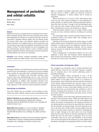
Malcolm A Buchanan
Wisam Muen
Peter Heinz
Abstract
Swelling of the tissues surrounding the eye is a relatively common presen-
tation in paediatric practice. Many of the mild, pre-septal cases of perior-
bital inflammation and infection are caused by insect bites, trauma and
conjunctivitis. Frequently infection spreads from the ethmoid sinuses
and invades orbital tissues. The distinction between pre-septal and
orbital involvement can be difficult based on clinical examination only,
and the research base supporting management of periorbital and orbital
cellulitis is limited. This review addresses the role of investigations and
the multi-disciplinary approach needed in order to establish accurate
diagnosis, appropriate treatment and prevention of serious complications
including blindness and venous sinus thrombosis.
Keywords acute sinusitis; computed tomography; orbital cellulitis;
periorbital cellulitis; pre-septal cellulitis; venous sinus thrombosis
Introduction
Periorbital cellulitis is an umbrella term for infections of the tissues
around the eye and represents a continuum of severity resulting
from a number of causes rather than a well-defined disease entity.
Management of children with periorbital infections often
involves input from ophthalmology and ear, nose and throat
(ENT) specialists. This in itself, and the lack of evidence base for
the management of children with periorbital infections pose
a number of challenges. This review attempts to present a prag-
matic approach to this relatively common paediatric problem.
Epidemiology and definitions
Very little reliable data are available on the incidence of peri-
orbital and orbital cellulitis. Based on local and published audit
figures, a paediatric emergency department seeing 20,000 chil-
dren per annum is likely to admit one child per month for
inpatient management. A similar number will be treated as
outpatients.
Before introduction of a vaccine in 1985, Haemophilus influ-
enzae was the most common pathogen to cause periorbital or
orbital cellulitis, an infection often associated with CNS infection.
Since then there has been a change in the microbiological spec-
trum, with the most common culture positive cases now being
due to Staphylococcus aureus and streptococcus species.
Whilst periorbital cellulitis occurs at all ages, children under
10 years are most commonly affected, including rare neonatal
cases.
The terminology used to classify periorbital infection refers to
anatomical extent of the disease rather than aetiology and is
summarized in Figure 1.
Infection of the structures behind the orbital septum is called
orbital cellulitis and is considered by some to be a different entity
altogether. In clinical practice the distinction between the two
can cause considerable diagnostic difficulty, and the authors
make no excuses for using the term periorbital cellulitis synon-
ymously for both.
Severe complications of periorbital cellulitis are rare if the
diagnosis is made early and treatment started appropriately;
these include visual impairment, orbital and intracranial abscess
formation and venous sinus thrombosis.
Clinical presentation and diagnostic pitfalls
Five categories are commonly used to classify periorbital and
orbital infection. These do not necessarily represent a progres-
sion of symptoms due to the various underlying causes of
periorbital infections.
Pre-septal cellulitis (which corresponds to type 1 of the
Chandler classification) is limited to the tissues anterior to the
orbital septum of the upper eyelid, which is an extension of
the periosteum of the frontal bone and fuses with the tarsal plates
of the eyelids. Unless breached, this structure seems to be an
effective barrier to further expansion of infection, which explains
why the majority of cases of pre-septal cellulitis are due to an
obvious local cause, e.g. dacryocystitis, stye, chalazion, or due to
infection stemming from breaches of the skin barrier such as
insect bites, infected varicella lesions and minor trauma, to name
the most common.
Orbital cellulitis, on the other hand, generally spreads from an
infection of the paranasal sinuses. Whilst pre-septal disease can
expand in an unrestricted fashion into the surrounding facial
tissues, the impact of inflammatory and infectious processes in
the confined space of the orbit can be more difficult to assess and
has the potential for lasting damage. The most important struc-
ture is the optic nerve which carries neuronal signals from the
retina to the optic chiasm and thus is essential in mediating the
sense of sight. Damage to the optic nerve can occur through
direct pressure, optic neuritis or vasculitis. Damage to the vessels
within the orbit can also result in ischaemia. This can cause
irreversible visual loss or even total blindness in the involved
orbit. Intracranial spread of the infection can cause sagittal sinus
thrombosis, meningitis and encephalitis which can lead to death.
Malcolm A Buchanan BSc (Hons) MBChB PhD FRCS (ORL-HNS) is a Specialist
Registrar in Otorhinolaryngology at Addenbrooke’s Hospital,
Cambridge University Hospitals NHS Foundation Trust, Cambridge, UK.
Conflict of interest: none.
Wisam Muen MRCS Ed FRCOphth is a Locum Consultant in Paediatric
Ophthalmology at Addenbrooke’s Hospital, Cambridge University
Hospitals NHS Foundation Trust, Cambridge, UK. Conflict of interest:
none.
Peter Heinz MD FRCPCH is Consultant in Acute and Emergency Paediatrics
at Addenbrooke’s Hospital, Cambridge University Hospitals NHS
Foundation Trust, Cambridge, UK. Conflict of interest: none.
OCCASIONAL REVIEW
PAEDIATRICS AND CHILD HEALTH 22:2 72 Ó 2012 Elsevier Ltd. All rights reserved.
2. As far as clinical signs are concerned, proptosis, conjunctival
injection and limitation of extraocular movements are highly
suggestive of an intraorbital process. Unfortunately, absence of
these features does not exclude the presence of intraorbital
infection, and depending on the child’s age and degree of eyelid
swelling, a clinical assessment can be unreliable if not impos-
sible. The child with systemic features, such as a high fever and
neurological compromise, is at high risk of having severe disease
with intracranial involvement. Although bacteria are the most
common cause of orbital infection, viral, parasitic and fungal
infections have also been reported. The latter are most common
in immunocompromised individuals, and early detection of such
infections is imperative as they can be associated with a high
mortality.
Differential diagnoses of periorbital cellulitis also include
autoimmune disease including thyroid eye disease, as well as
trauma and neoplasms; these are all very rare in children but
should be borne in mind if there is no response to treatment.
Investigations
Blood tests do not provide any diagnostic value per se but are
commonly performed and may be useful in monitoring response
to treatment. Although more severe disease increases the likeli-
hood of leucocytosis and raised inflammatory markers, the
negative predictive value of an absence of abnormal parameters
is not sufficiently good to be relied upon. Blood tests are also
poor indicators of the location and extent of infection. A number
of studies have confirmed that blood tests do not help to differ-
entiate between pre-septal cellulitis, intraorbital abscess and
orbital cellulitis. Positive blood cultures are found in no more
than a third of cases and seem to have very little bearing on the
crucial early management.
In the presence of ophthalmological signs such as proptosis,
reduced visual acuity and colour vision, ophthalmoplegia and/or
diplopia, urgent imaging should be acquired. Radiological
investigations have a role in assessing potential complications
arising from orbital cellulitis, namely presence of an abscess and
involvement of the orbital structures, paranasal sinuses and
intracranial structures. Imaging will help to define the exact
location and extent of the problem and aid in planning for
subsequent treatment. The question is when to request imaging
as it may involve significant exposure to radiation, and younger
children will possibly also require sedation.
In the absence of any systemic features and presence of mild
eyelid swelling only, reliable assessment of the normal range of
eye movement, acuity and colour vision can be made, and
there is no indication for immediate radiological assessment.
An unwell child, inability to make a reliable clinical assess-
ment and clinical suspicion of orbital involvement warrant
a high definition computed tomography (CT) scan taken in
coronal and axial planes. This will delineate any bony defect
Type 1
Pre-septal cellulitis: Inflammation does not
extend beyond the orbital septum
Type 2
Post-septal/orbital cellulitis: Inflammation
extends into the orbital tissues, with no
abscess formation
Type 3
Subperiosteal abscess: Abscess forms
deep to the periosteum of the orbit
Orbital septum
Inflammation, no pus
Inflammation, pus/abscess
Type 4
Orbital abscess: Abscess forms within the
orbit, with breach of the periosteum
Type 5
Cavernous sinus thrombosis
Figure 1 Chandler classification of periorbital infection.
OCCASIONAL REVIEW
PAEDIATRICS AND CHILD HEALTH 22:2 73 Ó 2012 Elsevier Ltd. All rights reserved.
3. within the orbit and sinuses (often a breach of the paper-thin
lamina papyracea separating the medial orbital wall from the
anterior ethmoid sinuses (Figure 2)), and will demonstrate the
presence of a subperiosteal or orbital abscess. It will also
confirm the presence of pus in the sinuses. Due to its inability
to provide bony definition, Magnetic Resonance Imaging (MRI)
is inferior to CT in assessing and planning the surgical
management of local complications of orbital sinusitis.
However, MRI is better than CT in assessing intracranial
complications and distinguishing pre-septal from post-septal
disease.
Given the difficulties in determining extent and severity of
periorbital cellulitis based on clinical examination alone, further
investigations and involvement of ophthalmologists and ENT
surgeons is usually called for in all but the mildest cases, i.e.
clinical suspicion of Chandler grade 2 and above.
Ophthalmology review
It is important to liaise closely with the ophthalmologist for all
patients needing admission, in order to allow for a fuller ocular
assessment and reach a consensus opinion on who needs to be
scanned urgently and who can wait. An urgent ophthalmology
review must be requested for all cases of orbital cellulitis.
The main ocular symptoms and signs are reduced vision,
orbital pain, lid swelling, chemosis and proptosis which may be
axial or non-axial, pain on eye movements and possible
diplopia. The patient may not necessarily complain of diplopia
laminae
papyraceae
medial recti
nasal septum
clear posterior
ethmoid air cells
clear sphenoid
sinuses
subperiosteal
abscess
lateral recti
opacified
anterior and
posterior
ethmoid sinuses
optic nerves
Figure 2 Axial scan demonstrating right subperiosteal abscess with resultant lateral displacement of the ipsilateral medial rectus and proptosis,
secondary to anterior and posterior ethmoid sinus inflammation.
OCCASIONAL REVIEW
PAEDIATRICS AND CHILD HEALTH 22:2 74 Ó 2012 Elsevier Ltd. All rights reserved.
4. if swelling of the lids has occluded the visual axis of the affected
eye.
It is important for the doctor seeing a child with periorbital
cellulitis to assess the following:
can the lids be opened?
is the conjunctiva involved (hyperaemia, chemosis,
discharge)?
if possible obtain visual acuities for each eye. This should
be treated as a game with the child being asked to see as
many letters/characters as possible. The more they can see
with each eye, the higher the score. Some mobile phone
applications exist for assessing visual acuity of children,
however the accuracy of these apps has not been fully
assessed.
are the extraocular movements grossly normal? This can
be assessed by moving a toy or object of interest in the
main positions of gaze. It is most important to assess how
well the affected eye adducts (movement towards the
nose) and elevates, as these are often the first to be
restricted in cases of orbital cellulitis.
are the pupillary responses normal? It is important to try
and look for a relative afferent pupillary defect (RAPD)
through using the swinging light torch test.
Ishihara colour plates can be used in age appropriate
children (those who are verbal) to assess optic nerve
function.
it is also important to try and visualize the optic disc for
signs of swelling.
how well is the patient systemically?
The presence of any of the above ocular signs is an indication
of orbital cellulitis and would necessitate immediate admission
and treatment with intravenous antibiotics as well as imaging.
Close monitoring may be necessary in such patients, with
hourly checks of optic nerve function, including visual acuities,
pupillary response and colour vision. This is not always possible
in the very young child, however specialized tests in the
orthoptic department to assess vision and eye movements can be
carried out, but this may not be available in the emergency
situation.
The role for intravenous steroids in cases where optic nerve
involvement is suspected remains controversial and there is no
strong evidence base to support it. Instruction should be given to
the ward staff to starve the patient, and the ophthalmologist will
assess the patient and liaise with an orbital and ENT surgeon
regarding the need for abscess drainage or possible emergency
orbital decompression.
ENT review
An ENT surgeon should be involved in the clinical assessment of
a child who fits categories Chandler 2 and upwards. The nose
should be examined to assess for the presence of mucopus. In
a co-operative child, a more detailed assessment can be made
endoscopically, and a swab taken for bacteriological analysis.
Whether to treat subperiosteal abscess medically or surgically
is an area of controversy in the literature. The decision to operate
should be made in the absence of improvement with intravenous
antibiotics, and on the basis of the eye, namely reduction in
visual acuity, colour vision, ophthalmoplegia and/or proptosis.
Surgical treatment should address the underlying rhinosinu-
sitis and its associated complication(s). Drainage of a sub-
periosteal or orbital abscess is performed externally or
endoscopically. The latter approach is technically more difficult
in paediatric cases, especially with inflammation and bleeding,
and is usually reserved for experienced endoscopic surgeons. It is
avoided in the presence of intracranial complications, frontal
bone osteomyelitis (Pott’s puffy tumour) or orbital complications
with acute visual compromise. The sinuses should be drained
and irrigated endoscopically.
Surgical treatment of intracranial complications should
involve collaboration with a neurosurgeon. Intracranial compli-
cations are more likely in the presence of frontal rhinosinusitis
than the other sinuses, and external trephine drainage of the
frontal sinus should be performed. Any intracranial abscess
should be drained.
Pott’s puffy tumour necessitates abscess drainage and
removal of the infected bone, followed by 6 weeks of antibiotic
treatment.
Medical treatment
Antibiotic treatment is the mainstay of medical management of
periorbital infections. The following recommendations should be
regarded as a rough guide only and we would advise obtaining
local microbiology advice before commencing ‘blind’ antibiotic
treatment of periorbital infections.
Mild, pre-septal infections will often respond very well to oral
treatment covering both staphylococci and streptococci as
a priority, given the typical pre-septal route of infection which
involves breaching the skin barrier. Parenteral treatment with
once daily intravenous ceftriaxone for ambulatory patients is
commonly used in paediatric emergency departments and
assessment units. At least once daily review and robust safety
netting are advised for any patient discharged home on antibiotic
treatment, and escalation of management to imaging and review
of antibiotic therapy may be required if there is clinical deterio-
ration or no improvement within 24e48 h.
Any infection suspected or with the potential to affect post-
septal tissues will require treatment with intravenous antibiotics
with broad spectrum cover including anaerobes from the outset.
The duration of treatment will vary and depend on clinical
response, the extent of the infection and presence of complica-
tions. Usually a treatment course would be for 10e14 days.
There is currently no evidence to support the routine use of
nasal decongestants or oral steroids. An overview of the approach
to the child with periorbital swelling is outlined in Figure 3.
Surgical treatment
The primary aim of surgery is drainage of the subperiosteal or
intraorbital abscess. This can be performed under general
anaesthetic, either externally or endoscopically.
In the external approach, a curvilinear incision is made
halfway between the medial canthus of the eye and the nasion of
the nose. Dissection proceeds along the periosteum. The anterior
ethmoidal artery will be identified and ligated. As dissection
continues, the abscess cavity is encountered, the pus drained,
and the cavity irrigated. A hole should be made in the lamina
papyracea to drain the abscess cavity medially into the anterior
OCCASIONAL REVIEW
PAEDIATRICS AND CHILD HEALTH 22:2 75 Ó 2012 Elsevier Ltd. All rights reserved.
5. ethmoid sinuses. A corrugated drain is usually placed into the
cavity, to drain any residual blood and pus, and brought out
externally through the skin incision. The drain prevents the
abscess from re-collecting, and is usually removed on the ward
24e48 h later.
In the endoscopic approach there is no scar, and the operation
is performed entirely through the nasal cavity. In the presence of
acute inflammation and bleeding, access can be difficult. This
approach is usually reserved for a more experienced endoscopic
surgeon, typically a rhinologist. The lamina papyracea is
breached to enable the subperiosteal or intraorbital abscess to
drain into the nose, and subsequent irrigation to be performed.
Provided a sufficiently large hole is fashioned, it is not usually
necessary to insert a drain.
In both the above approaches, there is a risk of blindness in an
already inflamed orbit, due to pressure from retraction, too
vigorous irrigation, or haemorrhage into the orbit. The surgeon
should therefore be prepared to undertake a lateral canthotomy if
haemorrhage occurs, to release the intraorbital pressure on the
optic nerve.
The secondary aim of surgery is to open up and washout the
sinuses. The anterior ethmoid sinuses will have already been
opened by draining the abscess. The ostia (drainage channels) of
the maxillary sinuses should be widened endoscopically, and
irrigated through an infra-meatal antrostomy.
Follow-up and prognosis
Review of children discharged home from hospital is particularly
important in the early stages of the illness in order to assess
response to treatment. Review in hospital or by the child’s
General Practitioner should be arranged within 24e48 h. Follow-
up after inpatient treatment will depend on the extent of the
infection, complications and treatment modalities and will be
guided by the relevant teams involved in the patient’s care.
If treated promptly, the prognosis for return of normal vision
following orbital cellulitis þ/À abscess is excellent, and patients can
usuallybe discharged withina fewdays following surgery.Follow-up
is advised for any patient who underwent surgical treatment, to
exclude early complications. Residual or recurrent disease and
intracranial sepsis are possible, and usually equate to inadequate
surgical drainage. There is a small risk of diplopia following surgery
which usually resolves. Late complications include residual visual
defect (diplopia, reduced acuity), scarring or webbing from an
external drainage approach, and enophthalmos.
Periorbital swelling:
Clinical assessment in the Emergency Department/Paediatric Assessment Unit
Child at risk of orbital cellulitis
• Age under 3 months
• Febrile and/or unwell
• Immune-compromise (steroid treatment, HIV infection)
• Inability to open the eye (and as a result to assess clinically)
• Clinical features suggesting orbital involvement:
– Red eye, proptosis, ophthalmoplegia, reduced visual
acuity/colour vision, pain on eye movement
• Features of sinus disease:
– blocked or runny nose, facial pain (frontal headache
and local tenderness)
Well child with pre-septal cellulitis
minimal disease (Chandler type 1)
• Age over 3 months
• Systemically well (afebrile)
• No immune-compromise
• Minimal swelling of tissues, enabling eye opening and
assessment of globe
• Clear evidence of pre-septal infection
– No features of orbital involvement (see over)
– Skin breach as likely cause of infection (insect bite,
dacryocystitis)
• No suggestion of sinus disease (blocked nose, pain)
Out-patient management
• Trial of oral antibiotics (oral Co-Amoxiclav) or once daily
intravenous Ceftriaxone (follow local recommendations
and guidelines)
• Clinical review within 24 hours
• Safety netting: written and verbal advice for carers
• Review following completion of antibiotic course by
General Practitioner
Out-patient review within 24 hours
No improvement/features of concern on re-assessment:
• As per child at risk of orbital cellulitis
In-patient management
• Admit to hospital
• Blood tests: Full blood count, C-reactive protein and blood
cultures
• ENT and Ophthalmology assessments
• Consider imaging (CT scan or MRI if intracranial involvement
suspected)
• Intravenous antibiotic treatment as per local guideline
• Surgical management as indicated involving possibly
– ENT
– Ophthalmology
– Neurosurgery
• Follow-up by the most appropriate team, depending on
surgical interventions, complications and clinical course as
in-patient
Figure 3 Management flowchart.
OCCASIONAL REVIEW
PAEDIATRICS AND CHILD HEALTH 22:2 76 Ó 2012 Elsevier Ltd. All rights reserved.
6. For frontal sinus involvement, an elective ventilation or
frontal sinus obliteration procedure can be considered, to prevent
recurrence of sinusitis.
Uncomplicated, pre-septal cellulitis which is responding to
treatment does not require long term follow-up. A
FURTHER READING
Baring DEC, Hilmi OJ. An evidence based review of periorbital cellulitis.
Clin Otolaryngol 2011; 36: 57e64.
Beech TJ, Robinson A, McDermott AL, Sinha A. Paediatric periorbital
cellulitis and its management. Rhinology 2007; 45: 47e9.
Chandler JR, Langenbrunner DJ, Stevens ER. The pathogenesis of orbital
complications in acute sinusitis. Laryngoscope 1970; 80: 1414e28.
Georgakopoulos CD, Eliopoulou MI, Stasinos S, Exarchou A,
Pharmakakis N, Varvarigou A. Periorbital and orbital cellulitis: a 10-
year review of hospitalized children. Eur J Ophthalmol 2010 Nov-Dec;
20: 1066e72.
Howe L, Jones NS. Guidelines for the management of periorbital
cellulitis/abscess. Clin Otolaryngol 2004; 29: 725e8.
Jones NS, Walker JL, Bassi S, Jones T, Punt J. The intracranial complica-
tions of rhinosinusitis. Can they be prevented? Laryngoscope 2002;
112: 59e63.
Lang EE, Curran AJ, Patil N, Walsh RM, Rawluk D, Walsh MA. Intracranial
complications of acute frontal rhinosinusitis. Clin Otolaryngol 2001;
26: 452e7.
Marshall AH, Jones NS. Osteomyelitis of the frontal bone secondary to
frontal rhinosinusitis. J Laryngol Otol 2000; 114: 944e6.
Reynolds DJ, Kodsi SR, Rubin SE, Rodgers IR. Intracranial infection
associated with preseptal and orbital cellulitis in the pediatric patient.
J AAPOS 2003; 7: 413e7.
Rudloe TF, Harper MB, Prabhu SP, Rabhar R, Vanderveen D, Kimia AA.
Acute periorbital infections: who needs emergent imaging? Pediatrics
2010; 125: e719e26.
Tsimpida M, Gore DM, Petrou P, Reddy MA. Protocol-guided management
of paediatric peri-orbital cellulitis: an audit of multidisciplinary care.
J Eval Clin Pract 2011; 17: 503e6.
Practice points
C Clinical assessment of periorbital cellulitis is challenging
except in the mildest of cases
C In the majority of cases of mild pre-septal cellulitis (Chandler
classification type 1) oral antibiotic treatment will be sufficient
to achieve full resolution
C An early detection of extent and severity of infection is
required to prevent long-term morbidity
C Proptosis, conjunctival injection, decreased optic nerve
function and impaired extraocular movements are red flags
C Management typically requires multi-disciplinary input
C CT scanning should include sinuses, orbita and intracranial
structures
C Antibiotic treatment is broad spectrum including anaerobes
OCCASIONAL REVIEW
PAEDIATRICS AND CHILD HEALTH 22:2 77 Ó 2012 Elsevier Ltd. All rights reserved.