Anatomy of inner ear
•Télécharger en tant que PPTX, PDF•
39 j'aime•1,627 vues

To explain the anatomy of the inner ear, ultrastructure of the organ of Corti, mechanism of hearing

Recommandé
Contenu connexe
Tendances
Tendances (20)
Similaire à Anatomy of inner ear
Similaire à Anatomy of inner ear (20)
Dernier
Dernier (20)
Anatomy of inner ear
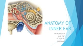
- 1. ANATOMY OF INNER EAR Dr PRAWAL S D M.S. ENT 1st YEAR PG KIMS HUBLI
- 2. EMBRYOLOGY Involves three main phases: Development(4th to 8th week) Growth (8th to 16th week) Ossification (16th to 24th week)
- 3. DEVELOPMENT OF INNER EAR OTIC PLACODES OTIC PITS OTOCYST
- 6. Otic vesicle or the otocyst sinks into the mesenchymal cells of developing head These mesenchymal cells helps in the development of the outer bony capsule of the inner ear
- 7. Otocyst differentiates into 1. Dorsal utricular portion – utricle, semicircular canal and endolymphatic duct 2. Ventral saccular portion – saccule and cochlear duct All of these structures together forms the membranous labyrinth
- 8. DEVELOPMENT OF SACCULE AND COCHLEAR DUCT
- 10. DEVELOPMENT OF ORGAN OF CORTI Initially , epithelial cells of the cochlear duct are alike They form two ridges : The inner ridge , and the outer ridge The outer ridge forms one row of inner hair cells and three or four rows of outer hair cells Hair cells are the sensory cells of auditory system Tectorial membrane , a fibrillar gelatinous substance attached to the spiral limbus that rests with its tip on the hair cells The sensory cells and tectorial membrane together constitutes the ORGAN OF CORTI
- 12. ANATOMY OF INNER EAR Inner ear is present in the petrous part of the temporal bone It is present between middle ear laterally and the internal acoustic meatus medially
- 13. BONY LABYRINTH 1. VESTIBULE 2. SEMICIRCULAR CANAL 3. COCHLEA MEMBRANOUS LABYRINTH 1. COCHLEAR DUCT 2. UTRICLE AND SACCULE 3. SEMICIRCULAR DUCT 4. ENDOLYMPHATIC SAC AND DUCT
- 15. BONY LABYRINTH VESTIBULE Central chamber of the labyrinth In lateral wall lies the oval window Inner aspect of the medial wall presents two recesses, a spherical recesses for saccule, an elliptical recesses for utricle Opening of aqueduct of vestibule is present below it through which passes the endolymphatic duct Posterior superior part of vestibule are the five opening of the semicircular canal
- 17. Semicircular canal They are three in number lateral, posterior and superior All lie in planes at right angle to one another The non ampullated ends of the posterior and superior semicircular canals unite to form a common channel called the crus commune
- 20. BONY COCHLEA The bony cochlea is a coiled tube making 2.5 to 2.75 turns round the central pyramid of bone called the modiolus The base of the modiolus is directed towards the internal acoustic meatus and transmits vessels and nerves to the cochlea Around the modiolus winding spirally like the thread of a screw is a thin plate of bone called osseous spiral lamina, which gives attachment to the basilar membrane
- 21. The bony cochlea contains three compartments Scala vestibuli Scala media Scala tympani
- 25. MEMBRANEOUS LABYRINTH COCHLEAR DUCT It is a blind coiled tube Made up of three walls 1. The basilar membrane 2. The reissner’s membrane 3. The striae vascularis – contains vascular epithelium and concerned with secretion of endolymph
- 30. UTRICLE AND SACCULE Utricle lies in the posterior part of the bony vestibule Receives the five opening of semicircular canal Connected to saccule via utriculosaccular duct The macula of the utricle lies mainly in the horizontal plane on the inferior surface of the utricle and plays an important role in determining orientation of the head when the head is upright The macula of the saccule is located mainly in a vertical plane and signals head orientation when the person is lying down.
- 32. SEMICIRCULAR DUCTS They are three in number The ampullated end of each duct contains a thickened ridge of neuroepithelium called crista ampullaris
- 36. ENDOLYMPHATIC DUCT AND SAC Formed by the union of two ducts one form the utricle and another from the saccule Passes through the vestibular aqueduct The terminal part is dilated to form endolymphatic sac
- 38. INNER EAR FLUID AND THEIR CIRCULATION PERILYMPH Fills the space between the bony and the membranous labyrinth Resembles extracellular fluid is rich in sodium ions It communicates with CSF through the aqueduct of cochlea which opens into scala tympani
- 39. TWO VIEWS REGARDING THE FORMATION OF PERILYMPH It is the filtrate of blood serum and is formed by capillaries of the spiral ligament It is a direct continuation of CSF and reaches the labyrinth via aqueduct of cochlea
- 40. ENDOLYMPH Fills the entire membranous labyrinth and resembles intracellular fluid, being rich in potassium ions Secreted by the secretory cells of the stria vascularis of the cochlea and by the dark cells
- 41. TWO VIEWS REGARDING ITS FLOW LONGITUDINAL : Endolymph from the cochlea reaches saccule, utricle and endolymphatic duct and get absorbed through endolymphatic sac which lies in the subdural space RADIAL : Endolymph is secreted by stria vascularis and also gets absorbed via stria vascularis
- 42. ORGAN OF CORTI
- 43. It was discovered by Alfonso Giacomo Gaspare Corti Components of organ of corti Hair cells : Important receptor cells of hearing and transduce sound energy into electrical energy , Inner hair cells are arranged in a single row , inner cells are richly supplied by afferent cochlear fibers and are probably more important in the transmission of auditory impulse, outer hair cells are more in number and are arranged in 3 – 4 layers and mainly receives efferent innervation from olivary complex and are concerned with modulating the function of inner hair cells
- 50. Tunnel of corti : Formed by inner and the outer rods , it contains fluid called cortilymph Supporting cells : Dieters’ cells and cells of hansen Tectorial membrane : Contains gelatinous matrix with delicate fibers, The shearing force between the hair cells and tectorial membrane produces the stimulus to hair cells
- 54. MECHANISM OF HEARING MECHANICAL CONDUCTION OF SOUNDS IMPEDENCE MATCHING MECHANISM : Lever action of the ossicles and hydraulic action of tympanic membrane
- 55. FUNCTIONAL ANATOMY OF COCHLEA BASILAR MEMBRANE Basilar membrane is a fibrous membrane Made of 20,000 to 30,000 basilar fibers Length of basilar membrane increases from base 0.04mm to apex 0.5mm Diameter of the fibers decreases Hundred fold decrease in overall stiffness
- 56. TRANSMISSION OF SOUND WAVE IN COCHLEA – TRAVELING WAVE Initial effect of the soundwave entering the oval window causes the basilar membrane at the base of the cochlea to move in direction of the round window However due to elastic tension which is built in basilar membrane initiates a fluid wave that travel along the basilar membrane which travel towards the helicotrema
- 57. PATTERN OF VIBRATION – Each wave is weak at the onset but becomes stronger when it reaches that portion of the basilar membrane that has a natural resonant frequency equal to the respective sound frequency AMPLITUDE PATTERN OF VIBRATION
- 61. FUNCTION OF ORGAN OF CORTI Organ of corti is the receptor organ which generate the nerve impulse in response to vibration of basilar membrane Auditory impulses are mainly transmitted by inner hair cells Excitation of the hair cells - stereocilia, project upward from the hair cells and either touch or are embedded in the surface gel coating of the tectorial membrane Bending in one direction causes depolarization and bending in other direction causes hyperpolarization
- 62. Hair cell receptor potentials and excitation of auditory nerve fibers The stereocilia are stiff structures because each has a rigid protein framework. They become progressively longer on the side of the hair cell away from the modiolus The tops of the shorter stereocilia are attached by thin filaments to the back sides of their adjacent longer stereocilia. Therefore, whenever the cilia are bent in the direction of the longer ones, the tips of the smaller stereocilia are tugged outward from the surface of the hair cell.
- 63. This causes a mechanical transduction that opens 200 to 300 cation- conducting channels, allowing rapid movement of positively charged potassium ions from the surrounding scala media fluid into the stereocilia, which causes depolarization of the hair cell membrane. Hair cells releases excitatory neurotransmitter ENDOCHOCLEAR POTENTIAL : An electrical potential of about +80 mv exists between endolymph and perilymph
- 66. THANK YOU