Assessing Urinary Tract Function and Diagnosing Related Problems
•Télécharger en tant que PPT, PDF•
14 j'aime•11,471 vues

The document discusses diagnostic tests used to evaluate urinary tract function. It describes various urine tests including urinalysis, urine culture and sensitivity, urine osmolality, 24-hour urine collections for creatinine clearance and catecholamines. Imaging tests are also summarized such as abdominal ultrasound, intravenous pyelogram, renal scan, cystoscopy and renal biopsy. The goals are to identify parameters for assessing upper and lower urinary tract status and describe studies used to determine urinary tract function.

Recommandé
Contenu connexe
Tendances
Tendances (20)
Similaire à Assessing Urinary Tract Function and Diagnosing Related Problems
Similaire à Assessing Urinary Tract Function and Diagnosing Related Problems (20)
Dernier
Dernier (20)
Assessing Urinary Tract Function and Diagnosing Related Problems
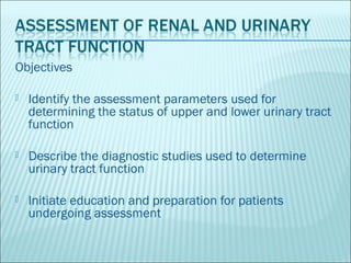
- 1. Objectives Identify the assessment parameters used for determining the status of upper and lower urinary tract function Describe the diagnostic studies used to determine urinary tract function Initiate education and preparation for patients undergoing assessment
- 2. Obtaining a urologic health history requires excellent communication skills because many patients are embarrassed or uncomfortable discussing genitourinary function or symptoms.
- 3. Px’s chief concern or reason for seeking health care, the onset of the problem & it’s effect on the px’s quality of life Location, character & duration of pain (if present) & its relationship t voiding; factors that precipitate pain and those that relieve it Hx of UTI, including past tx or hospitalization for UTI
- 4. Fever or chills Previous renal or urinary dx tests or use of indwelling catheters Dysuria & when it occurs during voiding (at initiation or termination of voiding) Hesitancy, straining, or pain during, after urination
- 5. Urinary Incontinence (stress intolerance, urge incontinence, overflow incontinence or functional incontinence) Hematuria or change in color, volume of urine Nocturia and its date of onset Renal calculi (kidney stones), passage of stones or gravel in urine
- 6. Female px: number & type (vaginal or cesarean) of deliveries; use of forceps; vaginal infxn, discharge or irritation; contraceptive practices Presence or history of genital lesions or STD’s Habits: use of tobacco, alcohol, or recreational drugs Any prescription & over-the-counter medications (including those prescribed for renal or urinary problems
- 7. Gradual kidney dysfunction can be insidious in its presentation, although fatigue is a common symptom. Fatigue, shortness of breath, and exercise intolerance all result from the condition known as “anemia of chronic dse” Hgb / Hct are quantified to detect anemia however Hgb level is more significant it’s the one responsible for circulating oxygen
- 9. Problems Associated with Changes in Voiding Problem Definition Possible Etiology Frequency Frequent voiding – more than Infection, obstruction of lower urinary tract leading to residual urine and every 3 hours overflow, anxiety diuretics, BPH, urethral stricture, diabetic neuropathy Urgency Strong desire to void Infection, chronic prostatitis, urethritis, obstruction of lower urinary tract leading to residual urine and overflow, anxiety, diuretics, BPH, urethral stricture, diabetic neuropathy Dysuria Painful or difficult voiding Lower urinary tract infection, inflammation of bladder or urethra, acute prostatitis, stones, foreign bodies, tumors in bladder Hesitancy Delay, difficulty in initiating BPH, compression of urethra, outlet obstruction, neurogenic bladder voiding Nocturia Excessive urination at night Decreased renal concentrating ability, ♥ failure, diabetis mellitus, incomplete bladder emptying, excessive fluid intake at bedtime, nephritic syndrome, cirrhosis with ascites Incontinence Involuntary loss of urine External urinary sphincter injury, obstetric injury, lesions of bladder neck, detrusor dysfunction, infection, neurogenic bladde, medications neurologic abnormalities Enuresis Involuntary voiding during sleep Delay in functional maturation of central NVS (bladder control usually achieved by 5 years of age) obstructive dse of lower urinary tract, genetic factors, failure to concentrate urine, UTI, psychological stress Polyuria Increased volume of urine voided DM, diabetes insipidus, use of of diuretics, excess fluid intake, lithium toxicity, some forms of kidney dse (hypercalmemic and hypokalemia nephropathy) Oliguria Urine output less than Acute or chronic renal failure, complete obstruction 400mL/day Anuria Urine output less than 50mL/day Acute or chronic renal failure, complete obstruction Hematuria Red blood cells in the urine Cancer of genitourinary tract, acute glomerulonephritis, renal stones, renal tuberculosis, blood dyscrasia, trauma, extreme exercise, rheumatic fever, hemophilia, leukemia, sickle cell trait or disease Proteinuria Abnormal amounts of protein in Acute and chronic renal disease, mephrotic syndrome, vigorous exercise, the urine heat stroke, severe ♥ failure, diabetic neuropathy, multiple myeloma
- 10. Gastrointestinal symptoms may occur with urologic conditions because of shared autonomic and sensory innervation and renointestinal reflexes. Common s/sx: N/V, diarrhea, abdominal discomfort, abd distention,. Urologic symptoms can mimic appendicits, PUD, cholecystitis, thus making diagnosis difficult especially in elderly because of decreased neurologic innervation to this area.
- 11. Identifying Characteristics of Genitourinary Pain TYPE LOCATION CHARACTER ASSOCIATED S/SX POSSIBLE ETIOLOGY KIDNEY Costovertebral angle, may Dull constant ache; if n/v, diaphoresis, pallor, Acute obstruction, kidney extend to umbilicus sudden distention of signs of shock stone, blood clot, acute capsule, pain is severe, pyelonepritis, trauma sharp, stabbing and colicky in nature BLADDER Suprapubic area Dull, continous pain, may Urgency, pain at the end Overdistended bladder, be intense with voiding, of voiding, painful infection, interstitial may be severe if bladder is straining cystitis; tumor full URETERAL Costovertebral angle, Severe, sharp, stabbing n/v, paralytic ileus Ureteral stone, edema or flank, lower abdominal pain, colicky in nature stricture, blood clot area, testis or labium PROSTATIC Perineum and rectum Vague discomfort, feeling Suprapubic tenderness, Prostatic cancer, acute or of fullness in perineum, obstruction to urine flow, chronic prsotatitis vague back pain frequency, urgency, dysuria, nocturia URETHRAL Male: along penis to Pain variable, most severe Frequency, urgency, Irritation of bladder neck, meatus; female: urethra to during and immediately dysuria, nocturia, urethral infection of urethra, meatus after voiding discharge trauma, foreign body in lower urinary tract
- 12. Aging affects the way the body absorbs, metabolizes, and excretes drugs thus placing the elderly patient at risk for adverse reactions, including compromised renal function Structural or functional abnormalities that occur with aging may prevent complete emptying of the bladder. This may be due to decrease bladder wall contractility due to myogenic or neurogenic causes or structurally related to bladder outlet obstrcution as in BPH.
- 13. URINALYSIS – a urine test for evaluation of the renal system and for determining renal disease
- 14. Changes in Urine Color and possible Causes Urine Color Possible Cause Colorless to pale yellow Dilute urine due to diuretics, alcohol consumption, diabetes insipidus, glycosuria, excess fluid intake, renal dse Yellow to milky white Pyuria, infection, vaginal cream Bright yellow Multiple vitamin preparation Pink to red Hgb breakdown, RBC, gross blood, menses, bladder or prostate surgery, beets, blackberries, medications (phenyton, rifampicin, phenothiazine, cascara, senna products) Blue, blue green Dyes, methylene blue, pseudmona species organisms, medications Orange to amber Concentrated urine due to dehydration, fever, bile, excess bilirubin or carotene, medications Brown to black Old RBC, urobilirogen, bilirubin, melanin, porphyrin, extremely concentrated urine due to dehydration, medications
- 15. A urine test that measures the ability of the kidneys to concentrate urine NV: 1.016 to 1.022 (may vary depending on the lab) An increase in the result may indicate insufficient fluid intake, decreased renal perfusion or increased ADH A decrease in result (less concentrated urine) occurs with increased fluid intake or DI.
- 16. Urine test that identifies the presence of microorganisms and determines the specific antibiotics to treat the existing microorganisms appropriately.
- 17. Evaluates how well the kidneys remove creatinine from the blood The urine specimen for the creatinine clearance is usually collected for 24 hours, but shorter periods such as 8 to 12 hours could be prescribed.
- 18. A 24 hour urine collection sample is tested to diagnose gout and kidney dse
- 19. The test is a 24 hour urine collection to diagnose pheocromocytoma, a tumor of the adrenal gland The test determines urinary catecholamine levels in the urine
- 20. May be performed for evaluating urinary frequency, inability to urinate or amount of residual urine (the amount of urine remaining in the bladder after voiding)
- 21. Performed to delienate the size, shape and position of the kidneys and to reveal any abnormalities such as calculi in the kidneys or urinary tract, hydronephrosis (distention of the pelvis of the kidney) cysts, tumors, or kidney displacement by abnormalities in surrounding tissues
- 22. Used in evaluating genitourinary masses, neprhrolithiasis, chronic renal infxn, renal or urinary tract trauma, metastatic disease and soft tissue abnormalities
- 23. Requires injection of isotope into the circulatory system. Hypersensitivity to the isotope is rare Nuclear scans are used to evaluate acute and chronic renal failure, renal masses and blood flow before and after kidney transplantation.
- 24. Intravenous urography includes test includes tests such as excretory urography, intravenous pyelography (IVP) and infusion drip pyelography. Used as the initial assessment of any suspected urologic problem, especially lesions in the kidneys and ureters. It also provide a rough estimate of renal function.
- 25. Catheters are advanced into renal pelvis by means of cystoscopy. It is usually performed if Intravenous urography provides inadequate visualization of the collecting systems. It may also be used before extracorporeal shock wave lithotripsy or in px with urologic cancer who need to follow up and are allergic to intravenous contrast.
- 26. Aids in evaluating vesicoureteral reflux (backflow of urine from the bladder into one or both ureters) and assessing the px for bladder injury
- 27. Uses fluoroscopy to visualize the lower urinary tract and assess urine storage in the bladder. A urethral catheter is inserted and a contrast agent in instilled into the bladder. When the bladder is full and the patient feels the urge to void, the catheter is removed and the px voids.
- 28. Renal angiogram/renal arteriogram provides an image of the renal arteries. The femoral or axillary are the preferred sites. Use to evaluate renal blood flow in suspected renal trauma, to differentiate renal cysts from tumors and to evaluate hypertension. It is used for preoperatively for tyransplantaion.
- 29. Endourology or urologic endoscopic procedures can be performed in one of two ways; using a cystoscope inserted into the urethra, or percutaneously through an incision. Used to directly visualize the urethra and bladder. The cystoscope also permits the urologist to obtain a urine specimen from each kidney to evaluate its function.
- 30. Cup forceps can be inserted through the cystoscope for biopsy. Calculi may be removed from the urethra, bladder and ureter using cystoscopy.
- 31. Brush biopsy techniques provide specific information when abnormal x-ray findings of the ureter or renal pelvis raise questions about whether the defect is a tumor, a stone, a blood clot, or an artifact. First a cystoscopic exam, then a ureteral catheter is introduced, follwed bya biopsy brush that is passed through the catheter.
- 32. Used in diagnosing and evaluating the extent of kidney dse. Indications for biopsy include unexplained acute renal failure, persistent proteinuria or hematruria, transplant rejection and glomerulonephritis . Obtained either percutaneously (needle biopsy) or by open incision through a small flank incision.
- 33. Uroflowmetry – is the record of the volume of urine passing through the urethra per time unit (milliliter per second). The px is advised to arrive for the test with a strong urge to void but not have an overly full bladder. It is combined with electromyographic measurement of the external urethral sphincter via surface wire or needle electrodes placed at th level of the sphincter, on eother side of the urethra.
- 34. Cystometrography – graphic recording of the pressures in the bladder filling and emptying. It is the major dx portion of urodynamic testing.
- 35. Involves placement of electrodes in the pelvic floor musculature or over the area of the anal sphincter to evaluate the neuromuscular function of the lower tract. It is performed simultaneously with CMG
- 36. Consideres optimal urodynamic evaluation. This test combines a study of the filling and voiding phases of the CMG and EMG with a simultaneous visualization of the lower urinary tract via a radiopaque filling and detailed assessment of the voiding dysfunction which may be due in part to anatomic dysfunction.
- 38. For some patients, contrast agents are neprhotoxic and allergenic. The following guidelines can help the nurse and other care givers respond quickly in the event of a problem.
- 39. Have emergency equipment and medications available in case of the patient has an anaphylactic reaction to the contrast agent. Emergency supplies include epinephrine, corticosteroids, and vasopressors, oxygen and airway and suction equipment
- 40. Thank you