
Recommandé
Contenu connexe
Tendances
Tendances (20)
En vedette
En vedette (18)
Similaire à Ppt parietal lobe
Similaire à Ppt parietal lobe (20)
Dernier
Dernier (20)
Ppt parietal lobe
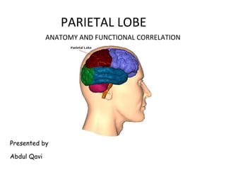
- 1. PARIETAL LOBE Presented by Abdul Qavi ANATOMY AND FUNCTIONAL CORRELATION
- 2. INTRODUCTION The parietal cortex is considered as one of the most complex region of human brain which is responsible for the integration of various stimuli . Have undergone a major expansion in the course of human evolution, largely in the inferior parietal region Receives, correlates, analyze primary sensory information to interpret stimulus and aid in discrimination and recognition.
- 3. Lobe of the hand
- 4. Defining thelobes central (rolandic) sulcus sylvyan (lateral) sulcus frontal lobe temporal lobe occipital lobe parietal lobe
- 5. BOUNDARIES OF THE PARIETAL LOBE
- 6. BOUNDARIES OF THE PARIETAL LOBE – Anterior border - Central Fissure – Ventral border - Sylvian Fissure – Dorsally by the cingulate gyrus – Posterior border - Parieto-occipital sulcus
- 7. SULCI AND GYRI ON THE SUPERO LATERAL SURFACE superolateral medial
- 8. SULCI AND GYRI ON THE VARIOUS SURFACE OF PARIETAL LOBE SUPERO LATERAL SURACE • Post central gyrus( area 1,2,3) • Superior parietal lobule (area 5,7) • Inferior parietal lobule • Supra marginal gyrus - lies around the upturned end of sylvian fissure. • Angular gyrus – lies around the upturned end of superior temporal gyrus. MEDIAL SURFACE • Supra splenial sulcus – separate the precuneus from cingulate gyrus • Precuneus - lies between parieto occipital sulcus and paracentral lobule • Isthmus – Separates the splenium of corpus callosum from calcarine sulcus
- 11. Parietal lobe sulci and gyri Post central sulcus – posterior boundary of somatosensory cortex. Intraparietal sulcus behind post central sulcus which divides the parietal lobe into sup. & inf. Parietal lobule Posterior end of sylvian fissure curves upwards to terminates into inf.parietal lobule – surrounding cortex supramarginal gyrus[SMG 40]Parietal lobe
- 12. Parietal lobe sulci and gyri • Posterior end of sup. Temporal sulcus – angular gyrus[AG 39] • SMG & AG =Ecker’s inf Parietal Lobule • Ecker’s IPL & post. Third of first temporal gyrus constitute the wernicke’language area • 3,1,2-primary sensory areas • 5- somatosensory association area • 7-somatosensory or somatosensory/visual
- 13. Parietal Topography •Postcentral Gyrus (1,2,3) •Superior Parietal Lobule (5 , 7) •Supramarginal Gyrus (40) •Angular Gyrus ( 39)
- 14. Subdivisions of the Parietal Lobes Functional zones Anterior zone -1,2,3, •Somatosensory cortex Posterior zone -remaining areas •Posterior parietal cortex von Economo: Posterior parietal areas •PE (5) •PF(7b) •PG -Polymodal and asymmetric larger in right hemisphere
- 15. • Visual processing areas – Intraparietal sulcus (cIPS) • Control of saccadic eye movements – Saccade - involuntary abrupt and rapid small movements made by the eyes when changing the fixation point • Visual control of grasping – Parietal reach regions (PRR) • Visually guided grasping movements Subdivisions of the Parietal Lobes
- 17. Somatosensory strip To area PE -Tactile recognition To motor regions -sensory information about limb position and movement •Area PE is somatosensory –Inputs from the somatosensory strip –Outputs to primary motor cortex, supplementary motor cortex, premotor regions, and area PF •Area PF Input from somatosensory, primary motor cortex, premotor cortex, and small visual input through area PG •Area PG –Receives complex connections including visual, somesthetic, proprioceptive, auditory, vestibular, oculomotor, and cingulate connections –Parieto-temporo-occipital crossroads –Part of the Dorsal Stream •Close relation between the posterior parietal connections and the prefrontal Connections of the Parietal Lobes
- 18. Connections of the Parietal Lobes
- 19. • Anterior zones - process somatic sensations and perceptions • Posterior zones - integrate information from vision with somatosensory information for movement • Spatial Map in the Brain? A Theory of Parietal Lobe Function
- 20. NP/MGH NEURO-IMAGING NORMAL CORTICAL ANATOMY • The Central Sulcus • Sagittal • Axial • Coronal
- 21. NP/MGH The Central SulcusThe Central Sulcus
- 22. NP/MGH • superior frontal sulcus - pre CS sign • sigmoidal Hook sign • pars bracket sign • Bifid post-CS sign • thin postcentral gyrus sign • intraparital sulcus - post-CS • midline sulcus sign The Central Sulcus (CS)* *Naidich & Brightbill. Int J Neurorad 1996;2:313-338*Naidich & Brightbill. Int J Neurorad 1996;2:313-338
- 23. NP/MGH • Superior frontal sulcus - preCS sign – the posterior end of the superior frontal sulcus joins the precentral sulcus in 85% The Central Sulcus (CS) Precentral sulcus Superior frontal sulcus Precentral gyrus Central sulcus Superior frontal gyrus Superior frontal sulcus Precentral sulcus Precentral gyrus
- 24. NP/MGH • pars bracket sign – The paired pars marginalis form a “bracket” to each side of the interhemispheric fissure at or behind the central sulcus (96%). The Central Sulcus (CS) Precentral sulcus Superior frontal sulcus Precentral gyrus Central sulcus Pars bracket Paracentral lobule
- 25. NP/MGH Sigmoid “Hook” – hooklike configuration of the posterior surface of the precentral gyrus – the “hook” corresponds to the motor hand area. – The “hook” is well seen on CT (89%) and MRI (98%). The Central Sulcus (CS)The Central Sulcus (CS) Precentral sulcus Central sulcus
- 26. NP/MGH • Bifid post-CS sign – the post-CS is bifid (85%). – The bifid post-CS encloses the lateral end of the pars marginalis (88%). The Central Sulcus (CS) Precentral sulcus Precentral gyrus Central sulcus Postcentral sulcus Pars bracket
- 27. NP/MGH Central sulcus Postcentral sulcus Central sulcus Central sulcus Postcentral sulcus Postcentral sulcus Pars bracketPars bracket
- 28. NP/MGH Thin post-CG sign – the postcentral gyrus is thinner than the precentral gyrus (98%). The Central Sulcus (CS)The Central Sulcus (CS) Precentral gyrus Postcentral gyrus
- 29. NP/MGH Intraparietal Sulcus (IPS) and the post-CS – in axial MRI, the IPS intersects the post-CS (99%). The Central Sulcus (CS)The Central Sulcus (CS) Pars bracket IPS Postcentral sulcus IPS Pars bracket
- 31. NP/MGH SFS-preCS sign Hook sign Pars bracket sign Bifid post-CS sign Thin postcentral gyrus sign IPS - postCS sign The Central Sulcus (CS)The Central Sulcus (CS)
- 33. NP/MGH Superior Temporal gyrus Middle Temporal gyrus Inferior Temporal gyrus Fusiform gyrus
- 34. NP/MGH Superior occipital gyrus Intra-occipital sulcus Middle occipital gyrus Cingulate gyrus Parieto-occipital fissure Calcarine sulcus Cuneus Middle temporal gyrus Superior temporal sulcus Superior temporal gyrus Insula Inferior frontal gyrus, pars orbitalis Superior frontal gyrus Middle frontal gyrus Inferior frontal gyrus, pars opercularis Lateral fissure Lateral fissure Inferior parietal gyrus
- 35. NP/MGH Middle occipital gyrus Superior temporal gyrus Intra-occipital sulcus Superior frontal gyrus Central sulcus Superior occipital gyrus Parieto-occipital sulcus Superior temporal sulcus Lateral fissure Inferior parietal gyrus Postcentral gyrus Lateral fissure Middle frontal gyrus Inferior frontal gyrus
- 36. NP/MGH Postcentral sulcus Superior frontal sulcus Central sulcus Intraparietal sulcus Superior frontal gyrus Middle frontal gyrus Superior parietal gyrus Centrum semiovale Parietooccipital sulcus Precuneus Angular gyrus Central sulcus Inferior frontal gyrus Supramarginal gyrus Postcentral sulcus
- 37. NP/MGH Central sulcus Postcentral sulcus Superior frontal sulcus Precentral sulcus Pars marginalis Intraparietal sulcus Superior frontal gyrus Middle frontal gyrus Precuneus Paracentral lobule Superior parietal gyrus
- 39. NP/MGH Olfactory bulb Gyrus rectus Medial Orbital gyrus Inferior Frontal gyrus Superior Frontal gyrus Middle Frontal gyrus Interhemispheric Fissure Inferior Frontal gyrus
- 40. NP/MGH Superior Frontal gyrusSuperior Frontal sulcus Middle Frontal gyrus Superior Temporal Sulcus Sylvian Fissure Amygdala Precentral sulcus Anterior commissure Cingulate sulcus Superior Temporal gyrus Middle Temporal gyrus Inferior Temporal gyrus Precentral gyrus
- 41. NP/MGH Paracentral lobule Superior Temporal gyrus Middle Temporal gyrus Inferior Temporal gyrus Central Sulcus Postcentral gyrus Cingulate gyrusIntraparietal sulcus Fusiform gyrus Collateral sulcus Parahippocampal gyrus Supramarginal gyrus Intraparietal sulcus
- 42. NP/MGH Superior Temporal gyrus Middle Temporal gyrus Inferior temporal gyrus Fusiform gyrus Central sulcus Paracentral lobule
- 43. NP/MGH Lingual gyrus Calcarine sulcus Superior parietal lobule precuneus Cingulate gyrus Tentorium cerebelli Fusiform gyrus Inferior parietal lobule Middle occipital gyrus Inferior occipital gyrus Lingual gyrus Collateral sulcus
- 45. NP/MGH Subcallosal gyrus Gyrus rectus Parietooccipital sulcus Fastigium, fourth ventricle Cingulate gyrus Calcarine sulcus Marginal ramus of Cingulate sulcus precuneus Paracentral lobule Cingulate sulcusSuperior frontal gyrus Cuneus Lingual gyrus
- 46. NP/MGH Parietooccipital sulcus Calcarine sulcus Superior parietal lobule Marginal ramus of Cingulate sulcus Central sulcus Precentral sulcus Precuneus Corona radiata Superior frontal gyrus Lingual gyrus Inferior occipital gyrus
- 47. NP/MGH Central sulcus Inferior Temporal gyrus Middle Temporal gyrus uperior Temporal gyrus
- 48. NP/MGH Gyrus rectus Parietooccipital sulcus Cingulate gyrus Calcarine sulcus Lingual gyrus Marginal ramus of Cingulate sulcus Superior parietal lobule Cingulate sulcus Caudothallamic groove Precuneus Central sulcus Cuneus Precentral gyrus Frontomarginal gyrus Superior frontal gyrus
- 49. NP/MGH Inferior Temporal gyrus Superior Temporal sulcus Superior Temporal gyrus Anterior occipital sulcus Superior frontal sulcus Precentral sulcus Central sulcus Postcentral sulcus Angular gyrus Lateral fissure, posterior segment Inferior frontal gyrus, pars orbitalis Middle Temporal gyrus Inferior occipital gyrus Middle occipital gyrus Inferior frontal gyrus, pars triangularis
- 50. BLOOD SUPPLY
- 51. BLOOD SUPPLY
- 52. Primary somatosensory area Location : Post central gyrus(ant parietal lobule) on lateral surface and dorsal aspect of paracentral lobule on medial serface. Broadman area (3 ,1, 2) Representation : contralatral half of body inverted Function: initial reception center for afferent impulses, especially for tactile, pressure, and position sensations. necessary for discriminating finer, more critical grades of sensation and for recognizing intensity. Afferent connections: VP nucleaus of thalamus Outputs: primary motor cortex, contralateral S1,association somatosensory cortex(area 5 & 7), thalamus Deficit: Postural sensation (proprioception), passive movement (kinesthesis), Tactile sensation, Two point discrimination, Astereognosis,High sensory thresholds Functional areas
- 54. Secondary Somatosensory area Location : superior lip of lateral fissure (parietal operculam) Representation :contralateral side dominant, Bilateral representation Afferent: Intralaminar nuclei and posterior group of nuclei of thalamus Function: not well described, ? Involved in less discriminative aspects of sensation. Lesions: none ascribed, rarely inability to appreciate pain(asymbolia)
- 55. Somato-sensory association area: Location : superior parietal lobule. broadmann’s area(5,7) Function : interpretation; similarities and differences, spatial relationships and 2D qualities, variations in form and weight, and localization of sensation • Area 5-, Manipulation of objects Tool use/body image • Area 7- Integration of visual and somato-sensory stimuli, Hand-eye coordination, reaching and grasping,, • Afferent: primary somato-sensory area • Deficit : Impair gnostic (knowing, recognition) aspects of sensation , stereognosis, graphesthesia, two-point discrimination, and tactile localization , poor hand eye coordination, (appreciation of primary sensations remains, but assoc. functions impaired)
- 56. Inferior parietal lobule • Location: supramarginal gyrus (40) and angular gyrus (39) • Function:. Left hemisphere – language ,maths, reading, writing, understanding of symbols. Right hemisphere—visuo-spatial orientation. • Lesions Aphasia, agnosia, and apraxia and visuspatial defects • A deeply placed parietal lesion may cause either an inferior quadrantic or hemianopic visual field defect
- 57. Clinical assesment and testing
- 58. Post-Central Gyrus,Dominant or Non-Dominant 1. Impaired Postural sensation (proprioception), passive movement (kinesthesis). 2. Astereognosis 3. Impaired Two point discrimination 4. Agraphesthesia 5. Weight discrimination
- 59. Inability to discriminate size and shape of objects and identify them by touch alone. Tests Patient identifies by touch such common objects as a coin, paperclip, pencil, or key (each hand tested separately) Patient judges the relative size of a series of coins Patient judges the texture of a series of objects, such as cloth, wire, sandpaper Astereognosis (tactile agnosia)
- 60. Astereognosis
- 61. Graphesthesia Ability to recognise letters or numbers written on skin with pencil,or dull pin Testing is often done over the finger pads, palms, or dorsum of the feet Letters or numbers about 1 cm in height are written on the finger pads, larger elsewhere clear figures as 8, 4 5 used first, more difficult 6, 9 ,3 can be used as finer tests Tactile movement sense, directional cutaneous kinesthesia- Ability to tell the direction of movement of a light scratch stimulus drawn for 2 cm to 3 cm across the skin which may be a sensitive indicator of function of the posterior columns and primary somatosensory cortex Loss of graphesthesia or the sense of tactile movement with intact peripheral sensation implies a cortical lesion, particularly when the loss is unilateral.
- 62. Two point discrimination Ability to differentiate, eyes closed, cutaneous stimulation by one point from stimulation by two points. Instruments: two-point discriminator, electrocardiogram calipers,compass, paper clip bent into “v,” adjusting the two points to different distances. Method Either one-point or two-point stimuli are delivered randomly, and the minimal distance that can be discerned as two points is determined. The result is taken as the minimum distance between two points that can be consistently felt separately. Normal 2-point discrimination - 1 mm (tip of the tongue), 2 mm to 3 mm ( lips), 2 mm to 4 mm ( fingertips), 4 mm to 6 mm (dorsum of the fingers), 8 mm to 12 mm( palm), 20 mm to 30 mm( back of the hand), and 30 mm to 40 mm ( dorsum of the foot). The findings on the two sides of the body must always be compared.
- 63. Superior Parietal Lobule,Dominant or Non-Dominant cannot reach for objects (optic ataxia) -Balint syndrome Poor visual guidance of hands, fingers, eyes, and limbs, head (hard time catching a ball) Hard time directing movement in space (trouble flying a kite) Hard time distinguishing left from right
- 64. Dominant inferior parietal lobule 1. Acalculia 2. Agraphia 3. Left-right confusion 4. Finger agnosia 5. Conductive aphasia 6. Alexia 7. Ideomotor apraxia Gerstmann’s syndrome
- 65. Ideomotor apraxia: failure to perform previously learned motor acts accurately. Results from left hemisphere lesion Usually affects both sides, may be worse on right side Can affect the face (buccofacial) and/or the limbs Tests Carrying out motor acts to command: Buccofacial (blow out a match, protrude tongue, drink through a straw) Limb (salute, use a toothbrush, flip a coin, hammer a nail, comb hair,,snap fingers, kick a ball, crush out a cigarette) Whole body commands(stand like a boxer, swing a baseball bat)
- 66. 1. wernicke area 2. Arcuate fasciculus 3. Lt premotor area 4. Lt motor cortex 5. Corpus callosum 6. Rt premotor area 7. Rt motor cortex Ideomotor apraxia:
- 67. Ideational apraxia: Able to carryout individual components of a complex motor act but can not perform the entire sequence properly leading to a goal. Results from left hemisphere lesion ( temporo-parietal) also seen in generalised cognitive impairment. Tests Carrying out complex motor acts to command: Opening tooth paste, taking tooth brush from holder, and placing toothpaste on brush. How to mail a letter How to drive a car.
- 68. Results from left hemisphere supramarginal gyrus lesion if the underlying arcuate fasciculus is cut Fluent speech with word finding pauses Severely defective repetition Paraphasia in repetition and in spontaneous speech Normal comprehension and reading Impaired writing, spontaneous and to dictation, errors in spelling, word choice, Naming may be mildly impaired Tests Repetition of words, phrases, & sentences Write to dictation (letters, words, sentences) Ask patient to write sentences describing a Job, the weather, or a picture Confrontation naming of objects, clothing, body parts, parts of objects, colors Conduction aphasia
- 69. Finger agnosia: Inability to recognize, name, and point to individual fingers on self and others Usually associated with lesion of dominant hemisphere Lt handed pts may have finger agnosia with lesions of either hemisphere Limited clinical utility for localisation Tests • Non verbal finger recognition: pt eyes closed, touch pt finger, then ask pt to point same finger on examiner hand • Identification of named fingers on examiner’s hand: examiner’s hand placed in various positions. Ask pt “point to my middle finger” • Verbal identification (naming) of finger on self and examiner: hand placed in various positions, ask pt “what is the name of this finger”
- 70. Right-left disorientation Inability to distinguish right from left on self or env. More common with left hemisphere lesion Normal population (9%males, 17% females ) can have difficulty in rt - lt testing Tests • Identification on self(show me your rt foot), • Crossed commands on self(With your rt hand touch your lt shoulder) • Identification on examiner(point to my lt elbow) • Crossed command on examiner(with ur rt hand point to my lt eye)
- 71. Acalculia Loss of ability to understand & order numbers More severe with left hemisphere lesion Also note errors in borrowing, alignment , error to particular calculation, Tests Verbal examples(addition, subtraction, multiplication, and division) Eg. 4+6, 8-5, 9*7, 9 /3 Verbal complex problems (allow 20 sec for response) Eg. 14+17, 43-38, 21*5, 128/8 Written complex problems(allow 30 sec for response) 108 605 108 559 +79 -86 *36 /43
- 72. Calculation errors Rt hemispheric lesion with lt neglect Rt parietal bleed , poor alignment, calculation errors Alzeimers ds, rote multiplication good but basic arithmatic disturbed
- 73. Diagnosed when pt demonstrate basic language errors, gross spelling errors, or use of paragraphias (word or syllable substitution) Test First, ask patient to write letters and numbers to dictation. Second , ask the pt write names of common objects or body parts Third , if pt can successfully write single words , ask them to write sentence describing his job , whether, or picture from magazine Agraphia Results from damage to the angular gyrus itself and renders the patient unable to understand the written words and write. Pt are not appreciably aphasic but anomia may be present Alexia
- 74. Non-dominant inferior parietal lobule 1. Constructional apraxia 2. Dressing apraxia 3. Contralateral Neglect 4. Topographic disorientation 5. Phonagnosia- 6. Amusia . 7. Somatoperceptual disorders(Asomatognosia, Anosagnosia) 8. Sensory extinction or inattention
- 75. Inability to draw or construct 2 or 3D figures or shapes in presence of normal strength, coordination, sensation , comprehension. More common and severe with right non dominant parietal lesion than left. Tests Reproduction drawings (both 2D and 3D drawings as vertical diamond, 2D cross, 3D block, 3D pipe, triangle within triangle are used). Scoring done from poor (0) to excellent (3) Drawings to command(clock with numbers and hands, daisy in flowerpot, house with 2 sides and roof). Constructional apraxia
- 76. Constructional apraxia Scoring Interpretation Poor (0) Non recognizable,gross distortion Fair(1) Mod distorted or rotated 2D and loss 3Dimensionality Good(2) Minimal distortion Excellent(3) Perfect or near perfect Rating 0 is 100% probability and Rating 1 is 80% prob of brain damage Vertical diamond
- 77. Constructional apraxia Reproduction drawings 2D cross test 3 D cube test 3 D pipe test Triangle within triangle
- 78. Drawings to command Constructional apraxia clock Flower pot House in perspective
- 79. Constructional apraxia Block designs Common errors Rt lt rotation Near far rotation Figure ground or color reversal
- 80. Interpretation Specific errors pathognomic of brain damage (non retarded, age > 10 yrs) 1. Rotation by >45 degree 2. Perseveration or repitition of figure 3. FragmentatIon of design Constructional apraxia
- 81. Dressing apraxia Unable to properly clothe themselves Most often leaves lt side partly undressed MC with Rt nondominant parietal lesions Associated with impaired tactile and visuospatial coordination Considered as part of neglect syndrome
- 82. Contralateral Neglect and denial Neglect for visual, auditory, and somesthetic stimulation on one side of the body or space Examples: 1.pt draw clock ,house flower with missing lt side 2.If pt asked to read foot ball or ice cream he will read “ ball” and“cream” 3.May shave only the right side of his face 4.May not use one side of body even if no weakness May be associated with denial disorder 1.Anosognosia-Unawareness or denial of illness in presence of obvious disability
- 83. Sensory extinction or inattention Loss of the ability to perceive two simultaneous sensory stimuli Double simultaneous light touch stimuli at homologous sites on the two sides of the body . Extinction can also be done on one side. In general the more rostral area is the dominant one; (the face hand test). It may be normal to extinguish the hand stimulus. Most commonly occurs with lesions of the inferior parietal lobule but may also occur with lesions of the temporoparietaloccipital junction, thalamus, and mesencephalic reticular formation .These areas have shown activation in attentional tasks Lesions causing hemispatial neglect are similar to those causing inattention and extinction
- 84. Sensory extinction or inattention
- 85. Topographic disorientation Inability to find way to familiar environments, localize places on maps, and find his way to new environment Evaluation History obtained from family- 1.Does pt lost at neighbourhood, or home? 2.Has pt lost travelling less frequent location? 3.Does pt have difficulty orienting new environment? Localizing places on maps Ask pt to draw map of India, if pt can’t draw, doctor should draw map Ask pt to locate cities on map eg. Delhi, mumbai, calcutta 1.Are cities located in appropriate states, ? 2.Are cities located on one half of map(either east or west)? Ability to orient self in hospital environment ask nurses staff regarding pt capacity to find their bed, ward, bathroom
- 86. Clinical syndromes Either hemisphere 1. Cortical -sensory syndrome & sensory extinction 2. Total hemi anesthesia may occur 3. Mild hemiparesis, unilateral muscular atrophy in children, hypotonia, slowness of movements, hemiataxia, pseudoathetosis of opposite side 4. Homonymous hemianopia, visual inattention , anosognosia, hemineglect (with right>left lesion) 5. Abolition of optokinetic nystagmus with target moving towards
- 87. Right hemisphere Left Hemisphere Topographic disorientation Visuospatial disorders Gerstman’s syndrome (Angular gyrus) Acalculia, Finger agnosia, Lt/rt disorientation, Agraphia Hemi inattention Tactile agnosia (bimanual asteriognosis) Anosognosia Bilateral Ideomotor & ideational apraxia Constructional apraxia, /dressing apraxia Disorder of language especially alexia
- 88. Take home message Both parietal lobes have equal processing capabilities for light touch, tactile localization, 2-point discrimination, joint position sense, passive movement sense, and stereognosis. Language and sequential analysis ability are strongly lateralized to the left inferior parietal lobe Spatial abilities are strongly lateralized than language. Both parietal lobes have substantial spatial abilities, with the right being superior Lesions to the parietal lobe are seldom localized to one particular quadrant (e.g. inferior, superior), or even restricted to the parietal lobe. Even after assessment of clinical symptom and signs it is difficult to ascertain all signs to particular area of the parietal lobe.
- 91. 1- Which one is not a part of parietal lobe a) Angular gyrus b) Gyrus rectus c) Supramarginal gyrus d) Precuneus Ans: b) Gyrus rectus
- 92. 2- Supramarginal gyrus corresponds to Brodmans area- a) 39 b) 40 c) 44 d) 42 Ans: b) 40
- 93. 3- Sigmoid Hook sign denotes- a) Central sulcus b) Precentral sulcus c) Calcarine sulcus d) Parieto-occipital sulcus Ans: a) Central sulcus
- 94. 4- All are functions of parietal lobe except- a) Stereognosis b) Proprioception c) Two point discrimination d) Prosody Ans: d) Prosody
- 95. 5- Inferior quadrantanopia occurs in lesion of- a) Frontal lobe b) Occipital lobe c) Parietal lobe d) Temporal lobe Ans: c) Parietal lobe
- 96. 6- Normal two point discrmination for the ‘tip of tongue’ is- a) 2-3 mm b) 4-6 mm c) 1 mm d) 6-8 mm Ans: a) 2-3 mm
- 97. 7- Gerstman syndrome include all except- a) Finger agnosia b) Agraphia c) Acalculia d) Aphasia Ans: d) Aphasia
- 98. 8- Conduction aphasia occurs in lesion of- a) Cuneus b) Paracentral lobule c) Angular gyrus d) Arcuate facsiculus Ans: d) Arcuate facsiculus
- 99. 9- Which one is not seen in lesion of non- dominant inferior parietal lobule lesion - a) Ideomotor apraxia b) Dressing apraxia c) Constructional apraxia d) Atopographia Ans: a) Ideomotor apraxia
- 100. 10- Unawareness or denial of illness (hemiplegia) is called as- a) Anosognosia b) Asomatognosia c) Anosodiaphoria d) Autotopagnosia Ans: a) Anosognosia
Notes de l'éditeur
- to correlate the perception of the stimulus with memory of past stimuli that were identical or similar, and with knowledge Theparietal lobe functions to analyze and synthesize the individual varieties of sensation; stimuli to interpret the stimulus and aid in discrimination and recognition . .It is not concerned with thecruder sensations, such as recognition of pain and temperature, which are subserved by the thalamus about related. The parietal cortex receives, correlates, synthesizes, and refines the primary sensory information. Thecortex is important in the discrimination of the finer or more critical grades of sensation, such as therecognition of intensity, the appreciation of similarities and differences, and the evaluation of the gnostic, or perceiving and recognizing, aspects of sensation. It is also important in localization, in the recognition ofspatial relationships and postural sense, in the appreciation of passive movement, and in the recognition of differences in form and weight and of two-dimensional qualities. These elements of sensation are more thansimple perceptions, and their recognition requires integration of the various stimuli into concrete concepts aswell as calling forth engrams. Cortical sensory functions are perceptual and discriminative, rather than thesimple appreciation of information from the stimulation of primary sensory nerve endings. The cortical modalities of greatest clinical relevance include stereognosis, graphesthesia, two-point discrimination, sensory attention, and other gnostic or recognition functions. The loss of these varieties of combined sensation may be considered a variety of agnosia, or the loss of the power to recognize the meaning of sensory stimuli. The primary modalities must be relatively preserved before concluding that a deficit in combined sensation is due to a parietal lobe lesion. Only when the primary sensory modalities are normal can the unilateral failure to identify an object by feel be termed astereognosis, and be attributed to a central nervous system (CNS) lesion. A patient with severe carpal tunnel syndrome and numb fingers may not be able to identify a small object by feel; this finding is not astereognosis anymore than the inability to recognize sound from a deaf ear is auditory agnosia or the inability to recognize objects visually from a blind eye is visual agnosia. With parietal lobe lesions, the ability to interpret peripheral sensory information is most likely to be affected, although the primary sensory modality itself may also be impaired. Knowing this will help us to better understand and differentiate the sensory deficits caused by lesions at the cortical level from those caused by peripheral lesions. Cerebral sensory functions are those which involve the primary sensory areas of the cortex to perceive the stimulus, and the sensory association areas to interpret the meaning of the stimulus and place it in context.. The term combined sensation describes perception that involves integration of information from more than one of the primary modalities forthe recognition of the stimulusCortical sensory processing is primarily a function of the parietal lobes
- the parietal lobe are also considered a "lobe of the hand" and receives sensory sensations from the bones, tendons, muscles, and skin of the hand, and guides the movement of the hand in visual-space. Therefore, the ability to reach for and manipulate a tool, open and remove the cap from a bottle and pour the contents into a glass, are made possible by the parietal lobe in association with the frontal motor areas and the visual cortex.
- The parietal lobe lies posterior to the central sulcus, anterior to the occipital lobe and superior to the temporal lobe. An imaginary line drawn between the parieto-occipital sulcus and the preoccipital notch separates the parietal and occipital lobes. An imaginary line extending from the Sylvian fissure to the midpoint of the preceding line separates the parietal lobe above from the temporal lobe
- The parietal lobe consists of the following five principal parts: the postcentral gyrus, the superior parietal lobule, the inferior parietal lobule, the precuneus, and the posterior portion of the paracentral Lobule. The postcentral gyrus (areas 1, 2, and 3) is the primary sensory cortex; it lies between the central sulcus and the postcentral sulcus. The superior parietal lobule is a somatosensory association area that lies posterior to the trunk and upper-extremity segments of the postcentral gyrus. The inferior parietal lobule lies posterior to the face and tongue segmentsof the postcentral gyrus, and it has the following two major components: the supramarginal gyrus, which capsthe upturned end of the Sylvian fissure; and the angular gyrus, which is at the end of the parallel superior temporal sulcus . The inferior parietal lobule is association cortex for somatosensory, visual, and auditory functions. The precuneus is an area of the cortex just anterior to the occipital lobe on the medial hemispheric surface.
- The lateral surface of the parietal lobe (Fig. 726) is cleft by a well-marked furrow, the intraparietal sulcus of Turner, which consists of an oblique and a horizontal portion. The oblique part is named the postcentral sulcus, and commences below, about midway between the lower end of the central sulcus and the upturned end of the lateral fissure. It runs upward and backward, parallel to the central sulcus, and is sometimes divided into an upper and a lower ramus. It forms the hinder limit of the posterior central gyrus. From about the middle of the postcentral sulcus, or from the upper end of its inferior ramus, the horizontal portion of the intraparietal sulcus is carried backward and slightly upward on the parietal lobe, and is prolonged, under the name of the occipital ramus, on to the occipital lobe, where it divides into two parts, which form nearly a right angle with the main stem and constitute the transverse occipital sulcus. The part of the parietal lobe above the horizontal portion of the intraparietal sulcus is named the superior parietal lobule; the part below, the inferior parietal lobule. The posterior central gyrus (gyrus centralis posterior; ascending parietal convolution; postcentral gyre) extends from the longitudinal fissure above to the posterior ramus of the lateral fissure below. It lies parallel with the anterior central gyrus, with which it is connected below, and also, sometimes, above, the central sulcus. The superior parietal lobule (lobulus parietalis superior) is bounded in front by the upper part of the postcentral sulcus, but is usually connected with the posterior central gyrus above the end of the sulcus; behind it is the lateral part of the parietoöccipital fissure, around the end of which it is joined to the occipital lobe by a curved gyrus, the arcus parietoöccipitalis; below, it is separated from the inferior parietal lobule by the horizontal portion of the intraparietal sulcus. The inferior parietal lobule (lobulus parietalis inferior; subparietal district or lobule) lies below the horizontal portion of the intraparietal sulcus, and behind the lower part of the postcentral sulcus. It is divided from before backward into two gyri. One, the supramarginal, arches over the upturned end of the lateral fissure; it is continuous in front with the postcentral gyrus, and behind with the superior temporal gyrus. The second, the angular, arches over the posterior end of the superior temporal sulcus, behind which it is continuous with the middle temporal gyrus. The medial surface of the parietal lobe (Fig. 727) is bounded behind by the medial part of the parietoöccipital fissure; in front, by the posterior end of the cingulate sulcus; and below, it is separated from the cingulate gyrus by the subparietal sulcus. It is of small size, and consists of a square-shaped convolution, which is termed the precuneus or quadrate lobe.
- The lateral surface of the parietal lobe (Fig. 726) is cleft by a well-marked furrow, the intraparietal sulcus of Turner, which consists of an oblique and a horizontal portion. The oblique part is named the postcentral sulcus, and commences below, about midway between the lower end of the central sulcus and the upturned end of the lateral fissure. It runs upward and backward, parallel to the central sulcus, and is sometimes divided into an upper and a lower ramus. It forms the hinder limit of the posterior central gyrus. From about the middle of the postcentral sulcus, or from the upper end of its inferior ramus, the horizontal portion of the intraparietal sulcus is carried backward and slightly upward on the parietal lobe, and is prolonged, under the name of the occipital ramus, on to the occipital lobe, where it divides into two parts, which form nearly a right angle with the main stem and constitute the transverse occipital sulcus. The part of the parietal lobe above the horizontal portion of the intraparietal sulcus is named the superior parietal lobule; the part below, the inferior parietal lobule. The posterior central gyrus (gyrus centralis posterior; ascending parietal convolution; postcentral gyre) extends from the longitudinal fissure above to the posterior ramus of the lateral fissure below. It lies parallel with the anterior central gyrus, with which it is connected below, and also, sometimes, above, the central sulcus. The superior parietal lobule (lobulus parietalis superior) is bounded in front by the upper part of the postcentral sulcus, but is usually connected with the posterior central gyrus above the end of the sulcus; behind it is the lateral part of the parietoöccipital fissure, around the end of which it is joined to the occipital lobe by a curved gyrus, the arcus parietoöccipitalis; below, it is separated from the inferior parietal lobule by the horizontal portion of the intraparietal sulcus. The inferior parietal lobule (lobulus parietalis inferior; subparietal district or lobule) lies below the horizontal portion of the intraparietal sulcus, and behind the lower part of the postcentral sulcus. It is divided from before backward into two gyri. One, the supramarginal, arches over the upturned end of the lateral fissure; it is continuous in front with the postcentral gyrus, and behind with the superior temporal gyrus. The second, the angular, arches over the posterior end of the superior temporal sulcus, behind which it is continuous with the middle temporal gyrus.
- The medial surface of the parietal lobe (Fig. 727) is bounded behind by the medial part of the parietoöccipital fissure; in front, by the posterior end of the cingulate sulcus; and below, it is separated from the cingulate gyrus by the subparietal sulcus. It is of small size, and consists of a square-shaped convolution, which is termed the precuneus or quadrate lobe.
- The primary sensory (somesthetic) cortex (areas 3, 1, and 2) occupies all but the lowest part of the postcentral gyrus, continuing onto the medial surface into the adjoining part of the paracentral lobule. In the depth of the central sulcus, area 3 abuts area 4. layers. Area 5a—the preparietal area, in the upper part of the parietal lobe justposterior to area 2—contains large, deep pyramidal cells, some as large as the smaller Betz cells in area 4. Area 5b, the superior parietal area, occupies a large part of the superiorparietal lobule, extending over the medial surface of the hemisphere to include the precuneus. Area 7, the inferior parietal area, constitutes the major portion of the inferior parietal lobule; it includes the supramarginal and angular gyri. The superior and inferior parietal lobules are sensory association areas. They connect with the postcentral gyrus by means of the association pathways, and they receive fibers from the nuclei lateralis dorsalis and posterior Brodman classification somatosensory cortex(3 1 2) , sensory association cortex(5 7), inf parietal lobule SMG40, AG 39
- The parietal lobes have undergone a major expansion in the course of human evolution, largely in the inferior parietal region. This increase in size has made comparisons of various areas in the human brain with those in the monkey brain confusing, especially because Brodmann did not identify areas 39 and 40 in the monkey. Whether monkeys actually have regions homologous to areas 39 and 40 is debatable. One solution to this problem is to consult another anatomist, Constantin von Economo. On von Economo’s maps, in which parietal areas are called PA (parietal area A), PB, and so forth, are three posterior parietal areas (PE, PF, PG) that von Economo described in both humans and monkeys (Figure 14.1C). If we use this system, area PF is equivalent to area 7b and PE to area 5 in Felleman and van Essen’s flat map of cortical areas in the macaque (see Figure10.17). Similarly, area PG in the monkey includes areas 7a, VIP, LIP, IPG, PP, MSTc, and MSTp. These PG areas are primarily visual
- VPL and VPM act to relay impulses received from the ascending trigeminothalamic, spinothalamic, and medial lemniscus pathways to cortical layer IV via internal capsule. This input is somatotopically organized and the entire body surface comes to be spatially represented in the parietal neocortex. Again, however, subdivision within these thalamic nuclei (the posterior and medial ventral posterior complex) project to the association and insular regions of the parietal lobe--the latter of which is concerned with pain perception.
- The somesthetic area is the initial reception center for afferent impulses, especially for tactile, pressure, and position sensations. It is necessary for discriminating finer, more critical grades of sensation and for recognizing intensity. Stimulation produces paresthesias on the opposite side of the body, with tactile and pressure sensations, numbness, tingling, sensations of constriction and movement, and occasional thermal sensations, but rarely pain. Such sensations may precede or accompany Jacksonian convulsions as part of a seizure; the spread of the sensory disturbance follows the same general pattern as in the motor area. Laterality imp. Rt lesions causes b/l asterognosis, while lt lesion affect only rt hand Primary somatosensory area I Location : post central gyrus(ant parietal lobule) on lateral surface and dorsal aspect of paracentral lobule on medial serface. Broadman area (3 ,1, 2) Representation : contralatral half of body inverted Cytoarchitecture: granular cortex Function: cutaneous(pain touch temprature) and proprioceptive sensation(position, vibration, 2 point discrimination) Afferent connections: VP nucleaus of thalamus which in turn receive afferent from medial lemniscus , spinothalamic, and trigeminothalamic tracts Outputs: primary motor cortex, contralateral S1,association somatosensory cortex(area 5 & 7), thalamus Deficit:Postural sensation (proprioception), Appreciation of passive movement (kinesthesis), Tactile sensation, Two point discrimination, Agraphism, Astereognosis, Sensory +/- visual inattention, High sensory thresholds Area 1 Cutaneous or proprioceptive Area 2 & 3A Proprioceptive Area 3B Cutaneous Electric stimulation produce sensation on corrosponding contralat half of body A crude form of pain and temp persist
- The sensory homunculus represents contralat half of body. it is similar but not identical to the motor homunculus . Disproportionately large presentation of face hand thumb index finger, leg and foot on medial surface.face and tongue are represented b/l
- Efferent : Ipsilateral primary somatosensory area, ipsilat primary motor and supplementary motor area.
- Medial parietal lobe lesion a/w loss of sense of direction. Precuneus lesion a/w severe disruption of somesthetic processing The sensory association areas are essential for the synthesis and interpretation of impulses, appreciation of similarities and differences, interpretation of spatial relationships and two-dimensional qualities, evaluations of variations in form and weight, and localization of sensation. Overactivity of these areas causes minimal symptoms, e.g., vague parasthesias or hyperesthesias on the opposite side of the body. Destructive lesions affect mainly the gnostic (knowing, recognition) aspects of sensation. Simple appreciation of primary sensations remains, but associative functions are impaired. These deficits are discussed further in Chapter 10 and Chapter 35. Parietal lobe lesions produce abnormalities of higher-level sensory functions, which require association cortex: stereognosis, graphesthesia, two-point discrimination, and tactile localization. Patients with nondominant parietal lobe lesions may display various forms of apraxia, hemi-inattention, hemineglect, and denial of disability, culminating in the striking syndrome of anosognosia, in which patients may deny owning their contralateral limbs (see Ch. 10). Detection threshold Is not altered, pt recognize touch but cant localize Somatosensory area I–area 3,1,2(primary sensory area) area 3a and 2 -Deep sensation/ integeration area 3b -Cutaneous response area 1 -Intervening projections/sensory perception. FUNCTIONS are , Tactile localisation , barroception, steriognosis. It receives input from the ventro postero lateral nucleus of thalamus – inturn from spino thalamic, medial lamniscus, trigemino thalamic tract. Discriminative functions are , spatial recognition, recognition of intensity of stimuli, recognition of similarities and differences between the stimuli. Somatosensory area II for somatotopic representation of body .The representation of the body in this area corresponds to that in the motor area in that contralateral half of body upside down . Somatosensory association area combines information from multiple points in the primary somatosensory area to decipher its meaning .
- The inferior parietal lobule—especially the angular and supramarginal gyri and the areas in close proximity to the occipital and temporal lobes—are functionally associated with the visual and auditory systems. The angular and supramarginal gyri of the dominant hemisphere are important in relation to language and related functions. Lesions in these areas may cause aphasia, agnosia, and apraxia; these are discussed in Chapter 10. The optic radiations course through the deep parietal lobe to reach the visual cortex. A deeply placed parietal lesion may cause either an inferior quadrantic or hemianopic visual field defect. Parietal lesions have been reported to cause contralateral muscular atrophy and trophic skin changes. Deafferentation may produce hypotonia, slowness of movement—especially of the proximal muscles—ataxia, and pseudoathetoid movements of the opposite side of the body (Figure 30.4). Focal motor seizures and partial paralysis involving the contralateral parts of the body can occur with parietal lesions. These may be due to impaired communication with areas 6 and 4, or they may indicate that the parietal lobes also possess some motor function. Wernick area involve SMG and AG, sup temporal gyrus, post end of middle temporal gyris
- pseudoathetosis can occur due to loss of positiion sense Post Central Gyrus This is the primary projection area for somatosensory and kinesth-etic/proprioceptive inputs. It is topographically organized, and important body areas for tactile analysis (hands, face, mouth, tongue) receive disproportionate representation. The sensory feedback loops that project into this region are intimately related to precise motor control as well. Thus, both sensory and motor impairment are possible sequelae of injury to the post central gyrus. A. Kinesthetic Basis of Movement Eyes closed - patient is to position hand to match position of other. Passive finger detection. Two point threshold. Von Frey Hair threshold. Vibration sense. With lesion most severe changes are distal, coarse sensations return first. Height discrimination. Pinpoint vs. head. Touch area on skin, have patient point to area on contralateral side. Fasten a button. Tie a shoelace. Localized lesion by deficit interactions. It is best to avoid hairy skin because the sensory stimulation due to hair motion may be confused with the test stimulus; hairy skin is exceptionally sensitive to touch. Twopoint discrimination is considered both a delicate tactile modality and a more complex sensation requiring cortical interpretation. More detailed and quantitative evaluation can be accomplished using Semmes-Weinstein filaments, an asthesiometer, or von Frey hairs. These methods employ filaments of different thicknesses to deliver stimuli of varying, graded intensity. The sense of motion, also known as the kinetic or kinesthetic sense, or the sensation of active or passive movement, consists of an awareness of motion of various parts of the body. The sense of position, or posture, is awareness of the position of the body or its parts in space. These sensations depend on impulses arising as a result of motion of the joint and of lengthening and shortening of the muscles. Motion and position sense are usually tested together, by passively moving a part and noting the patient's appreciation of the movement and recognition of the direction, force, and range of movement; the minimum angle of movement the patient can detect; and the ability to judge the position of the part in space. It is often useful simply to ask the patient to report when he first detects movement, then move the digit up and down in tiny increments, gradually increasing the excursion until the patient is aware of the motion. Quick movements are more easily detected than very slow ones. Healthy young individuals can detect great toe movements of about 1 mm; in the fingers virtually invisible movements at the distal interphalangeal joint are accurately detected. There is some rise in the threshold for movement and position sense with advancing age Post Central Gyrus Test Items: Eyes closed - match one hand to position set by examiner. Used in "Luria Battery". Passive finger detection, two point threshold, Von Frey Hairs ,Finger Agnosia - used in Halstead Reitan Battery In-Between test, two point finger test, matchbox test, finger-tip number writing, Rey's skinwriting. Vibration sense - tuning fork. Weight discrimination. Pinpoint vs. head - use a pin. Also see Talland, 1965 - cited in. Tactile Completion Test. Touch area and have patient point to contralateral area. Fine motor tasks - see also Grooved pegboard, motor steadiness, finger or stylus mazes. sensory threshold is the level of strength a stimulus must reach to be detected. Psychologists study sensory thresholds to learn how humans and animals process sensory information. An absolute threshold is the lowest level of strength necessary for detection. For example, when sounds are just loud enough to hear (but no louder), they occur at the absolute threshold. Absolute thresholds vary according to sensory adaptation (diminished sensitivity to stimulus after prolonged exposure). Animals often have different absolute thresholds than people. The differential threshold is the point of lowest intensity at which one can tell that a stimulus has strengthened. A closely related topic is subliminal perception, the unconscious detection of a brief, subtle stimulus; For clinical testing, the tuning fork is struck and placed on a bony prominence, usually the dorsum of the great toe interphalangeal joint initially, and held there until the patient no longer feels the vibration. A frequent problem is failure to adequately instruct the patient in the desired response. The novice examiner strikes the tuning fork, touches it to the patient's great toe, and says, “Do you feel that?” A deceptive problem lies in the definition of “that.” A patient with absent vibratory sensation may feel the touch of the handle of the tuning fork, The best control is an approximately age-matched normal, such as the patient's spouse. If patient and examiner are about the same age, the examiner can compare the patient's perception of vibration with his own.
- Stereognosis is the perception, understanding, recognition, and identification of the form and nature of objects by touch. Inability to do this is astereognosis. Astereognosis can be diagnosed only if cutaneous and proprioceptive sensations are intact; if these are significantly impaired, the primary impulses cannot reach consciousness for interpretation. There are several steps in object recognition. First, the size is perceived, followed by appreciation of shape in two dimensions, form in three dimensions, and finally identification of the object. These steps may be analyzed individually. Size perception is tested by using objects of the same shape but different sizes; shape perception with objects of simple shape (circle, square, triangle), cut out of stiff paper or plastic; and form perception by using solid geometric objects (cube, pyramid, ball). Finally, recognition is evaluated by having the patient identify only by feel simple objects placed in his hand (e.g., key, button, coin, comb, pencil, safety pin, paper clip). For more refined testing the patient may be asked to differentiate coins, identify letters carved from wood or fiberboard, or count the number of dots on a domino. Obviously, stereognosis can be tested only in the hands. If weakness or incoordination prevents the patient from handling the test object, the examiner may rub the patient's fingers over the object. When stereognosis is impaired, there may be a delay in identification, or a decrease in the normal exploring movements as the patient manipulates the unknown object. Stereognosis testing normally compares the two hands, and any deficit will be unilateral. Inability to recognize objects by feel with either hand, if the primary modalities are intact, is tactile agnosia. Recognition of texture is a related type of combined sensation in which the patient tries to recognize similarities and differences between objects of varying textures, such as cotton, silk, wool, wood, glass, and metal Astereognosis: impairment of somatosensory discrimination Left hemisphere lesion: both hands affected Right hemisphere lesion: deficit - left hand Finger agnosia: inability to recognize, name, and point to individual fingers on self and others (left hemisphere lesion). Right-left disorientation Can't distinguish right from left on self or env. More common with left hemisphere lesion
- Graphesthesia (traced figure discrimination) is the ability to recognize letters or numbers written on the skin with a pencil, dull pin, or similar object. It is a fine, discriminative variety of cutaneous sensation. Testing is often done over the finger pads, palms, or dorsum of the feet. Letters or numbers about 1 cm in height are written on the finger pads, larger elsewhere. Easily identifiable, dissimilar numbers should be used (e.g., 3 and 4 rather than 3 and 8). It really does not seem to matter whether the numbers are written as the patient would “read” them or “upside down;” and, despite the temptation, it is not necessary to “erase” between stimuli. Loss of this sensory ability is known as agraphesthesia or graphanesthesia. Even minimal impairment of primary sensory modalities may cause agraphesthesia. A related function is the ability to tell the direction of movement of a light scratch stimulus drawn for 2 cm to 3 cm across the skin (tactile movement sense, directional cutaneous kinesthesia), which may be a sensitive indicator of function of the posterior columns and primary somatosensory cortex. Loss of graphesthesia or the sense of tactile movement with intact peripheral sensation implies a cortical lesion, particularly when the loss is unilateral.
- Two types : static and moving Static two-point-instrument is held in place for a few seconds on the site Moving two-point on a finger pad, the discriminator would be pulled from the crease of the DIP joint toward the tip of the finger over several seconds. Discrimination for two moving points is better than for two stationary points. Two-point, or spatial, discrimination is the ability to differentiate, eyes closed, cutaneous stimulation by one point from stimulation by two points. The best instrument for testing is a two-point discriminator designed for the purpose. Commonly used substitutes are electrocardiogram calipers, a compass, or a paper clip bent into a “V,” adjusting the two points to different distances. There are two types of two-point discrimination: static and moving. To test static two-point, the test instrument is held in place for a few seconds on the site to be tested. To test moving two-point on a finger pad, the discriminator would be pulled from the crease of the distal interphalangeal joint toward the tip of the finger over several seconds. Either one-point or two-point stimuli are delivered randomly, and the minimal distance that can be discerned as two points is determined. Accurate instructions are vital. It is best to start with a two-point stimulus, points relatively far apart (“this is two points”), then a single point (“this is one point”), and then two points close together (“this is two so close it feels like one”). Then one- and two-point stimuli are varied randomly, bringing the points closer and closer until the patient begins to make errors. Normal two-point discrimination is about 1 mm on the tip of the tongue, 2 mm to 3 mm on the lips, 2 mm to 4 mm on the fingertips, 4 mm to 6 mm on the dorsum of the fingers, 8 mm to 12 mm on the palm, 20 mm to 30 mm on the back of the hand, and 30 mm to 40 mm on the dorsum of the foot. Greater separation is necessary for differentiation on the forearm, upper arm, torso, thigh, and leg Two-point discrimination requires keen tactile sensibility. The pathway is mainly through the posterior columns and medial lemniscus. Loss of two-point discrimination with preservation of other discriminatory tactile and proprioceptive sensation may be the most subtle sign of a lesion of the opposite parietallobe.
- Poor tactile recognition Poor knowing of limb position
- Supramarginal Gyrus A. This sensory association area integrates kinesthetic memories with auditory commands. Lesions may produce: Ideomotor apraxia: disruption of organization of complex actsthat is not caused by weakness, sensory loss, abnormality of tone or posture, abnormal movements, intellectual deterioration, or poor comprehension Results from left hemisphere lesion Usually affects both sides, may be worse on right side Can affect the face (buccofacial) and/or the limbs Conduction aphasia: results from left hemisphere lesion if the underlying arcuate fasciculus is cut Severely defective repetition Paraphasia in repetition and in spontaneous speech Normal comprehension Impaired writing, spontaneous and to dictation, errors in spelling, word choice, syntax
- Geschwind model motor command is first processed in wernicks area. transmission of the information through the arcuate fasciculus (2) to the left premotor area (3), and subsequent transmission of the information across the corpus callosum (5) to the right premotor area (6). Apraxic syndromes may occur with lesions along these pathways (see text). Number 4 = left motor cortex; 7 = right motor cortex.
- Supramarginal Gyrus A. This sensory association area integrates kinesthetic memories with auditory commands. Lesions may produce: Ideomotor apraxia: disruption of organization of complex actsthat is not caused by weakness, sensory loss, abnormality of tone or posture, abnormal movements, intellectual deterioration, or poor comprehension Results from left hemisphere lesion Usually affects both sides, may be worse on right side Can affect the face (buccofacial) and/or the limbs Conduction aphasia: results from left hemisphere lesion if the underlying arcuate fasciculus is cut Severely defective repetition Paraphasia in repetition and in spontaneous speech Normal comprehension Impaired writing, spontaneous and to dictation, errors in spelling, word choice, syntax
- Conduction aphasia can occur with lesion of insula, contagious auditory cortex,underlying white matter
- Linguistic msg to be written originates in poswt language area then IPL, then frontalmotor .all pt with apahsia show some agraphia Alexia with agraphia ; pt are not appreciably aphasic but anomia may be present MC acquired alexia is asso with aphasia In literate pt exam can begin with sentence writing Aphasic pt who can’t write spontanously may show residual writing ability when asked to write dictation Simple name writing may be preserved in even in presence of gross aphasia
- Patients with brain damage may experience the loss of ability to produce musical sounds while sparing speech, much like aphasics lose speech selectively but can sometimes still sing.
- High level nonverbal cognitive fn, Needs integration of occ. Parietal, frontal
- Esign arranged in ascending order of difficulty, take four blocks, blocks sides are either red white or either half red half white make designs like this picture. If pt fail then make it ask pt to make other design
- During recovery patients go through allesthesia, begin to respond to the neglected stimuli as if they were on the other side of the body or space, and then simultaneous extinctionLesions causing hemispatial neglect are similar to those causing inattention and extinction. Lesions of the right inferior parietal lobule are especially apt to cause this syndrome. The severity of neglect is increased with both the size of the lesion and the degree of prior diffuse cortical damage[283]. Sensory extinction and hemispatial neglect may also occur with lesions of the insula [293], striatum and internal capsule[204], or with lesions affecting the caudate nucleus, lenticular nuclei, and surrounding white matter tracts (i.e., lesions that disrupt the cortico-striato-nigral-collicular pathway)
- Lsion causingSensory extinction or inattention is loss of the ability to perceive two simultaneous sensory stimuli. Testing for tactile extinction uses double simultaneous light touch stimuli at homologous sites on the two sides of the body. Extinction occurs when one of the stimuli is not felt. Extinction can also be done on one side, touching the face and hand simultaneously. In general the more rostral area is the dominant one; when face and hand are stimulated, there is extinction of the hand percept (the face-hand test). It may be normal to extinguish the hand stimulus. The most subtle abnormality is for a hand stimulus on the normal side to extinguish a face stimulus on the abnormal side, but such testing pushes the limit of usefulness of the technique. The severity of extinction can be approximately quantitated by increasing the intensity of the stimulus on the abnormal side. Using one fingertip on the normal side, a patient with mild extinction will extinguish a two-fingertip stimulus on the abnormal side, but a one-fingertip/three-fingertip set will be felt as bilateral stimuli. With severe extinction, it may require a whole hand stimulus or even a firm squeeze on the abnormal side for the patientto appreciate that the stimulation was bilateral. Similar testing can be done with pinprick. Tactile extinction is most likely to occur with a lesion of the parietal lobe, but has been reported with lesionsinvolving the thalamus or sensory radiations. Double simultaneous stimulation above and below the presumed level of a spinal cord lesion in which there is relative but not absolute sensory loss may aid in demonstratingthe level of the lesion. If only the upper stimulus is perceived, the lower is moved more rostrally until theintensity of both is equal; this may indicate the segmental level of the lesion. Two stimuli are applied simultaneously to opposite sides of the body A failure to report a stimulus on one side is referred to as extinction
- Parietal lesions have been reported to cause contralateral muscular atrophy and trophic skin changes. Deafferentation may produce hypotonia, slowness of movement—especially of the proximal muscles—ataxia, and pseudoathetoid movementsof the opposite side of the body (Figure 30.4). Focal motor seizures and partial paralysis involving thecontralateral parts of the body can occur with parietal lesions. These may be due to impaired communicationwith areas 6 and 4, or they may indicate that the parietal lobes also possess some motor function.