
Recommandé
Contenu connexe
Tendances
Tendances (20)
Similaire à Stroke
Similaire à Stroke (20)
Plus de rahulverma1194
Plus de rahulverma1194 (20)
Dernier
Dernier (20)
Stroke
- 5. Emerging Risk Factors • Helicobacter pylori • HIV/AIDs • CMV/EBV/HSV • Increased CRP • Hyperhomocysteinaemia • RAS CVD and major risk actorsCVD and major risk actors • Tobacco • Diet • Alcohol • Physical Activity Behavioural Risk Factors Non-modifiable Risk Factors •Age •Sex •Race •Genes • SCD • CADASIL • MELAS • Migraine with aura Genetic factors: MODIFIABLE RISK FACTORS •Hypertension •DM •dyslipidaemia •Obesity / Overweight •OCP •Migraine •polycythemia
- 9. Blood supply to the brain
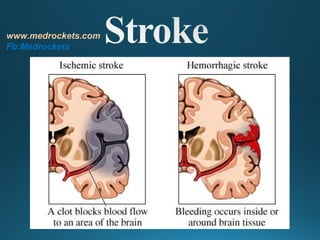
- 12. CLASSIFICATION OF STROKE Stroke Primary Hemorrhagic (20% of Strokes) Primary Ischemic (80% of Strokes) Thrombotic 50% Embolic 30% Intracerebral Hemorrhage 15% Subarachnoid Hemorrhage 5% www.medrockets.com Fb:Medrockets
- 16. Normal ml/100g/mi n 50 – 55 25 20 15 8 Ischemia Edema Loss of Na/K+ electrical pump ↑lactate activity failure; ↓ ATP Penumbra Infarction Cell Death
- 19. Stroke Warning Signs CLINICAL FEATURES OF STROKE www.medrockets.com Fb:Medrockets
- 29. Brain swelling Ventricular compression Focal cortical effacement www.medrockets.com Fb:Medrockets
- 30. Ischemic stroke Hemorrhage stroke Craniocerebral / cervical trauma Meningitis/encephalitis Intracranial mass •Tumor •Subdural hematoma Seizure with persistent neurological signs Migraine with persistent neurological signs Metabolic •Hyperglycemia (nonketotic hyperosmolar coma) •Hypoglycemia •Post-cardiac arrest ischemia www.medrockets.com Fb:Medrockets
- 36. Brain tissue ischemia Cerebral arterial stenosis/occlusion LAA/CE/SVD/others Decreased CBF Cerebral autoregulation (endothelial function etc)
- 37. First stroke blood pressure glucose smoking lipids mass popl. strategy hypertension TIA Atrial fibrillation other vascular disease high risk strategy stroke mortality acute treatment Secondary prevention recurrent stroke Stroke related disability Rehabilitation
Notes de l'éditeur
- Once the brain cells die from a lack of oxygen, the part of the body that part of the brain controls is affected through paralysis, language, motor skills, or vision. These symptoms usually persists for stroke survivors making routine daily functions extremely difficult.
- Noninvasive vascular tests include magnetic-resonance angiography, computed tomography angiography, and carotid ultrasound. Invasive vascular tests include conventional cerebral angiography and measurement of lesions. However, invasive measures can be risky; they should be undertaken only when the risk of stroke or death is less than 1%.
- Cranial computerised tomography (CT) is widely available and not only reliably distinguishes between haemorrhage and ischemic stroke or subarachnoid haemorrhage but can also rule out many other brain diseases. Signs of early ischemia can sometimes be detected as early as two hours after stroke onset, but this may be difficult even for the trained examiner, in particular in very early studies. Early infarct signs include sulcus effacement, swelling of the basal ganglia and the hyper-intense middle cerebral artery sign. Early signs of extensive infarction with intracranial midline shifts indicate a very serious event and a high risk, both for secondary haemorrhage and large malignant oedema formation and may justify repeated imaging after a short interval. Parenchymal haemorrhage can be identified almost immediately either in deep structures in patients with hypertension or in atypical areas in patients without hypertension or under adequate treatment, usually due to cerebral amyloid angiopathy. Infratentorial haemorrhage or cerebellar infarcts can be identified similar to supratentorial lesions, but smaller haemorrhages/ischemic infarcts in particular in the brain stem may easily be missed. In addition, CT may detect subarachnoid blood in the majority of cases with subarachnoid hemorrhage. Sometimes haemorrhages may even be interpreted as primary, but indeed are secondary to ischemic events. involvement of clearly defined vascular territories are indicative of such conditions, which are easier to identify in MRI studies. Brain haemorrhages tend to grow in the first 6-12 hours after stroke in about 40%-50% of all patients even without clinical deterioratio, which makes a second early CT study necessary. CT angiography (CTA) is a reliable tool to obtain information on extra- and intracranial arterial patency, and its use in clinical practice often adds value to the diagnostic work-up (Schellinger et al. 2003).