SlideShare une entreprise Scribd logo
1  sur  13
Télécharger pour lire hors ligne
Reviews                                                                         THE ANATOMICAL RECORD 290:770–782 (2007)


                   An In Vivo Look at Vertebrate Liver
                    Architecture: Three-Dimensional
                     Reconstructions from Medaka
                            (Oryzias latipes)
             RON C. HARDMAN,* DAVE C. VOLZ, SETH W. KULLMAN, AND DAVID E. HINTON
                   Duke University, Nicholas School of the Environment and Earth Sciences,
                                           Durham, North Carolina


                                                     ABSTRACT
                           Understanding three-dimensional (3D) hepatobiliary architecture is
                      fundamental to elucidating structure/function relationships relevant to
                      hepatobiliary metabolism, transport, and toxicity. To date, factual infor-
                      mation on vertebrate liver architecture in 3 dimensions has remained
                      limited. Applying noninvasive in vivo imaging to a living small fish ani-
                      mal model we elucidated, and present here, the 3D architecture of this
                      lower vertebrate liver. Our investigations show that hepatobiliary archi-
                      tecture in medaka is based on a polyhedral (hexagonal) structural motif,
                      that the intrahepatic biliary system is an interconnected network of
                      canaliculi and bile-preductules, and that parenchymal architecture in
                      this lower vertebrate is more related to that of the mammalian liver than
                      previously believed. The in vivo findings presented advance our compara-
                      tive 3D understanding of vertebrate liver structure/function, help clarify
                      previous discrepancies among vertebrate liver conceptual models, and
                      pose interesting questions regarding the ‘‘functional unit’’ of the verte-
                      brate liver. Anat Rec, 290:770–782, 2007. Ó 2007 Wiley-Liss, Inc.

                      Key words: liver; hepatobiliary; biliary; liver architecture;
                                 comparative hepatology; fish; 3-dimensional struc-
                                 ture; liver structure and function


   Several conceptual models emerged in the 19th and            of the liver has a physiological rather than morphologi-
20th centuries to describe structure/function relation-         cal basis (emphasizing afferent and efferent sinusoidal
ships of the vertebrate liver; lobular mammalian liver          flow within the parenchyma), and attempts to describe
models, and a tubular liver model to describe the lower         metabolic variance along a periportal to centrolobular
vertebrate livers of birds, fish, reptiles, and amphibians.      gradient (Jungermann, 1988). In the past 20 years ‘‘pri-
The mammalian ‘‘classic,’’ ‘‘modified,’’ and ‘‘portal’’ lobule   mary’’ and ‘‘secondary’’ lobule concepts have also
models describe morphological features encountered in           emerged (Matsumoto et al., 1979; Saxena et al., 1999;
two-dimensional (2D) single sectional views of the liver
(e.g. histological preparations, electron micrographs) and
attempt to characterize the relationship between vascu-           Grant sponsor: NIH (NCRR); Grant number: 1 RO1 RR018583-
lature, biliary passageways, and the hepatocellular com-        02; Grant sponsor: National Institute of Health (NIH/NCI); Grant
partment (Kiernan, 1833; Mall, 1906; Elias and Bengels-         number: R21CA106084-01A1.
dorf, 1952; Rappaport, 1958; Fig. 1). While these models          *Correspondence to: Ron Hardman, Duke University, Nicholas
share similarities, discrepancies exist in describing hep-      School of the Environment and Earth Sciences, Durham, NC,
atobiliary structure/function. For instance, Kiernan’s          27708. Fax: 919-684-8741, Phone: 919-741-0621.
classic lobule is a hexagonal structure with portal tracts        Received 11 July 2006; Accepted 20 February 2007
at the hexagon corners and a central hepatic venule,            DOI 10.1002/ar.20524
whereas Mall’s portal lobule places the portal tract as         Published online 21 May 2007 in Wiley InterScience (www.
the central axis of the model. Rappaport’s acinar model         interscience.wiley.com).

Ó 2007 WILEY-LISS, INC.
Fig. 1. A: The ‘‘classic’’ and ‘‘portal’’ lobule, and liver ‘‘acinus’’    chitectural motifs: fractal branching in portal tracts (larger bile ductules
characterize mammalian hepatobiliary structure/function relationships.       and ducts) and a polyhedral (hexagonal) motif at the canalicular level
A ‘‘tubular’’ concept has been used to describe the lower vertebrate         of organization. The polyhedral architectural motif (1) appears to order
livers of birds, fish, reptiles, and amphibians. a: The tubular concept in    the arrangement of the interconnected canalicular network and indi-
transverse view; two rows of hepatocytes form a tubule lumen (TL) at         vidual cells, and (2) may provide maximal structural/functional relation-
their apical membranes, while basal membranes border sinusoidal or           ships. The latter is suggested given hexagonal organization is the least
intercellular/intertubular space. b–d: Findings from three-dimensional       perimeter way for space filling, meaning hepatocellular (metabolic)
(3D) reconstructions reveal ‘‘tubular’’ architecture is more complex         space is maximized, while minimal material is used for construction
than previously considered. The tubule lumen is actually an intercon-        and maintenance of the hepatocellular membranes, and consequently
nected network of branching canalicular and bile preductule passage-         in the creation of canaliculi. Because blood to bile distance is mini-
ways. Findings from 3D reconstructions also suggest a common/                mized (Figs. 3, 4), transport is maximized. Lastly, the presence of a
shared structural/functional unit (indicated by brown oval in both con-      hexagonal motif in both the ordered arrangement of the canalicular
ceptual models) among lower and higher vertebrate livers. This func-         network, and in the organization of the hepatic lobule, suggest a hex-
tional unit is comprised of (1) a ‘‘portal tract/hilus,’’ a single conduit   agonal structural motif may provide structural integrity to the organ
containing two afferent blood supplies (hepatic portal and arterial ves-     system in total through the cellular and tissue level of organization.
sels), and efferent bile duct(s); (2) a ‘‘primary efferent vascular con-     Similarly, fractal branching of the blood and bile conduits in the portal
duit,’’ central vein/hepatic vein/terminal hepatic vein; and (3) an anas-    tracts confers maximal afferent and efferent transport efficiency
tomosing hepatic muralium (predominantly monolayered in mammals,             through these fluid compartments. Numbered segments in the hilus
bilayered in lower vertebrates), perfused by a canalicular network and       and bile duct regions indicate the branching order (fourth order)
sinusoidal bed, that bridges these two. B: Our investigations, in con-       observed in medaka. These underlying structural principles may serve
junction with extant mammalian liver information, suggest the verte-         to order the system toward maximal structural integrity and metabolic
brate hepatobiliary system to be constructed on two fundamental ar-          and transport efficiency.
772                                                 HARDMAN ET AL.

Teutsch et al., 1999). These concepts, based on 3D recon-         The 3D in vivo findings we present here not only elu-
structions of serial sections of human (Matsumoto et al.,      cidate hepatobiliary architecture of the lower vertebrate
1979) and rodent liver (Teutsch et al., 1999) describe         liver phenotype in medaka, but may help better our
cone-shaped units observed at the tissue level of organiza-    comparative understanding of various conceptual models
tion (branching of portal tracts). For instance; secondary     applied to vertebrate liver structure/function.
lobules (synonymous with the ‘‘classic’’ lobule), comprised
of six to eight primary lobules, have a central terminal he-
patic vein and six portal tracts at the periphery. Although               MATERIALS AND METHODS
each of these models remain valuable for communicating
                                                                  For decades, various color mutant strains of medaka
normalcy and disease, they are incomplete in conceptu-
                                                               (Oryzias latipes), acquired from natural and commer-
ally describing vertebrate hepatobiliary structure and
                                                               cially available populations, have been maintained in
function, particularly in 3 dimensions, and, understand-
                                                               the Laboratory of Freshwater Fish Stocks at Nagoya
ably, disparities remain in applying these models across
                                                               University, Japan. Cross-breeding from these stocks was
vertebrate species. As such, lobular and acinar models of
                                                               used to produce a stable ‘‘transparent’’ strain of medaka
the liver have never been completely accepted (Rappa-
                                                               (STII), homozygous recessive for all four pigments (iri-
port, 1958; Lamers et al., 1989). Moreover, none of these
                                                               diophores, leucophores, xanthophores, melanophores;
conceptual models have seen successful application to
                                                               Wakamatsu et al., 2001). STII medaka allow high reso-
lower vertebrate hepatobiliary structure/function. Such
                                                               lution (<1 mm) noninvasive in vivo imaging of internal
discrepancies likely belie inadequacies in the conceptual
                                                               organs and tissues at the subcellular level. Using laser
models themselves, or rather, our lack of comparative
                                                               scanning confocal microscopy (LSCM) and fluorescent
understanding of vertebrate liver architecture.
                                                               probes to elucidate the hepatobiliary system, we imaged
   Much less is known about the nonmammalian verte-
                                                               liver structure and function at various stages of develop-
brate liver. The prevailing model for these livers is the
                                                               ment in over a hundred individual living medaka. Of
hepatic tubule, which emerged from the predominance
                                                               these, 15 medaka were used for 3D reconstructions pre-
of observations of a two hepatocyte thick parenchyma
                                                               sented here (3 medaka at 8 days postfertilization [dpf], 4
and the fact that the vast majority of studies in lower
                                                               medaka at 12 dpf, 3 medaka at 30 dpf, 3 at 40 dpf, 2
vertebrate liver over the past century have shown no
                                                               medaka at 60 dpf). With widefield and confocal fluores-
clear lobular formation (Elias and Bengelsdorf, 1952;
                                                               cence microscopy, salient features of the organ system
McCuskey et al., 1986; Hampton et al., 1988; Rocha
                                                               such as canaliculi, space of Disse, endothelial cells, bili-
et al., 1994; Hinton and Couch, 1998). The current tubu-
                                                               ary epithelial cells, red blood cells, and hepatocytes and
lar concept describes of two rows of hepatocytes (in lon-
                                                               their nuclei, were clearly resolved in vivo (Fig. 2). Confo-
gitudinal section), the adjacent apical membranes of
                                                               cal stacks from in vivo imaging of the hepatobiliary sys-
which form a tubule lumen (bile passageway), into
                                                               tem were used for 3D reconstructions, and from these,
which bile is actively secreted (Figs. 1, 2). Basal mem-
                                                               architectural, morphometric, and volumetric analyses
branes of hepatocytes face sinusoidal or intertubular
                                                               were made.
space. Of interest; the ‘‘tubular’’ formation is widely held
                                                                  In vivo investigations included medaka embryos, lar-
as a common hepatobiliary architecture found among all
                                                               vae, and juveniles, from organogenesis (50 hours post-
vertebrate species; it is the shared predominant pheno-
type in all embryonic vertebrates, to include humans,          fertilization) through 60 days. Medaka were exposed by
and has been used to characterize the adult phenotype          means of aqueous bath (238C) to the fluorescent probes
for amphibians, birds, reptiles, and fishes (Elias, 1949;       Bodipy C5 Ceramide, Bodipy C5 HPC and fluorescein
Arias, 1988). While the hepatic ‘‘tubule’’ formation pre-      isothiocyanate to facilitate in vivo investigation of hepa-
dominates among lower vertebrates throughout their life        tobiliary structure/function. Each of the fluorophores
span, mammalian liver undergoes transition from a              saw hepatobiliary uptake and transport and excretion
tubular to laminar (muralium) architecture that is com-        into the gut lumen. Exposed cohorts were typically com-
plete in man by age 5 (Arias, 1988). ‘‘Tubule’’ formations     prised of 10 individual medaka. At various time points,
(denoting more than one row of hepatocytes) are also           individual medaka were removed from exposure cohorts,
observed in liver regeneration in mammalian species af-        sedated with 10 mM tricaine-methane sulfonate (MS-
ter severe injury, such as after hepatectomy and from          222), mounted in aqueous medium (Embryo Rearing Me-
marked acute exposure to toxins/toxicants (Van Eyken           dium for dechorionated embryos and hatchlings and de-
et al., 1989; Vandersteenhoven et al., 1990).                  ionized water for 20 dpf larvae and older) on depression
   From the brief review above it can be understood that       well glass slides with cover slip, and imaged live using
our comparative understanding of the vertebrate liver is       brightfield, and widefield and confocal fluorescence mi-
increasingly important. Not only is the tubular liver          croscopy. Time points of study varied from 10 min to 2
structure the most ubiquitous hepatic phenotype in the         hr depending on the fluorophore used and the aspect of
world, a better comparative understanding of the verte-        the hepatobiliary system being studied. For instance,
brate liver enhances our ability to interpret and commu-       Bodipy C5 HPC and fluorescein isothiocyanate accumu-
nicate normalcy and disease across the variety of animal       lation in the hepatic parenchyma is first observed at 10
models employed in research. By example, small fish ani-        min after exposure, with maximal saturation of the fluo-
mal models such as medaka and zebrafish are proving             rophore in the hepatic parenchyma occurring at 30
increasingly invaluable to the study of vertebrate devel-      min. Time points between 15 and 45 min post fluoro-
opment, carcinogenesis, and for investigation of molecular     phore exposure were commonly used for in vivo studies.
mechanisms of disease and toxicity (Wittbrodt et al.,             Imaging systems: Confocal fluorescence microscopy
2002; Shima and Mitani, 2004; Berghmans et al., 2005;          was performed with a Zeiss 510 Meta system and Zeiss
Alestrom et al., 2006).                                        LSM 5 Axiovision image acquisition software, an Argon
VERTEBRATE LIVER ARCHITECTURE                                                             773


   Fig. 2. Noninvasive in vivo imaging of hepatobiliary structure/func-     were observed circulating through sinusoids (S/r) abutting the basal
tion in living STII medaka. A: STII medaka embryo 6 days postfertiliza-     membranes of hepatocytes. E2: Single section from in vivo confocal
tion (dpf), left lateral view. The fluorescent cytochrome P4503A metab-      image stack showing conservative b-Bodipy C5 Phosphocholine
olite resorufin (red) in transport through the intrahepatic biliary net-     (green fluorescence) in transport from sinusoid (S) to canalicular space
works (IHBPs) of the embryonic liver (L), and concentration in the gall     (IHBP), imaged in vivo 30 min. Post administration of the fluorophore
bladder (GB). Otic vesicle (Ov), yolk sac (Y). B: Tubule-like formations,   to medaka (aqueous bath). E3: Composite of E1 and E2, localizing flu-
seen in transverse section, in the developing parenchyma, observed in       orophore concentrate to IHBPs. E4: Surface map of region of interest
vivo at 6 dpf. C: Concentrative transport of resorufin from hepatocyte       (ROI) of inset (white square) in frame E2; illustrating concentrative
into the tubule lumen (TL/IHBP). D1: STII medaka, 30 dpf, left lateral      transport of the fluorophore from sinusoidal space (S) to bile space
view, brightfield microscopy. Green algae can be seen transiting the         (IHBP). The ROI, showing a 17-mm span from blood to bile (across he-
gut lumen. D2: An in vivo confocal image of liver from the region of in-    patocyte), was quantitatively assessed, and revealed b-Bodipy C5
terest (grey square) in frame (D1), showing fluorophore transport            Phosphocholine concentration to be 66 times greater in the biliary
(FITC) through IHBPs. E1–E4: In vivo confocal imaging of blood to bile      passageway than in sinusoidal space. These studies demonstrated in
transport. E1: Two rows of hepatocytes and their nuclei (HN) are seen       vivo evaluation of concentrative blood to bile transport of fluorescent
in longitudinal section. Red blood cells (elongated ovate structures)       probes was possible.


and HeNe laser, Carl Zeiss C-apochromatic 40x/1.2, and                      tomical imaging was performed using a Nikon SZM
C-apochromatic 10x/0.45. For wide-field fluorescence mi-                      1500 dissecting microscope with a Nikon DXM 1200 digi-
croscopy, a Zeiss Axioskopp with DAPI/TRITC/FITC fil-                        tal capture system (brightfield). Regarding software,
ter cubes was used. Excitation/emission parameters for                      image analysis and compilation was performed with
filter cube sets were: DAPI/UV (Ex 360–380 nm/Em All                         EclipseNet (Nikon, USA), Adobe Photoshop (Adobe,
Vis 400 nm); FITC (Ex 450–490 nm/Em 515–565 nm);                           Inc.), Amira 3D (Mercury Computer Systems, Berlin),
TRITC (Ex 528–552 nm/Em 578–632 nm). Gross ana-                             ImageJ (V1.32j), IP Lab software (Scanalytics, Inc., ver-
774                                                HARDMAN ET AL.

sion 3.55), and Zeiss Image Browser (Carl Zeiss). Fluo-      likely imparts structural integrity, and metabolic and
rescent probes used were 7-benzyloxyresorufin (10–50          transport efficiency, features provided by higher-order
mM); b-Bodipy C5-HPC [BODIPY1 581/591 C5-HPC (2-             organizing principles such as hexagonal architecture,
(4,4-difluoro-5-   (4-phenyl-1,3- butadienyl)-4-bora-3a,4a-   not uncommon in biological systems (Mainzer, 1996).
diaza-s- indacene- 3-pentanoyl)-1-hexadecanoyl- sn-glyc-
ero-3-phosphocholine), (30 nM–10 mM)]; Bodipy FL C5-
                                                             Biliary System and Hepatocytes
ceramide [N-(4,4-difluoro-5,7-dimethyl-4-bora-3a,4a-diaza-
s-indacene-3- pentanoyl) sphingosine, (500 nM–5 mM)];           Linear segments of IHBPs (canaliculi) averaged 11 mm
and fluorescein isothiocyanate (1 nM–50 mM). Fluoro-          in length and 1.3 mm in diameter, and exhibited unary,
phores were acquired through Invitrogen/Molecular            binary, ternary, and quaternary branching. This finding
Probes (Carlsbad, CA). All fluorescent probes were admin-     should not be confused with fractal branching patterns
istered to STII medaka by means of aqueous bath in con-      (Turcotte et al., 1998); the biliary system in medaka
centration ranges given, at room temperature, under dark     does not appear (except for the localized area at the liver
conditions.                                                  hilus) to follow a fractal branching tree motif. Rather,
   All transmission electron microscopy (TEM) was per-       there is ubiquitous feedback/interconnectedness within
formed at the Laboratory for Advanced Electron and           the 3D biliary network (Fig. 5). Four to six hepatocytes
Light Optical Methods (LAELOM), College of Veterinary        were associated with each linear segment of IHBP
Medicine, North Carolina State University. Individual        (Fig. 6) and arranged such that a single hepatocyte
medaka were anesthetized and fixed in 4F:1G fixative           could contribute to two or three bile segments (canali-
(4% formaldehyde and 1% glutaraldehyde in a monoba-          culi/bile-preductules). The same hepatocellular/canalicu-
sic phosphate buffer with a final pH of 7.2–7.4 and a         lar relationship has also been described in rat (Motta
final osmolality of 176 mosmol). Thin sections (Spurr         and Fumagalli, 1975), where the same side of a hepato-
resin embedded) were then examined using a FEI/Phi-          cyte was observed to bound two or more bile canaliculi.
lips EM 208S Transmission Electron Microscope.               While hepatocytes had a mean diameter of 11.3 mm, they
                                                             showed varying morphology in vivo, apparently adaptive
                                                             to the space they were filling (e.g., at bifurcating or tri-
                   FINDINGS                                  furcating junctions). Hence, while the intrahepatic biliary
Polyhedral (Hexagonal) Architecture of the                   network showed a more ordered structure, hepatocellular
                                                             organization and morphology appeared much more adapt-
Intrahepatic Biliary System in Medaka
                                                             ive to space filling and parenchymal organization.
  The 3D reconstructions from in vivo imaging revealed          The IHBP network, composed of canaliculi and bile
intrahepatic biliary passageways (IHBP) in medaka to         preductules, occupied 95% of the liver corpus uni-
be predominantly an interconnected network of canali-        formly, with each area of livers examined (n ¼ 15) con-
culi and bile preductules, the organization of which         taining approximately equal volumes of IHBPs relative
appeared to be based on a polyhedral (hexagonal) struc-      to hepatocellular and vasculature volumes, regardless of
tural motif (Figs. 3–6). The polyhedral motif was most       how distal/proximal an observed volume of liver was
clearly observed in the arrangement of IHBPs, and was        from the liver hilus. Canaliculi were observed to merge
not evident in sinusoidal architecture. Empirical obser-     with bile preductules at unique morphological sites
vations found IHBP branching to be distinctly angular,       formed by junctional complexes between bile preductular
and statistical analysis of IHBP branching angles (in        epithelial cells (BPDECs, discussed following) and hepa-
3D) revealed clustering of angles into three main groups:    tocytes (Fig. 6). Together, canaliculi and bile preductules
120 degrees (74.9%), 90 degrees (14.8%), and 58 degrees      comprised the IHBP network. This interconnected net-
(10.3%). The majority of angles measured averaged            work of IHBPs was observed to feed three primary intra-
120 degrees, the interior angle of the hexagon (Fig. 4).     hepatic bile ducts (IHD), lined by cuboidal biliary epithe-
The 3D angle measurements were made by randomly              lia. IHDs merged at the liver hilus into a common he-
selecting intrahepatic biliary passageway bifurcation        patic duct that was associated with the cystic and
and trifurcation points in 3D reconstructions, achieved      common bile ducts (Fig. 5). Cuboidal and large squa-
by rotating the 3D model and randomly selecting              mous biliary epithelial cells (non-BPDECs) were
branching points. All angles of a bile segment branching     observed primarily localized to the hilar and peri-hilar
point were measured, inclusive of all 3D planes (Fig. 3).    regions of the liver.
Further 3D analyses revealed IHBP architecture to be
based, conceptually, on a tessellating (3D) hexagonal
                                                             Parenchymal Architecture
mesh (Carle et al., 2001). This hexagonal motif was also
apparent in the ratio between bile segment length and          Of interest, while ‘‘tubule’’ like formations were
blood to bile distance (Figs. 3, 4). When bile segment       observed (in vivo and in single sectional views) in em-
(canaliculi) length was considered to correspond to one      bryonic livers (3 dpf to 7 dpf), tubule formations were
side of a hexagon, the distance (radii) from hexagon side    not readily apparent in 3D reconstructions (n ¼ 15) of
(canaliculus) to hexagon center (sinusoid) appeared to       larval and older livers. Rather, parenchymal architec-
correlate to the hexagonal ratio given in Figure 3. A        ture in larval and later developmental stages of medaka
hexagonal architecture has also been described, from         appeared to be more consistent with an anastomosing,
empirical observations, in the fine structure of the bili-    predominantly dual layered, muralium. First, 3D recon-
ary system in rat (Mochizuki et al., 1988; Murakami          structions revealed the ‘‘tubular’’ lumen to be composed,
et al., 2001) and human (Yamamoto et al., 1990).             not of an extended linear biliary passageway that would
Although a discussion of this topic is beyond the scope of   create a classic ‘‘lumen,’’ but of a hexagonal network of
this study, the presence of a polyhedral structural motif    branching, interconnected canaliculi and preductules,
VERTEBRATE LIVER ARCHITECTURE                                                               775


   Fig. 3. Examples from three-dimensional (3D) reconstructions of           bular profile). In medaka, we found this structural arrangement of
medaka hepatobiliary system, illustrating the types of analyses that         branching canaliculi that comprises the ‘‘tubule lumen,’’ revealing ‘‘tu-
led to findings on polyhedral (hexagonal) architecture. Grayscale back-       bular’’ architecture more complex than previously considered (see Fig.
ground images are sections from in vivo confocal image stacks, from          1). F: 3D reconstruction of the relationship between IHBPs and sinu-
which 3D reconstructions were generated. In (A–D,F–H) the empty              soids. Between 2 sinusoids (red) can be seen a branching canalicular
space surrounding biliary passageways (green/gold), and between si-          network (green/gold). G–I: Examples of 3D morphometric analyses,
nusoids (red), is hepatocellular space (hepatocytes not rendered for         which suggest medaka parenchymal architecture to be more consist-
visual clarity). Colored dots in A–C are given for anatomical reference.     ent with an anastomosing muralium. 3D measurements of hepatocellu-
A–D: An isolated section of the intrahepatic biliary network, illustrating   lar space (between sinusoids) revealed a muralium-like architecture.
relationship of canaliculi (green) to sinusoids (red). A: Branching canal-   G: Example dimensions given are 196.3 mm in length, 98.2 mm in
iculi (intrahepatic biliary networks [IHBP]) are shown between two           height, and 24.7 mm in width. H: Same as G viewed in a horizontal
rows of hepatocytes (confocal grayscale image). B: Same as A with si-        longitudinal section (from the top). Sample dimensions are shown. I:
nusoids present, and C is AB viewed directly on the ‘‘z’’ axis (from        Single slice from in vivo confocal stack of hepatic parenchyma. Gray
top). C: Example of analyses of polyhedral structural motif, which was       arrowheads indicate sinusoids, black arrowheads hepatocyte nuclei.
evident in the relationship between blood to bile distance and canalic-      Hepatocellular compartment width (analyzed in vivo and in 3D) was
ular length (see Fig. 4). Bile segment length (canaliculus, S) correlated    found to be predominantly two cells thick, although occasionally up to
to calculated and measured values of blood to bile distance (r), when        eight cells in some areas. Average parenchymal width was 24.58 mm.
assessed with the hexagonal ratio [r ¼ 0.5 (S) 1.44]. D: Empirical           Parenchymal depth varied from 21.2 mm to  200 mm. Note that typi-
observations (3D) of angular canalicular and bile preductule branching       cal dimensions of 3D reconstructions were 100 mm in depth; hence,
led to statistical analysis of branching angles, which suggested the         depth dimension is limited. Parenchymal metrics were derived from
presence of a polyhedral structural motif (see Fig. 4). E: Schematic         127 measurements (3D) in medaka at 8, 12, 30, and 40 dpf. Space of
showing modified ‘‘tubular’’ concept of lower vertebrate liver (note          Disse (SD), Sinusoidal Endothelial Cell (SE), Red Blood Cell (R), Sinu-
IHBPs, and the hexagonal arrangement in relation to hepatocytes/tu-          soid (Sn, S1–S4).
776                                                             HARDMAN ET AL.


   Fig. 4. Morphometric and volumetric analyses (upper left graph) of       to calculated values, falling between the longest (R) and shortest (r)
random measures (341 observations) of three-dimensional (3D)                radii in the hexagon [r ¼ 0.5 (S) 1.44]. Where bile segment length (e.g.,
branching angles of bile segments in four individual livers (8–40 days      canaliculus) ¼ S, blood to bile distance ¼ r. Pearson’s correlation
postfertilization [dpf]) found branching angles to cluster into three       coefficient (0.049, P ¼ 0.6). The difference in the mean between calcu-
primary groups: 120 degrees (75%), 93 degrees (15%), and                  lated and measured r values was 0.67 mm, half the width of the canali-
64 degrees (10%). Upper right graph: Using the hexagonal ratio in          culus. In vivo volumetric investigations in three individual medaka at
the lower portion of the plot, a polyhedral structural motif was also       8 dpf, 12 dpf, and 30 dpf revealed consistent ratios between intrahe-
suggested in the relationship between blood to bile distance and can-       patic biliary passageway volume, blood (sinusoid) volume, hepatocel-
alicular length. Measured values of blood to bile distance (r) correlated   lular volume, and parenchymal volume.


which formed the inner parenchymal framework                                (because confocal stacks were 100 mm in depth, the
(Figs. 3–6). In other words, tubule lumens were found to                    maximum measurement for plate ‘‘height’’ was limited
be canalicular segments with an average length between                      to 100 mm). Third, the longest non-branching biliary seg-
10 and 13 mm. Second, hepatocellular muralium-like                          ments were observed to be 30 mm in length, and these
structures were apparent, which varied in height from                       were few in number (2–3% of bile segments measured).
20 mm to 100 mm, and width from 23 mm to 83 mm,                          For a tubular architecture to be present, tubules would,
observations consistent with an anastomosing muralium                       from these 3D investigations, be less than 30 mm in linear
VERTEBRATE LIVER ARCHITECTURE                                                               777


   Fig. 5. Three-dimensional (3D) architecture of liver hilus, bile duc-     was followed and the branching of the IHBPs mapped to a 2D sche-
tules/ducts, and canalicular network. A: In vivo image capture of the        matic. Each colored line corresponds to one bile segment (11 mm in
liver hilus at 30 days postfertilization (dpf). Three primary intrahepatic   length). Colors are for illustrative purposes only. The 90-degree angles
ducts (IHDs) conjoined a common hepatic duct (HD), which fed the             correlate to bile segment branching (not to scale or degree, for illus-
cystic duct (CD), gall bladder (GB), and common bile duct (CBD). The         trative purposes only). The schematic is used to illustrate the intercon-
liver hilus of medaka contained two afferent blood supplies (the he-         nectedness of the canaliculo-bile preductular network. For clarity, part
patic portal vein [HPV] and hepatic artery [not shown]), and hepatic         of the network was abbreviated by using dashed lines to illustrate
duct. Mucosal folds (MF) of the gut (lower left) are seen caudal to the      feedback and interconnectedness within the IHB network. Dashed
liver. B,C,F: The 3D reconstructions aided in elucidating the relation-      lines represent a branching network of canaliculi (as illustrated in col-
ship between the canaliculo-preductule network, bile ductules and            ored lines). The canaliculo-bile preductule network occupied 95% of
ducts, and liver hilus. Images C (horizontal longitudinal section) and F     the liver body (by area). D: Schematic showing overall biliary architec-
(sagittal view) show an example of 3D reconstruction of the canali-          ture of medaka liver. Bile ductules and ducts exhibited fourth order
culo-preductular network draining to a common IHD (one of three              fractal branching, and were largely found in hilar/peri-hilar region of
IHDs shown in B). E: A hand-traced schematic of an isolated section          the liver. Ductules terminated in a canaliculo-bile preductular network
of the canaliculo-preductular network (diamond-shaped region of inter-       (illustrated in E), that followed a polyhedral (hexagonal) architectural
est [ROI] indicated in C and F). One individual IHD (green arrowhead)        motif.


length and would be highly branched and interconnected                       tive correlary of mammalian progenitor/stem cells, given
(following biliary network architecture). Hence, 3D in vivo                  they share morphological characteristics ascribed to
observations found medaka parenchymal structure to                           mammalian oval cells (bipotential progenitor cells).
more resemble a predominantly two- to three-cell-thick he-                   BPDECs, like oval cells, are phenotypically distinct from
patic muralium, with an average width of 26 mm and                           hepatocytes, characterized by a high nuclear to cyto-
height that varied from 20 mm to  100 mm. It follows that                   plasm ratio with no basement membrane, and are inti-
medaka parenchymal architecture appeared highly analo-                       mately associated with the biliary system (Golding
gous to mammalian architecture; although predominantly                       et al., 1996; Fausto and Campbell, 2003). In vivo,
two to three hepatocytes thick (and in rare instances up to                  BPDECs were observed to form unique junctional com-
seven or eight cells thick), as opposed to the predominantly                 plexes with hepatocytes, at which were created IHBPs
one- to two-cell-thick mammalian muralium (Elias and                         termed bile preductules. These junctional complexes
Bengelsdorf, 1952).                                                          were morphologically distinct (Fig. 6). In single section
                                                                             view these unique morphological formations appeared as
                                                                             BPDECs surrounded by bile passageways on all sides
Biliary Epithelia
                                                                             (Fig. 6). 3D reconstructions from in vivo imaging
  3D investigations found BPDECs (Hinton and Pool,                           revealed BPDECs to occupy the ‘‘center’’ of these com-
1976; Hampton et al., 1988; Okihiro and Hinton, 1999)                        plexes, where one or more BPDECs formed multiple
to be located throughout the parenchyma of medaka liv-                       junctional complexes with surrounding hepatocytes (Fig.
ers studied (from 8 dpf to 60 dpf). BPDECs are the puta-                     6). Biliary passageways (bile preductules) formed at
778                                                             HARDMAN ET AL.


   Fig. 6. A–A2: Canaliculus/bile preductule and hepatocellular rela-       (BPD). The 3D reconstructions (B1,B2) elucidated the actual architec-
tionships. A1,A2: Three-dimensional (3D) reconstructions from confo-        ture of these canaliculo-bile preductule complexes, revealing bile pre-
cal image in A. Four to six hepatocytes (hepatocyte nuclei shown in         ductular epithelia form multiple junctional complexes with surrounding
purple, numbered) were observed in relation to a given canaliculus          hepatocytes, at which bile preductules are formed. In B1 and B2, the
(green). Circled red A indicates a bifurcating canalicular segment. A       empty space surrounding BPDEC is hepatocellular space (left
single hepatocyte was observed to contribute to one to three canali-        ‘‘empty,’’ or not rendered, for illustrative purposes). In vivo findings
culi. B1–B2: The 3D reconstructions of confocal image in (B), illustrat-    were corroborated with ultrastructural studies (C1,C2), which aided in
ing the structure of a bile preductule (BPD) junction. Red arrowheads       elucidating/validating in vivo observations. Frame C is a still image
in (B) indicate bile preductular epithelial cells (BPDECs) within the he-   from a 3D reconstruction illustrating the distribution of BPDECs
patic parenchyma. In 2D confocal sections BPDECs appeared to                (purple, indicated by red arrowhead) throughout the hepatic paren-
occupy the ‘‘center’’ of these canaliculo-bile preductule structures.       chyma. Junctional Complex/Bile Preductule (Jc/BPD), Intrahepatic Bili-
BPDECs formed unique junctional complexes with surrounding hepa-            ary Passageway (IHBP), Hepatic Nuclei (HN), Sinusoid/red blood cell
tocytes, which created biliary passageways, termed bile preductules         (S/r), Bile Preductular Epithelial Cell (BPDE/C).


these BPDEC/hepatocyte junctional complexes com-                            Theise et al., 1999; Fausto and Campbell, 2003; Knight
monly showed ternary or quaternary branching. While                         et al., 2005). While we have localized BPDECs to specific
BPDECs varied in morphology and size, showing ovate,                        locations within hepatic parenchyma, and these cells
triangular and ellipsoidal nuclei, the majority of                          share morphological characteristics consistent with
BPDECs were commonly found to be 6 mm in diameter.                         mammalian hepatic progenitor cells, BPDECs in medaka
Our in vivo findings on BPDECs in medaka are, inter-                         have only been partially characterized (Okihiro and Hin-
estingly, consistent with previous oval cell studies in                     ton, 2000). Cuboidal and squamous biliary epithelia
rodents and humans (Farber, 1956; Golding et al., 1996;                     (non-BDPEC), lining larger diameter intrahepatic biliary
VERTEBRATE LIVER ARCHITECTURE                                               779
passageways, were observed almost exclusively near               network of canaliculi and bile preductules (bile seg-
hilar and perihilar regions of the liver (Fig. 5).               ments) that averaged 11–13 mm in length (Fig. 5).
                                                                    Because 3D investigations reveal medaka hepatobili-
Morphometric and Volumetric Information                          ary architecture, representative of the lower vertebrate
                                                                 tubular phenotype, to more closely resemble a dual-lay-
  3D reconstructions enabled highly accurate volumetric          ered anastomosing muralium, akin to the single-cell-
and ratiometric analyses of biliary, parenchyma, vascu-          thick muralium described in mammals, important ques-
lature, and liver volumes (Fig. 4). We found prior ex            tions are raised. If hepatic tubules comprise a two-hepa-
vivo volumetric studies (Hess et al., 1973; Blouin et al.,       tocyte-thick muralium, how are they integrated into a
1977; Rocha et al., 1997; Hinton et al., 2001) in verte-         muralium-like structure? Although tubule formations
brate livers to correspond well with our in vivo findings.        may theoretically comprise a dual-layered muralium,
                                                                 this would necessitate an intertubular space, which
                                                                 raises another question; if tubules are present, are
                      DISCUSSION                                 tubules joined at the intertubular space? If so, does this
   Surprisingly, we did not observe a well-developed             not describe a muralium structure? These important
arborizing biliary ‘‘tree,’’ well characterized in mamma-        questions remain to be answered, given our findings
lian studies (Ludwig et al., 1998; Masyuk et al., 2001).         from in vivo 3D reconstructions suggest a dual-layered
This ‘‘tree’’ describes bile ducts of the liver hilus arboriz-   plate-like muralium predominates in larval and later
ing into more highly branched, and numerous, bile ducts          life stages of medaka, whereas tubule-like formations
and ductules of diminishing diameter, as the biliary sys-        were observed in the embryonic liver.
tem infiltrates more distal regions of liver (relative to            The tubular concept of the lower vertebrate liver was
the liver hilus). We attribute the overall lack of an arbo-      derived largely from 2D observations of histological and
rizing biliary tree in medaka liver to two factors: (1)          electron micrograph (EM) preparations. In histological
mammalian studies are describing portal tract arboriza-          sections, the fine structure of the biliary system is diffi-
tion, or interlobular stromal areas of the liver (meaning        cult to discern (canaliculi average 1–2 mm in diameter),
biliary tree arborization describes the tissue level of or-      and when resolved, we now know, due to a hexagonal ar-
ganization, lobule formation); (2) medaka liver is the ar-       chitectural formation, would show a random order of
chitectural analogue of the intralobular mammalian pa-           appearance in a 2D section. For instance, using histolog-
renchyma. The two points are discussed in detail below.          ical sections Rocha et al. (1994) described biliary passa-
   These findings raise important questions regarding             geways in trout as appearing randomly dispersed
current conceptual models of vertebrate liver architec-          throughout the liver; this would be an accurate 2D ob-
ture. In mammalian liver, hepatocytes have been de-              servation, or how the 3D hexagonal architecture of the
scribed as irregularly shaped polygonal cells that form,         canalicular network we have described would appear in
predominantly, a one- to two-cell-thick wall/plate-like          a 2D histological section. Consequently, from a 2D per-
structure (muralium), which anastomoses throughout               spective, a dual-layered muralium may appear, or be
the liver (Elias and Bengelsdorf, 1952). Observations of         interpreted as, a tubule-like formation. Hence, given the
dual layered hepatocytes in fish and other lower verte-           interconnected 3D biliary architecture we have eluci-
brate livers, and the lack of observed lobular formations        dated in this study, it may be that a ‘‘tubular’’ formation
in the majority of lower vertebrate livers studied               and dual layered plate-like muralium are perhaps, one
(McCuskey et al., 1986; Hampton et al., 1989; Rocha              and the same, and that discrepancies in understanding
et al., 1997; Hinton et al., 2001; Akiyoshi and Inoue,           muralium architecture in lower vertebrates have arisen
2004) led to the reasoning that lower vertebrate livers          from varied 2D viewpoints (and thereby varied interpre-
may be composed primarily of anastomosing ‘‘tubules.’’           tation), and a lack of 3D studies in vertebrate liver
In single section view (e.g. histological preparations with      structure at the canalicular level of organization.
longitudinal orientation), hepatic tubules appear as two            If fish and mammals share similar muralium architec-
rows of hepatocytes, the apical membranes of which               ture, what can explain the absence of ‘‘portal tracts’’ and
form a tubule lumen (bile passageway), and basal mem-            lobule formation in lower vertebrate livers? Addressing
branes of which border sinusoidal, intracellular, or inter-      these questions involves consideration of organ system
tubular space. As our 3D in vivo findings have shown,             ontogeny and the nature of the ‘‘functional unit’’ of the
the true structure of the ‘‘tubular’’ liver is more complex      vertebrate liver. Although this is beyond the scope of
than previously understood. Because one hepatocyte               this article, a brief discussion is warranted. First, while
may contribute to one to three canaliculi, ‘‘tubule              portal tracts/triads are well described in mammalian
lumens’’ are actually composed of an interconnected hex-         liver and integral to current mammalian liver concep-
agonally branching network of canaliculi and bile pre-           tual models (lobular, acinar), they are not often observed
ductules. Where ‘‘tubule-like’’ formations were observed         in lower vertebrates. In lower vertebrates, anatomical
in 2D transverse sectional views in the embryonic liver          structures that may be perhaps the evolutionary, or
(in vivo), ‘‘tubule’’ formations were very rarely encoun-        even functional, precursors to portal tracts have been
tered in larval and older fish, and were not readily appa-        described as venous biliary arteriolar tracts (VBAT), ve-
rent in 3D reconstructions. For a tubular architecture to        nous arteriolar tracts (VAT), venous biliary tracts (VBT),
be considered, tubules in medaka would, from our 3D in           biliary tracts (BT), and arteriolar tracts (AT) (Rocha
vivo investigations, average  29 mm in linear length (         et al., 1995; Hampton, 1988). These morphological fea-
three hepatocytes), and would be quite rare (2–3% of             tures bear some anatomical resemblance to the portal
bile segments measured were found to span up to 29               tract, although no semblance of lobule formation in
mm). The prevailing 3D structure of the biliary system           lower vertebrates has been found. Second, based on our
in medaka was a highly branched and interconnected               3D reconstructions, it can be hypothesized that the hep-
780                                                   HARDMAN ET AL.

atobiliary systems of both medaka and mammals share              ratively higher metabolic demand, typically higher body
a common fundamental functional unit: (1) a ‘‘portal             temperature, higher load bearing on the liver resulting
tract/hilus,’’ a single conduit containing two afferent          from gravity, and relatively larger mammalian liver
blood supplies (hepatic portal and arterial vessels), and        mass. It follows that higher vertebrates likely show
efferent bile duct(s); (2) a ‘‘primary efferent vascular         ‘‘lobulation’’ of the liver in support of greater organ mass
conduit,’’ central vein/hepatic vein/terminal hepatic vein;      and metabolic demand; the iteration of a single hepatic
and (3) anastomosing hepatic plates/cords, perfused by a         functional unit (e.g. HMS). In more massive mamma-
canalicular network and sinusoidal bed, that bridges             lian livers the formation of hexagonal lobules, organized
these two. A similar conceptual functional unit, the he-         by stromal tissue (portal tracts/triads), would impart
patic microcirculatory subunit (HMS), was proposed by            structural integrity to the organ (due to the physical
Ekataksin et al. (1997). This wedge-shaped unit is com-          properties of hexagonal packing; Weaire and Phelan,
posed of ‘‘base’’ (the portal tract/hilus), ‘‘apex’’ (the cen-   1994; Hales, 2001), much needed in an organ of such
tral vein/terminal hepatic vein/hepatic vein), and a con-        mass (3–5% of body weight in mammals), and one
tinuous system (muralium) of anastomosing hepatic                highly perfused with a liquid medium (the liver of mam-
plates (laminae hepatic) connecting the base and apex,           mals can store up 30% of total blood volume). It follows,
perfused by sinusoidal bed (labyrinthus hepatic). Hence,         from an ontological viewpoint, that appearance of
it can be considered that, in the lower vertebrate liver of      VBATs, VATs, VBTs, BTs, and ATs in larger piscine spe-
medaka; the liver hilus is the correlary of the portal           cies may be evidence of the emergence of ‘‘lobulation’’
tract, and hepatic vein the corollary of the central vein/       among lower vertebrates such as teleosts. Such a consid-
terminal hepatic vein. In between these is the laminae           eration begs more detailed investigation of larger fish
hepatic, the fine structure of which, up to this study, has       species, and reptilian and amphibian livers. Lastly, the
remained a mystery in medaka and other lower verte-              hexagonal structural motif appears to be conserved
brates.                                                          among all vertebrates; found not only in the organiza-
   One of the outstanding questions in human biliary             tion of the canalicular network (intralobular paren-
architecture has been; does each duct of Hering corre-           chyma), but also in the formation of the classic lobule
spond to one canaliculus? Or do canaliculi form a conflu-         (arborization of portal tracts).
ence before entering the canals of Hering (Saxena et al.,            Reaching a conceptual model of the ‘‘functional unit’’
1999)? Our 3D studies in medaka show the canalicular             of the liver has long been sought, and the spatial archi-
network to form a confluence before entering the hilar            tecture of the lower vertebrate liver, particularly the bil-
intrahepatic ducts, the anatomical corollary of the canals       iary system, has long been in question. Differences in
of Hering. Given the findings presented here and that:            extant conceptual models have arisen from how lower
bile canalicular diameter is conserved in vertebrate liv-        and higher vertebrate livers have been viewed at vari-
ers (1–2 mm); early observations by Elias and Bengels-           ous levels of biological organization (cellular, tissue,
dorf (1952) suggest a polygonal hepatocellular formation;        organ), in which anatomical plane, and whether in two
that a ‘‘chicken-wire’’ pattern of bile canaliculi has been      or three dimensions. Given various ways of viewing the
described in human liver (Ekataksin et al., 1995); hexag-        liver and the prior technological constraints (lack of 3D
onal architecture has also been described in the fine             and in vivo tools), understandably, discrepancies have
structure of the biliary system in rat (Murakami et al.,         appeared to exist between lower vertebrate and mamma-
2001) and human (Yamamoto et al., 1990); the tubular             lian hepatobiliary conceptual models. Such discrepancies
structure (dual layered parenchyma) appears to be con-           can be illustrated in a study by Akiyoshi and Inoue
served among all embryonic vertebrates; erosion cast             (2004), who investigated two hundred different teleost
studies by Murakami et al. (2001) show a hexagonal               species (histological preparations) and described varying
canalicular network feeding intrahepatic bile ducts (rat);       liver architecture among them (muralium as cord-like
bipotential progenitor-like cells in both medaka and             [one cell], tubular [two cells], and solid [two cells]).
mammals are closely associated with the biliary system,          Indeed, we observed all three structural forms in our in
it is not unlikely that all vertebrate livers share the          vivo and 3D investigations, each comprising the hepatic
same fundamental functional unit. Hence, it is interest-         parenchyma, although the muralium of medaka was
ing to consider the medaka hepatobiliary system, repre-          found to be predominantly two cells thick. Similar obser-
sentative of lower vertebrate architecture, as a single          vations have been made in developing mammalian liver
functional unit (akin to the HMS), while mammalian               by McCuskey et al. (2003). Hence, it appears the limits
hepatobiliary systems can be considered to be composed           of spatial observation/understanding permitted by single
of multiple functional units organized into hepatic              sectional views of the liver have led to these, while accu-
lobules (typically 1–2 mm; Fig. 1). In mammals, we               rate descriptions from a 2D viewpoint, discrepancies in
hypothesize that portal tracts arborize within the liver         interpretation, and thereby discrepancies in a compara-
to form the classic intrahepatic biliary/vascular trees,         tive understanding of the 3D architecture of vertebrate
giving rise to lobulation (multiple functional units).           liver. Such discrepancies can be understood considering
Hence, as conceptual models go, the primary architec-            the difficult task of extrapolating 2D information to 3D
tural differences between medaka and mammalian hepa-             architectural models. Particularly when 2D observations
tobiliary systems appears to arise at the tissue/organ           in single sectional views of the liver have accurately
level of organization (multiple lobules in mammals vs.           characterized the 3D structure we have elucidated here.
single lobule in medaka). These differences can be at-           It can be said that in one sense, prior observations of
tributable to the metabolic and structural demands/              lower vertebrate liver architecture that described a tu-
needs between lower and higher vertebrate hepatobili-            bular conceptual model were correct, in so far as they
ary systems (in the context of organ system ontogeny             were communicating the 2D morphology encountered in
and functional capacity), where mammals see a compa-             single sectional views of the liver.
VERTEBRATE LIVER ARCHITECTURE                                                          781
                          SUMMARY                                      Ekataksin W, Zou Z, Wake K, Chunhabundit P, Somana R, Nishida
                                                                         J, McDonnell D. 1995. HMS, Hepatic microcirculatory subunits in
   Collectively, these findings elucidate the 3D architec-                mammalian species. Intralobular grouping of liver tissue with
ture of the medaka hepatobiliary system and improve                      definition enhanced by dropout sinusoids. In: Cells of the Hepatic
our comparative understanding of vertebrate liver struc-                 Sinusoid. Leiden: Wisse E, Knook DL, Wake K, eds. The Kupffer
ture and function. These findings also present interest-                  Cell Foundation, The Netherlands.
ing questions regarding the ‘‘functional unit’’ of the ver-            Elias H. 1949. A re-examination of the structure of the mammalian
tebrate liver; where the hepatobiliary system in medaka                  liver. II. The hepatic lobule and its relation to the vascular and
                                                                         biliary systems. Am J Anat 85:379–456.
can be, as a conceptual model, considered a single func-
                                                                       Elias H, Bengelsdorf H. 1952. The structure of the liver of verte-
tional unit, akin to the individual lobule. We have also                 brates. Acta Anat 14:297–337.
addressed prior discrepancies among conceptual models                  Farber E. 1956. Similarities in the sequence of early histological
of vertebrate hepatobiliary system architecture, and the                 changes induced in the liver of the rat by ethionine, 2-acetyla-
findings presented should help with integration of prior                  mino-fluorene, and 30 -methyl-4-dimethylaminoazobenzene. Cancer
observations of vertebrate liver structure/function into a               Res 16:142–148.
more cohesive conceptual framework. In summary,                        Fausto N, Campbell JS. 2003. The role of hepatocytes and oval
in vivo and 3D findings in medaka show: (1) parenchy-                     cells in liver regeneration and repopulation. Mech Dev 120:117–
mal architecture is predominantly a two-cell-thick mura-                 130.
                                                                       Golding M, Sarraf C, Lalani EN, Alison MR. 1996. Reactive biliary
lium, although tubule-like formations were observed in
                                                                         epithelium: the product of a pluripotential stem cell compart-
embryonic livers; (2) the hepatic muralium is organized                  ment? Hum Pathol 27:872–884.
through a polyhedral (hexagonal) structural motif,                     Hales T. 2001. The honeycomb conjecture. Discrete Comput Geom
revealed in biliary architecture; (3) the intrahepatic bili-             25:1–22.
ary system is an interconnected network of canaliculi                  Hampton J, Lantz R, Goldblatt P, Lauren D, Hinton D. 1988. Func-
and bile preductules; (4) the canaliculo-preductular net-                tional units in rainbow trout (Salmo gairdneri, Richardson) liver:
work occupies the majority of the liver corpus (95%)                    II. The biliary system. Anat Rec 221:619–634.
uniformly, with equidiameter IHBPs (1–2 mm) observed                   Hampton JA, Lantz RC, Hinton DE. 1989. Functional units in rain-
throughout the liver; (5) larger bile ductules and ducts                 bow trout (Salmo gairdneri, Richardson) liver: III. Morphometric
                                                                         analysis of parenchyma, stroma, and component cell types. Am J
were observed predominantly at the liver hilus, conse-
                                                                         Anat 185:58–73.
quently an arborizing biliary tree was largely absent,                 Hess FA, Gnagi HR, Weibel ER, Preisig R. 1973. Morphometry of
seen only in the rudimentary branching of intrahepatic                   dog liver: comparison of wedge and needle biopsies. Eur J Clin
ducts from the hepatic duct; and (6) the livers of these                 Invest 3:451–458.
small fish are replete with BPDECs, the putative mam-                   Hinton DE, Couch JA. 1998. Architectural pattern, tissue and cellu-
malian correlative of bipotential progenitor/stem cells.                 lar morphology in livers of fishes: Relationship to experimentally-
                                                                         induced neoplastic responses. In: Fish Ecotoxicology. Braunbeck,
                                                                                                                         ¨
                                                                         T., D.E. Hinton, and B. Streit, eds., Birkhauser Verlag. Basel,
                 ACKNOWLEDGMENTS                                         Switzerland.
                                                                       Hinton D, Pool C. 1976. Ultrastructure of the liver in channel cat-
   Thanks to Dr. David Miller, Laboratory of Pharmacol-                  fish Ictalurus punctatus (Rafinesque). J Fish Biol 8:209–219.
ogy and Chemistry, National Institute of Environmental                 Hinton D, Segner H, Braunbeck T. 2001. Toxic Responses of the
Health Sciences, Research Triangle Park, for providing                   Liver. In: New Perspectives: Toxicology and the Environment,
access to their laser scanning confocal microscopy facil-                Target Organ Toxicity in Marine and Freshwater Teleosts.
                                                                         D. Schlenk and Benson, W. eds. Taylor  Francis, New York.
ity, and special thanks to the Duke University Inte-
                                                                       Jungermann K. 1988. Metabolic zonation of liver parenchyma.
grated Toxicology Program. This publication was made                     Semin Liver Dis 8:329–341.
possible by a grant from the National Center for                       Kiernan F. 1833. The anatomy and physiology of the liver. Philos
Research Resources, a component of the National Insti-                   Trans R Soc Lond 123:711–770.
tutes of Health. Its contents are solely the responsibility            Knight B, Matthews VB, Olynyk JK, Yeoh GC. 2005. Jekyll and
of the authors and do not necessarily represent the offi-                 Hyde: evolving perspectives on the function and potential of the
cial views of NCRR or NIH.                                               adult liver progenitor (oval) cell. Bioessays 27:1192–1202.
                                                                       Lamers WH, Hilberts A, Furt E, Smith J, Jonges GN, van Noorden
                                                                         CJ, Janzen JW, Charles R, Moorman AF. 1989. Hepatic enzymic
                                                                         zonation: a reevaluation of the concept of the liver acinus. Hepa-
                  LITERATURE CITED                                       tology 10:72–76.
Akiyoshi H, Inoue A. 2004. Comparative histological study of teleost   Ludwig J, Ritman EL, LaRusso NF, Sheedy PF, Zumpe G. 1998.
  livers in relation to phylogeny. Zoolog Sci 21:841–850.                Anatomy of the human biliary system studied by quantitative
Alestrom P, Holter JL, Nourizadeh-Lillabadi R. 2006. Zebrafish in         computer-aided three-dimensional imaging techniques. Hepato-
  functional genomics and aquatic biomedicine. Trends Biotechnol         logy 27:893–899.
  24:15–21.                                                            Mainzer K. 1996. Symmetries of nature: a handbook for philosophy
Arias IM. 1988. The Liver: Biology and Pathobiology. Irwin M.            of nature and science. Walter De Gruyter Inc., Berlin, Germany
  Arias, William B. Jakoby, Hans Popper, David Schachter, David        Mall F. 1906. A study of the structural unit of the liver. J Anat
  A. Shafritz, eds. Raven Press, Baltimore, Maryland.                    5:227–308.
Berghmans S, Jette C, Langenau D, Hsu K, Stewart R, Look T,            Masyuk TV, Ritman EL, LaRusso NF. 2001. Quantitative assess-
  Kanki JP. 2005. Making waves in cancer research: new models in         ment of the rat intrahepatic biliary system by three-dimensional
  the zebrafish. Biotechniques 39:227–237.                                reconstruction. Am J Pathol 158:2079–2088.
Blouin A, Bolender RP, Weibel ER. 1977. Distribution of organelles     Matsumoto T, Komori R, Magara T, Ui T, Kawakami M, Tokuda T,
  and membranes between hepatocytes and nonhepatocytes in the            Takasaki S. 1979. A study on the normal structure of the human
  rat liver parenchyma. A stereological study. J Cell Biol 72:441–       liver, with special reference to its angioarchitecture. Jikeikai Med
  455.                                                                   J 26:1–40.
Carle J, Myoupo J, Stojmenovic I. 2001. Higher dimensional honey-      McCuskey P, McCuskey RS, Hinton D. 1986. Electron microscopy of
  comb networks. J Intercon Netw 2:391–420.                              the hepatic sinusoids in rainbow trout. In: Cells of the Hepatic
782                                                         HARDMAN ET AL.

  Sinusoid 1. Kirn A, Knook DL, Wisse E., Eds. The Kupffer Cell         Rocha E, Monteiro RA, Pereira CA. 1997. Liver of the brown trout,
  Foundation, The Netherlands                                             Salmo trutta (Teleostei, Salmonidae): a stereological study at light
McCuskey RS, Ekataksin W, LeBouton AV, Nishida J, McCuskey                and electron microscopic levels. Anat Rec 247:317–328.
  MK, McDonnell D, Williams C, Bethea NW, Dvorak B, Koldovsky           Saxena R, Theise ND, Crawford JM. 1999. Microanatomy of the human
  O. 2003. Hepatic microvascular development in relation to the           liver-exploring the hidden interfaces. Hepatology 30:1339–1346.
  morphogenesis of hepatocellular plates in neonatal rats. Anat Rec     Shima A, Mitani H. 2004. Medaka as a research organism: past,
  A Discov Mol Cell Evol Biol 275:1019–1030.                              present and future. Mech Dev 121:599–604.
Mochizuki Y, Furukawa K, Mitaka T, Yokoi T, Kodama T. 1988. Po-         Teutsch HF, Schuerfeld D, Groezinger E. 1999. Three-dimensional
  lygonal networks, ‘‘geodomes’’, of adult rat hepatocytes in primary     reconstruction of parenchymal units in the liver of the rat. Hepa-
  culture. Cell Biol Int Rep 12:1–7.                                      tology 29:494–505.
Motta P, Fumagalli G. 1975. Structure of rat bile canaliculi as         Theise ND, Saxena R, Portmann BC, Thung SN, Yee H, Chiriboga
  revealed by scanning electron microscopy. Anat Rec 182:499–513.         L, Kumar A, Crawford JM. 1999. The canals of Hering and he-
Murakami T, Sato H, Nakatani S, Taguchi T, Ohtsuka A. 2001. Bili-         patic stem cells in humans. Hepatology 30:1425–1433.
  ary tract of the rat as observed by scanning electron microscopy      Turcotte D, Pelletier J, Newman W. 1998. Networks with side
  of cast samples. Arch Histol Cytol 64:439–447.                          branching in biology. J Theor Biol 193:577–592.
Okihiro M, Hinton D. 1999. Progression of hepatic neoplasia in          Van Eyken P, Sciot R, Desmet VJ. 1989. A cytokeratin immunohisto-
  medaka (Oryzias latipes) exposed to diethylnitrosamine. Carcino-        chemical study of cholestatic liver disease: evidence that hepatocytes
  genesis 206:933–940.                                                    can express ‘bile duct-type’ cytokeratins. Histopathology 15:125–135.
Okihiro MS, Hinton DE. 2000. Partial hepatectomy and bile duct liga-    Vandersteenhoven AM, Burchette J, Michalopoulos G. 1990. Char-
  tion in rainbow trout (Oncorhynchus mykiss): histologic, immuno-        acterization of ductular hepatocytes in end-stage cirrhosis. Arch
  histochemical and enzyme histochemical characterization of hepatic      Pathol Lab Med 114:403–406.
  regeneration and biliary hyperplasia. Toxicol Pathol 28:342–356.      Wakamatsu Y, Pristyazhnyuk S, Kinoshita M, Tanaka M, Ozato K.
Rappaport AM. 1958. The structural and functional unit in the             2001. The see-through medaka: a fish model that is transparent
  human liver (liver acinus). Anat Rec 130:673–689.                       throughout life. Proc Natl Acad Sci USA 98:10046–10050.
Rocha E, Monteiro RA, Pereira CA. 1994. The liver of the brown          Weaire D, Phelan R. 1994. Optimal design of honeycombs. Nature
  trout, Salmo trutta fario: a light and electron microscope study.       367:123.
  J Anat 185(Pt 2): 241–249.                                            Wittbrodt J, Shima A, Schartl M. 2002. Medaka--a model organism
Rocha E, Monoteiro R, Pereira C. 1995. Microanatomical organiza-          from the far East. Nat Rev Genet 3:53–64.
  tion of hepatic stroma of Brown Trout, Salmo trutta fario (Tele-      Yamamoto K, Itoshima T, Tsuji T, Murakami T. 1990. Three-dimen-
  ostei, Salmonidae): a qualitative and quantitative approach.            sional fine structure of the biliary tract: scanning electron micros-
  J Morphol 223:1–11.                                                     copy of biliary casts. J Electron Microsc Tech 14:208–217.

Contenu connexe

Similaire à An In Vivo Look at Vertebrate Liver

Book decussatio pyramid and optical chiasm as aninteresting example of evolu...
Book  decussatio pyramid and optical chiasm as aninteresting example of evolu...Book  decussatio pyramid and optical chiasm as aninteresting example of evolu...
Book decussatio pyramid and optical chiasm as aninteresting example of evolu...M. Luisetto Pharm.D.Spec. Pharmacology
 
HYPOTHESISThe evolution and conservation of left-right pat.docx
HYPOTHESISThe evolution and conservation of left-right pat.docxHYPOTHESISThe evolution and conservation of left-right pat.docx
HYPOTHESISThe evolution and conservation of left-right pat.docxadampcarr67227
 
Book decussatio pyramid and optical chiasm as an interesting example of evol...
Book decussatio pyramid and  optical chiasm as an interesting example of evol...Book decussatio pyramid and  optical chiasm as an interesting example of evol...
Book decussatio pyramid and optical chiasm as an interesting example of evol...M. Luisetto Pharm.D.Spec. Pharmacology
 
William bioreactor 2014
William bioreactor 2014William bioreactor 2014
William bioreactor 2014William
 
Extra credit instructionsAfter reading the two articles posted.docx
Extra credit instructionsAfter reading the two articles posted.docxExtra credit instructionsAfter reading the two articles posted.docx
Extra credit instructionsAfter reading the two articles posted.docxmydrynan
 
Age Related Histomorphological and Transmission Electron Microscopic Studies ...
Age Related Histomorphological and Transmission Electron Microscopic Studies ...Age Related Histomorphological and Transmission Electron Microscopic Studies ...
Age Related Histomorphological and Transmission Electron Microscopic Studies ...iosrjce
 
2. Evolutonary theory predicts that orgunisms should apgear in a secp.pdf
 2. Evolutonary theory predicts that orgunisms should apgear in a secp.pdf 2. Evolutonary theory predicts that orgunisms should apgear in a secp.pdf
2. Evolutonary theory predicts that orgunisms should apgear in a secp.pdfinfo639484
 
Liver anatomy and histology
Liver anatomy and histologyLiver anatomy and histology
Liver anatomy and histologyAliLaith5230
 
The kdney hlus Name tuncs of a kdney What helps kdney to f.pdf
The kdney hlus Name tuncs of a kdney What helps kdney to f.pdfThe kdney hlus Name tuncs of a kdney What helps kdney to f.pdf
The kdney hlus Name tuncs of a kdney What helps kdney to f.pdfAdilsalleppey
 
Ann decussatio pyramid and optical chiasm as an interesting example of evolut...
Ann decussatio pyramid and optical chiasm as an interesting example of evolut...Ann decussatio pyramid and optical chiasm as an interesting example of evolut...
Ann decussatio pyramid and optical chiasm as an interesting example of evolut...M. Luisetto Pharm.D.Spec. Pharmacology
 
Ojnbd decussatio pyramid and optical chiasm as an interesting example of evol...
Ojnbd decussatio pyramid and optical chiasm as an interesting example of evol...Ojnbd decussatio pyramid and optical chiasm as an interesting example of evol...
Ojnbd decussatio pyramid and optical chiasm as an interesting example of evol...M. Luisetto Pharm.D.Spec. Pharmacology
 
Ermak styela clava cell proliferation j exp zool 1975
Ermak styela clava cell proliferation j exp zool 1975Ermak styela clava cell proliferation j exp zool 1975
Ermak styela clava cell proliferation j exp zool 1975Thomas Ermak
 
Histological Characteristics of Alimentary canal in Fish and Blood Of Carps
Histological Characteristics of Alimentary canal in Fish and Blood Of CarpsHistological Characteristics of Alimentary canal in Fish and Blood Of Carps
Histological Characteristics of Alimentary canal in Fish and Blood Of CarpsMosharrof Hossain Anik
 

Similaire à An In Vivo Look at Vertebrate Liver (20)

Book decussatio pyramid and optical chiasm as aninteresting example of evolu...
Book  decussatio pyramid and optical chiasm as aninteresting example of evolu...Book  decussatio pyramid and optical chiasm as aninteresting example of evolu...
Book decussatio pyramid and optical chiasm as aninteresting example of evolu...
 
HYPOTHESISThe evolution and conservation of left-right pat.docx
HYPOTHESISThe evolution and conservation of left-right pat.docxHYPOTHESISThe evolution and conservation of left-right pat.docx
HYPOTHESISThe evolution and conservation of left-right pat.docx
 
Book decussatio pyramid and optical chiasm as an interesting example of evol...
Book decussatio pyramid and  optical chiasm as an interesting example of evol...Book decussatio pyramid and  optical chiasm as an interesting example of evol...
Book decussatio pyramid and optical chiasm as an interesting example of evol...
 
Urino genitial system of vertebrates
Urino genitial system of vertebratesUrino genitial system of vertebrates
Urino genitial system of vertebrates
 
William bioreactor 2014
William bioreactor 2014William bioreactor 2014
William bioreactor 2014
 
Extra credit instructionsAfter reading the two articles posted.docx
Extra credit instructionsAfter reading the two articles posted.docxExtra credit instructionsAfter reading the two articles posted.docx
Extra credit instructionsAfter reading the two articles posted.docx
 
Presentation1.pdf
Presentation1.pdfPresentation1.pdf
Presentation1.pdf
 
Age Related Histomorphological and Transmission Electron Microscopic Studies ...
Age Related Histomorphological and Transmission Electron Microscopic Studies ...Age Related Histomorphological and Transmission Electron Microscopic Studies ...
Age Related Histomorphological and Transmission Electron Microscopic Studies ...
 
2. Evolutonary theory predicts that orgunisms should apgear in a secp.pdf
 2. Evolutonary theory predicts that orgunisms should apgear in a secp.pdf 2. Evolutonary theory predicts that orgunisms should apgear in a secp.pdf
2. Evolutonary theory predicts that orgunisms should apgear in a secp.pdf
 
SICB Poster
SICB PosterSICB Poster
SICB Poster
 
Bio 169 Essay
Bio 169 EssayBio 169 Essay
Bio 169 Essay
 
Liver anatomy and histology
Liver anatomy and histologyLiver anatomy and histology
Liver anatomy and histology
 
The kdney hlus Name tuncs of a kdney What helps kdney to f.pdf
The kdney hlus Name tuncs of a kdney What helps kdney to f.pdfThe kdney hlus Name tuncs of a kdney What helps kdney to f.pdf
The kdney hlus Name tuncs of a kdney What helps kdney to f.pdf
 
Ann decussatio pyramid and optical chiasm as an interesting example of evolut...
Ann decussatio pyramid and optical chiasm as an interesting example of evolut...Ann decussatio pyramid and optical chiasm as an interesting example of evolut...
Ann decussatio pyramid and optical chiasm as an interesting example of evolut...
 
Ojnbd decussatio pyramid and optical chiasm as an interesting example of evol...
Ojnbd decussatio pyramid and optical chiasm as an interesting example of evol...Ojnbd decussatio pyramid and optical chiasm as an interesting example of evol...
Ojnbd decussatio pyramid and optical chiasm as an interesting example of evol...
 
Ermak styela clava cell proliferation j exp zool 1975
Ermak styela clava cell proliferation j exp zool 1975Ermak styela clava cell proliferation j exp zool 1975
Ermak styela clava cell proliferation j exp zool 1975
 
Vannote 1980
Vannote 1980Vannote 1980
Vannote 1980
 
Histological Characteristics of Alimentary canal in Fish and Blood Of Carps
Histological Characteristics of Alimentary canal in Fish and Blood Of CarpsHistological Characteristics of Alimentary canal in Fish and Blood Of Carps
Histological Characteristics of Alimentary canal in Fish and Blood Of Carps
 
Animals
AnimalsAnimals
Animals
 
Animals
AnimalsAnimals
Animals
 

Dernier

All Time Service Available Call Girls Marine Drive 📳 9820252231 For 18+ VIP C...
All Time Service Available Call Girls Marine Drive 📳 9820252231 For 18+ VIP C...All Time Service Available Call Girls Marine Drive 📳 9820252231 For 18+ VIP C...
All Time Service Available Call Girls Marine Drive 📳 9820252231 For 18+ VIP C...Arohi Goyal
 
Top Quality Call Girl Service Kalyanpur 6378878445 Available Call Girls Any Time
Top Quality Call Girl Service Kalyanpur 6378878445 Available Call Girls Any TimeTop Quality Call Girl Service Kalyanpur 6378878445 Available Call Girls Any Time
Top Quality Call Girl Service Kalyanpur 6378878445 Available Call Girls Any TimeCall Girls Delhi
 
Call Girls Bareilly Just Call 9907093804 Top Class Call Girl Service Available
Call Girls Bareilly Just Call 9907093804 Top Class Call Girl Service AvailableCall Girls Bareilly Just Call 9907093804 Top Class Call Girl Service Available
Call Girls Bareilly Just Call 9907093804 Top Class Call Girl Service AvailableDipal Arora
 
Call Girls Faridabad Just Call 9907093804 Top Class Call Girl Service Available
Call Girls Faridabad Just Call 9907093804 Top Class Call Girl Service AvailableCall Girls Faridabad Just Call 9907093804 Top Class Call Girl Service Available
Call Girls Faridabad Just Call 9907093804 Top Class Call Girl Service AvailableDipal Arora
 
Premium Call Girls Cottonpet Whatsapp 7001035870 Independent Escort Service
Premium Call Girls Cottonpet Whatsapp 7001035870 Independent Escort ServicePremium Call Girls Cottonpet Whatsapp 7001035870 Independent Escort Service
Premium Call Girls Cottonpet Whatsapp 7001035870 Independent Escort Servicevidya singh
 
Book Paid Powai Call Girls Mumbai 𖠋 9930245274 𖠋Low Budget Full Independent H...
Book Paid Powai Call Girls Mumbai 𖠋 9930245274 𖠋Low Budget Full Independent H...Book Paid Powai Call Girls Mumbai 𖠋 9930245274 𖠋Low Budget Full Independent H...
Book Paid Powai Call Girls Mumbai 𖠋 9930245274 𖠋Low Budget Full Independent H...Call Girls in Nagpur High Profile
 
Bangalore Call Girls Nelamangala Number 9332606886 Meetin With Bangalore Esc...
Bangalore Call Girls Nelamangala Number 9332606886  Meetin With Bangalore Esc...Bangalore Call Girls Nelamangala Number 9332606886  Meetin With Bangalore Esc...
Bangalore Call Girls Nelamangala Number 9332606886 Meetin With Bangalore Esc...narwatsonia7
 
College Call Girls in Haridwar 9667172968 Short 4000 Night 10000 Best call gi...
College Call Girls in Haridwar 9667172968 Short 4000 Night 10000 Best call gi...College Call Girls in Haridwar 9667172968 Short 4000 Night 10000 Best call gi...
College Call Girls in Haridwar 9667172968 Short 4000 Night 10000 Best call gi...perfect solution
 
Call Girls Bangalore Just Call 9907093804 Top Class Call Girl Service Available
Call Girls Bangalore Just Call 9907093804 Top Class Call Girl Service AvailableCall Girls Bangalore Just Call 9907093804 Top Class Call Girl Service Available
Call Girls Bangalore Just Call 9907093804 Top Class Call Girl Service AvailableDipal Arora
 
VIP Service Call Girls Sindhi Colony 📳 7877925207 For 18+ VIP Call Girl At Th...
VIP Service Call Girls Sindhi Colony 📳 7877925207 For 18+ VIP Call Girl At Th...VIP Service Call Girls Sindhi Colony 📳 7877925207 For 18+ VIP Call Girl At Th...
VIP Service Call Girls Sindhi Colony 📳 7877925207 For 18+ VIP Call Girl At Th...jageshsingh5554
 
Top Rated Bangalore Call Girls Mg Road ⟟ 9332606886 ⟟ Call Me For Genuine S...
Top Rated Bangalore Call Girls Mg Road ⟟   9332606886 ⟟ Call Me For Genuine S...Top Rated Bangalore Call Girls Mg Road ⟟   9332606886 ⟟ Call Me For Genuine S...
Top Rated Bangalore Call Girls Mg Road ⟟ 9332606886 ⟟ Call Me For Genuine S...narwatsonia7
 
Best Rate (Hyderabad) Call Girls Jahanuma ⟟ 8250192130 ⟟ High Class Call Girl...
Best Rate (Hyderabad) Call Girls Jahanuma ⟟ 8250192130 ⟟ High Class Call Girl...Best Rate (Hyderabad) Call Girls Jahanuma ⟟ 8250192130 ⟟ High Class Call Girl...
Best Rate (Hyderabad) Call Girls Jahanuma ⟟ 8250192130 ⟟ High Class Call Girl...astropune
 
Top Rated Hyderabad Call Girls Erragadda ⟟ 6297143586 ⟟ Call Me For Genuine ...
Top Rated  Hyderabad Call Girls Erragadda ⟟ 6297143586 ⟟ Call Me For Genuine ...Top Rated  Hyderabad Call Girls Erragadda ⟟ 6297143586 ⟟ Call Me For Genuine ...
Top Rated Hyderabad Call Girls Erragadda ⟟ 6297143586 ⟟ Call Me For Genuine ...chandars293
 
Night 7k to 12k Navi Mumbai Call Girl Photo 👉 BOOK NOW 9833363713 👈 ♀️ night ...
Night 7k to 12k Navi Mumbai Call Girl Photo 👉 BOOK NOW 9833363713 👈 ♀️ night ...Night 7k to 12k Navi Mumbai Call Girl Photo 👉 BOOK NOW 9833363713 👈 ♀️ night ...
Night 7k to 12k Navi Mumbai Call Girl Photo 👉 BOOK NOW 9833363713 👈 ♀️ night ...aartirawatdelhi
 
Call Girls Coimbatore Just Call 9907093804 Top Class Call Girl Service Available
Call Girls Coimbatore Just Call 9907093804 Top Class Call Girl Service AvailableCall Girls Coimbatore Just Call 9907093804 Top Class Call Girl Service Available
Call Girls Coimbatore Just Call 9907093804 Top Class Call Girl Service AvailableDipal Arora
 
The Most Attractive Hyderabad Call Girls Kothapet 𖠋 6297143586 𖠋 Will You Mis...
The Most Attractive Hyderabad Call Girls Kothapet 𖠋 6297143586 𖠋 Will You Mis...The Most Attractive Hyderabad Call Girls Kothapet 𖠋 6297143586 𖠋 Will You Mis...
The Most Attractive Hyderabad Call Girls Kothapet 𖠋 6297143586 𖠋 Will You Mis...chandars293
 
VIP Hyderabad Call Girls Bahadurpally 7877925207 ₹5000 To 25K With AC Room 💚😋
VIP Hyderabad Call Girls Bahadurpally 7877925207 ₹5000 To 25K With AC Room 💚😋VIP Hyderabad Call Girls Bahadurpally 7877925207 ₹5000 To 25K With AC Room 💚😋
VIP Hyderabad Call Girls Bahadurpally 7877925207 ₹5000 To 25K With AC Room 💚😋TANUJA PANDEY
 
Top Rated Bangalore Call Girls Ramamurthy Nagar ⟟ 9332606886 ⟟ Call Me For G...
Top Rated Bangalore Call Girls Ramamurthy Nagar ⟟  9332606886 ⟟ Call Me For G...Top Rated Bangalore Call Girls Ramamurthy Nagar ⟟  9332606886 ⟟ Call Me For G...
Top Rated Bangalore Call Girls Ramamurthy Nagar ⟟ 9332606886 ⟟ Call Me For G...narwatsonia7
 
Lucknow Call girls - 8800925952 - 24x7 service with hotel room
Lucknow Call girls - 8800925952 - 24x7 service with hotel roomLucknow Call girls - 8800925952 - 24x7 service with hotel room
Lucknow Call girls - 8800925952 - 24x7 service with hotel roomdiscovermytutordmt
 
Call Girls Dehradun Just Call 9907093804 Top Class Call Girl Service Available
Call Girls Dehradun Just Call 9907093804 Top Class Call Girl Service AvailableCall Girls Dehradun Just Call 9907093804 Top Class Call Girl Service Available
Call Girls Dehradun Just Call 9907093804 Top Class Call Girl Service AvailableDipal Arora
 

Dernier (20)

All Time Service Available Call Girls Marine Drive 📳 9820252231 For 18+ VIP C...
All Time Service Available Call Girls Marine Drive 📳 9820252231 For 18+ VIP C...All Time Service Available Call Girls Marine Drive 📳 9820252231 For 18+ VIP C...
All Time Service Available Call Girls Marine Drive 📳 9820252231 For 18+ VIP C...
 
Top Quality Call Girl Service Kalyanpur 6378878445 Available Call Girls Any Time
Top Quality Call Girl Service Kalyanpur 6378878445 Available Call Girls Any TimeTop Quality Call Girl Service Kalyanpur 6378878445 Available Call Girls Any Time
Top Quality Call Girl Service Kalyanpur 6378878445 Available Call Girls Any Time
 
Call Girls Bareilly Just Call 9907093804 Top Class Call Girl Service Available
Call Girls Bareilly Just Call 9907093804 Top Class Call Girl Service AvailableCall Girls Bareilly Just Call 9907093804 Top Class Call Girl Service Available
Call Girls Bareilly Just Call 9907093804 Top Class Call Girl Service Available
 
Call Girls Faridabad Just Call 9907093804 Top Class Call Girl Service Available
Call Girls Faridabad Just Call 9907093804 Top Class Call Girl Service AvailableCall Girls Faridabad Just Call 9907093804 Top Class Call Girl Service Available
Call Girls Faridabad Just Call 9907093804 Top Class Call Girl Service Available
 
Premium Call Girls Cottonpet Whatsapp 7001035870 Independent Escort Service
Premium Call Girls Cottonpet Whatsapp 7001035870 Independent Escort ServicePremium Call Girls Cottonpet Whatsapp 7001035870 Independent Escort Service
Premium Call Girls Cottonpet Whatsapp 7001035870 Independent Escort Service
 
Book Paid Powai Call Girls Mumbai 𖠋 9930245274 𖠋Low Budget Full Independent H...
Book Paid Powai Call Girls Mumbai 𖠋 9930245274 𖠋Low Budget Full Independent H...Book Paid Powai Call Girls Mumbai 𖠋 9930245274 𖠋Low Budget Full Independent H...
Book Paid Powai Call Girls Mumbai 𖠋 9930245274 𖠋Low Budget Full Independent H...
 
Bangalore Call Girls Nelamangala Number 9332606886 Meetin With Bangalore Esc...
Bangalore Call Girls Nelamangala Number 9332606886  Meetin With Bangalore Esc...Bangalore Call Girls Nelamangala Number 9332606886  Meetin With Bangalore Esc...
Bangalore Call Girls Nelamangala Number 9332606886 Meetin With Bangalore Esc...
 
College Call Girls in Haridwar 9667172968 Short 4000 Night 10000 Best call gi...
College Call Girls in Haridwar 9667172968 Short 4000 Night 10000 Best call gi...College Call Girls in Haridwar 9667172968 Short 4000 Night 10000 Best call gi...
College Call Girls in Haridwar 9667172968 Short 4000 Night 10000 Best call gi...
 
Call Girls Bangalore Just Call 9907093804 Top Class Call Girl Service Available
Call Girls Bangalore Just Call 9907093804 Top Class Call Girl Service AvailableCall Girls Bangalore Just Call 9907093804 Top Class Call Girl Service Available
Call Girls Bangalore Just Call 9907093804 Top Class Call Girl Service Available
 
VIP Service Call Girls Sindhi Colony 📳 7877925207 For 18+ VIP Call Girl At Th...
VIP Service Call Girls Sindhi Colony 📳 7877925207 For 18+ VIP Call Girl At Th...VIP Service Call Girls Sindhi Colony 📳 7877925207 For 18+ VIP Call Girl At Th...
VIP Service Call Girls Sindhi Colony 📳 7877925207 For 18+ VIP Call Girl At Th...
 
Top Rated Bangalore Call Girls Mg Road ⟟ 9332606886 ⟟ Call Me For Genuine S...
Top Rated Bangalore Call Girls Mg Road ⟟   9332606886 ⟟ Call Me For Genuine S...Top Rated Bangalore Call Girls Mg Road ⟟   9332606886 ⟟ Call Me For Genuine S...
Top Rated Bangalore Call Girls Mg Road ⟟ 9332606886 ⟟ Call Me For Genuine S...
 
Best Rate (Hyderabad) Call Girls Jahanuma ⟟ 8250192130 ⟟ High Class Call Girl...
Best Rate (Hyderabad) Call Girls Jahanuma ⟟ 8250192130 ⟟ High Class Call Girl...Best Rate (Hyderabad) Call Girls Jahanuma ⟟ 8250192130 ⟟ High Class Call Girl...
Best Rate (Hyderabad) Call Girls Jahanuma ⟟ 8250192130 ⟟ High Class Call Girl...
 
Top Rated Hyderabad Call Girls Erragadda ⟟ 6297143586 ⟟ Call Me For Genuine ...
Top Rated  Hyderabad Call Girls Erragadda ⟟ 6297143586 ⟟ Call Me For Genuine ...Top Rated  Hyderabad Call Girls Erragadda ⟟ 6297143586 ⟟ Call Me For Genuine ...
Top Rated Hyderabad Call Girls Erragadda ⟟ 6297143586 ⟟ Call Me For Genuine ...
 
Night 7k to 12k Navi Mumbai Call Girl Photo 👉 BOOK NOW 9833363713 👈 ♀️ night ...
Night 7k to 12k Navi Mumbai Call Girl Photo 👉 BOOK NOW 9833363713 👈 ♀️ night ...Night 7k to 12k Navi Mumbai Call Girl Photo 👉 BOOK NOW 9833363713 👈 ♀️ night ...
Night 7k to 12k Navi Mumbai Call Girl Photo 👉 BOOK NOW 9833363713 👈 ♀️ night ...
 
Call Girls Coimbatore Just Call 9907093804 Top Class Call Girl Service Available
Call Girls Coimbatore Just Call 9907093804 Top Class Call Girl Service AvailableCall Girls Coimbatore Just Call 9907093804 Top Class Call Girl Service Available
Call Girls Coimbatore Just Call 9907093804 Top Class Call Girl Service Available
 
The Most Attractive Hyderabad Call Girls Kothapet 𖠋 6297143586 𖠋 Will You Mis...
The Most Attractive Hyderabad Call Girls Kothapet 𖠋 6297143586 𖠋 Will You Mis...The Most Attractive Hyderabad Call Girls Kothapet 𖠋 6297143586 𖠋 Will You Mis...
The Most Attractive Hyderabad Call Girls Kothapet 𖠋 6297143586 𖠋 Will You Mis...
 
VIP Hyderabad Call Girls Bahadurpally 7877925207 ₹5000 To 25K With AC Room 💚😋
VIP Hyderabad Call Girls Bahadurpally 7877925207 ₹5000 To 25K With AC Room 💚😋VIP Hyderabad Call Girls Bahadurpally 7877925207 ₹5000 To 25K With AC Room 💚😋
VIP Hyderabad Call Girls Bahadurpally 7877925207 ₹5000 To 25K With AC Room 💚😋
 
Top Rated Bangalore Call Girls Ramamurthy Nagar ⟟ 9332606886 ⟟ Call Me For G...
Top Rated Bangalore Call Girls Ramamurthy Nagar ⟟  9332606886 ⟟ Call Me For G...Top Rated Bangalore Call Girls Ramamurthy Nagar ⟟  9332606886 ⟟ Call Me For G...
Top Rated Bangalore Call Girls Ramamurthy Nagar ⟟ 9332606886 ⟟ Call Me For G...
 
Lucknow Call girls - 8800925952 - 24x7 service with hotel room
Lucknow Call girls - 8800925952 - 24x7 service with hotel roomLucknow Call girls - 8800925952 - 24x7 service with hotel room
Lucknow Call girls - 8800925952 - 24x7 service with hotel room
 
Call Girls Dehradun Just Call 9907093804 Top Class Call Girl Service Available
Call Girls Dehradun Just Call 9907093804 Top Class Call Girl Service AvailableCall Girls Dehradun Just Call 9907093804 Top Class Call Girl Service Available
Call Girls Dehradun Just Call 9907093804 Top Class Call Girl Service Available
 

An In Vivo Look at Vertebrate Liver

  • 1. Reviews THE ANATOMICAL RECORD 290:770–782 (2007) An In Vivo Look at Vertebrate Liver Architecture: Three-Dimensional Reconstructions from Medaka (Oryzias latipes) RON C. HARDMAN,* DAVE C. VOLZ, SETH W. KULLMAN, AND DAVID E. HINTON Duke University, Nicholas School of the Environment and Earth Sciences, Durham, North Carolina ABSTRACT Understanding three-dimensional (3D) hepatobiliary architecture is fundamental to elucidating structure/function relationships relevant to hepatobiliary metabolism, transport, and toxicity. To date, factual infor- mation on vertebrate liver architecture in 3 dimensions has remained limited. Applying noninvasive in vivo imaging to a living small fish ani- mal model we elucidated, and present here, the 3D architecture of this lower vertebrate liver. Our investigations show that hepatobiliary archi- tecture in medaka is based on a polyhedral (hexagonal) structural motif, that the intrahepatic biliary system is an interconnected network of canaliculi and bile-preductules, and that parenchymal architecture in this lower vertebrate is more related to that of the mammalian liver than previously believed. The in vivo findings presented advance our compara- tive 3D understanding of vertebrate liver structure/function, help clarify previous discrepancies among vertebrate liver conceptual models, and pose interesting questions regarding the ‘‘functional unit’’ of the verte- brate liver. Anat Rec, 290:770–782, 2007. Ó 2007 Wiley-Liss, Inc. Key words: liver; hepatobiliary; biliary; liver architecture; comparative hepatology; fish; 3-dimensional struc- ture; liver structure and function Several conceptual models emerged in the 19th and of the liver has a physiological rather than morphologi- 20th centuries to describe structure/function relation- cal basis (emphasizing afferent and efferent sinusoidal ships of the vertebrate liver; lobular mammalian liver flow within the parenchyma), and attempts to describe models, and a tubular liver model to describe the lower metabolic variance along a periportal to centrolobular vertebrate livers of birds, fish, reptiles, and amphibians. gradient (Jungermann, 1988). In the past 20 years ‘‘pri- The mammalian ‘‘classic,’’ ‘‘modified,’’ and ‘‘portal’’ lobule mary’’ and ‘‘secondary’’ lobule concepts have also models describe morphological features encountered in emerged (Matsumoto et al., 1979; Saxena et al., 1999; two-dimensional (2D) single sectional views of the liver (e.g. histological preparations, electron micrographs) and attempt to characterize the relationship between vascu- Grant sponsor: NIH (NCRR); Grant number: 1 RO1 RR018583- lature, biliary passageways, and the hepatocellular com- 02; Grant sponsor: National Institute of Health (NIH/NCI); Grant partment (Kiernan, 1833; Mall, 1906; Elias and Bengels- number: R21CA106084-01A1. dorf, 1952; Rappaport, 1958; Fig. 1). While these models *Correspondence to: Ron Hardman, Duke University, Nicholas share similarities, discrepancies exist in describing hep- School of the Environment and Earth Sciences, Durham, NC, atobiliary structure/function. For instance, Kiernan’s 27708. Fax: 919-684-8741, Phone: 919-741-0621. classic lobule is a hexagonal structure with portal tracts Received 11 July 2006; Accepted 20 February 2007 at the hexagon corners and a central hepatic venule, DOI 10.1002/ar.20524 whereas Mall’s portal lobule places the portal tract as Published online 21 May 2007 in Wiley InterScience (www. the central axis of the model. Rappaport’s acinar model interscience.wiley.com). Ó 2007 WILEY-LISS, INC.
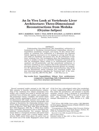
  • 2. Fig. 1. A: The ‘‘classic’’ and ‘‘portal’’ lobule, and liver ‘‘acinus’’ chitectural motifs: fractal branching in portal tracts (larger bile ductules characterize mammalian hepatobiliary structure/function relationships. and ducts) and a polyhedral (hexagonal) motif at the canalicular level A ‘‘tubular’’ concept has been used to describe the lower vertebrate of organization. The polyhedral architectural motif (1) appears to order livers of birds, fish, reptiles, and amphibians. a: The tubular concept in the arrangement of the interconnected canalicular network and indi- transverse view; two rows of hepatocytes form a tubule lumen (TL) at vidual cells, and (2) may provide maximal structural/functional relation- their apical membranes, while basal membranes border sinusoidal or ships. The latter is suggested given hexagonal organization is the least intercellular/intertubular space. b–d: Findings from three-dimensional perimeter way for space filling, meaning hepatocellular (metabolic) (3D) reconstructions reveal ‘‘tubular’’ architecture is more complex space is maximized, while minimal material is used for construction than previously considered. The tubule lumen is actually an intercon- and maintenance of the hepatocellular membranes, and consequently nected network of branching canalicular and bile preductule passage- in the creation of canaliculi. Because blood to bile distance is mini- ways. Findings from 3D reconstructions also suggest a common/ mized (Figs. 3, 4), transport is maximized. Lastly, the presence of a shared structural/functional unit (indicated by brown oval in both con- hexagonal motif in both the ordered arrangement of the canalicular ceptual models) among lower and higher vertebrate livers. This func- network, and in the organization of the hepatic lobule, suggest a hex- tional unit is comprised of (1) a ‘‘portal tract/hilus,’’ a single conduit agonal structural motif may provide structural integrity to the organ containing two afferent blood supplies (hepatic portal and arterial ves- system in total through the cellular and tissue level of organization. sels), and efferent bile duct(s); (2) a ‘‘primary efferent vascular con- Similarly, fractal branching of the blood and bile conduits in the portal duit,’’ central vein/hepatic vein/terminal hepatic vein; and (3) an anas- tracts confers maximal afferent and efferent transport efficiency tomosing hepatic muralium (predominantly monolayered in mammals, through these fluid compartments. Numbered segments in the hilus bilayered in lower vertebrates), perfused by a canalicular network and and bile duct regions indicate the branching order (fourth order) sinusoidal bed, that bridges these two. B: Our investigations, in con- observed in medaka. These underlying structural principles may serve junction with extant mammalian liver information, suggest the verte- to order the system toward maximal structural integrity and metabolic brate hepatobiliary system to be constructed on two fundamental ar- and transport efficiency.
  • 3. 772 HARDMAN ET AL. Teutsch et al., 1999). These concepts, based on 3D recon- The 3D in vivo findings we present here not only elu- structions of serial sections of human (Matsumoto et al., cidate hepatobiliary architecture of the lower vertebrate 1979) and rodent liver (Teutsch et al., 1999) describe liver phenotype in medaka, but may help better our cone-shaped units observed at the tissue level of organiza- comparative understanding of various conceptual models tion (branching of portal tracts). For instance; secondary applied to vertebrate liver structure/function. lobules (synonymous with the ‘‘classic’’ lobule), comprised of six to eight primary lobules, have a central terminal he- patic vein and six portal tracts at the periphery. Although MATERIALS AND METHODS each of these models remain valuable for communicating For decades, various color mutant strains of medaka normalcy and disease, they are incomplete in conceptu- (Oryzias latipes), acquired from natural and commer- ally describing vertebrate hepatobiliary structure and cially available populations, have been maintained in function, particularly in 3 dimensions, and, understand- the Laboratory of Freshwater Fish Stocks at Nagoya ably, disparities remain in applying these models across University, Japan. Cross-breeding from these stocks was vertebrate species. As such, lobular and acinar models of used to produce a stable ‘‘transparent’’ strain of medaka the liver have never been completely accepted (Rappa- (STII), homozygous recessive for all four pigments (iri- port, 1958; Lamers et al., 1989). Moreover, none of these diophores, leucophores, xanthophores, melanophores; conceptual models have seen successful application to Wakamatsu et al., 2001). STII medaka allow high reso- lower vertebrate hepatobiliary structure/function. Such lution (<1 mm) noninvasive in vivo imaging of internal discrepancies likely belie inadequacies in the conceptual organs and tissues at the subcellular level. Using laser models themselves, or rather, our lack of comparative scanning confocal microscopy (LSCM) and fluorescent understanding of vertebrate liver architecture. probes to elucidate the hepatobiliary system, we imaged Much less is known about the nonmammalian verte- liver structure and function at various stages of develop- brate liver. The prevailing model for these livers is the ment in over a hundred individual living medaka. Of hepatic tubule, which emerged from the predominance these, 15 medaka were used for 3D reconstructions pre- of observations of a two hepatocyte thick parenchyma sented here (3 medaka at 8 days postfertilization [dpf], 4 and the fact that the vast majority of studies in lower medaka at 12 dpf, 3 medaka at 30 dpf, 3 at 40 dpf, 2 vertebrate liver over the past century have shown no medaka at 60 dpf). With widefield and confocal fluores- clear lobular formation (Elias and Bengelsdorf, 1952; cence microscopy, salient features of the organ system McCuskey et al., 1986; Hampton et al., 1988; Rocha such as canaliculi, space of Disse, endothelial cells, bili- et al., 1994; Hinton and Couch, 1998). The current tubu- ary epithelial cells, red blood cells, and hepatocytes and lar concept describes of two rows of hepatocytes (in lon- their nuclei, were clearly resolved in vivo (Fig. 2). Confo- gitudinal section), the adjacent apical membranes of cal stacks from in vivo imaging of the hepatobiliary sys- which form a tubule lumen (bile passageway), into tem were used for 3D reconstructions, and from these, which bile is actively secreted (Figs. 1, 2). Basal mem- architectural, morphometric, and volumetric analyses branes of hepatocytes face sinusoidal or intertubular were made. space. Of interest; the ‘‘tubular’’ formation is widely held In vivo investigations included medaka embryos, lar- as a common hepatobiliary architecture found among all vae, and juveniles, from organogenesis (50 hours post- vertebrate species; it is the shared predominant pheno- type in all embryonic vertebrates, to include humans, fertilization) through 60 days. Medaka were exposed by and has been used to characterize the adult phenotype means of aqueous bath (238C) to the fluorescent probes for amphibians, birds, reptiles, and fishes (Elias, 1949; Bodipy C5 Ceramide, Bodipy C5 HPC and fluorescein Arias, 1988). While the hepatic ‘‘tubule’’ formation pre- isothiocyanate to facilitate in vivo investigation of hepa- dominates among lower vertebrates throughout their life tobiliary structure/function. Each of the fluorophores span, mammalian liver undergoes transition from a saw hepatobiliary uptake and transport and excretion tubular to laminar (muralium) architecture that is com- into the gut lumen. Exposed cohorts were typically com- plete in man by age 5 (Arias, 1988). ‘‘Tubule’’ formations prised of 10 individual medaka. At various time points, (denoting more than one row of hepatocytes) are also individual medaka were removed from exposure cohorts, observed in liver regeneration in mammalian species af- sedated with 10 mM tricaine-methane sulfonate (MS- ter severe injury, such as after hepatectomy and from 222), mounted in aqueous medium (Embryo Rearing Me- marked acute exposure to toxins/toxicants (Van Eyken dium for dechorionated embryos and hatchlings and de- et al., 1989; Vandersteenhoven et al., 1990). ionized water for 20 dpf larvae and older) on depression From the brief review above it can be understood that well glass slides with cover slip, and imaged live using our comparative understanding of the vertebrate liver is brightfield, and widefield and confocal fluorescence mi- increasingly important. Not only is the tubular liver croscopy. Time points of study varied from 10 min to 2 structure the most ubiquitous hepatic phenotype in the hr depending on the fluorophore used and the aspect of world, a better comparative understanding of the verte- the hepatobiliary system being studied. For instance, brate liver enhances our ability to interpret and commu- Bodipy C5 HPC and fluorescein isothiocyanate accumu- nicate normalcy and disease across the variety of animal lation in the hepatic parenchyma is first observed at 10 models employed in research. By example, small fish ani- min after exposure, with maximal saturation of the fluo- mal models such as medaka and zebrafish are proving rophore in the hepatic parenchyma occurring at 30 increasingly invaluable to the study of vertebrate devel- min. Time points between 15 and 45 min post fluoro- opment, carcinogenesis, and for investigation of molecular phore exposure were commonly used for in vivo studies. mechanisms of disease and toxicity (Wittbrodt et al., Imaging systems: Confocal fluorescence microscopy 2002; Shima and Mitani, 2004; Berghmans et al., 2005; was performed with a Zeiss 510 Meta system and Zeiss Alestrom et al., 2006). LSM 5 Axiovision image acquisition software, an Argon
  • 4. VERTEBRATE LIVER ARCHITECTURE 773 Fig. 2. Noninvasive in vivo imaging of hepatobiliary structure/func- were observed circulating through sinusoids (S/r) abutting the basal tion in living STII medaka. A: STII medaka embryo 6 days postfertiliza- membranes of hepatocytes. E2: Single section from in vivo confocal tion (dpf), left lateral view. The fluorescent cytochrome P4503A metab- image stack showing conservative b-Bodipy C5 Phosphocholine olite resorufin (red) in transport through the intrahepatic biliary net- (green fluorescence) in transport from sinusoid (S) to canalicular space works (IHBPs) of the embryonic liver (L), and concentration in the gall (IHBP), imaged in vivo 30 min. Post administration of the fluorophore bladder (GB). Otic vesicle (Ov), yolk sac (Y). B: Tubule-like formations, to medaka (aqueous bath). E3: Composite of E1 and E2, localizing flu- seen in transverse section, in the developing parenchyma, observed in orophore concentrate to IHBPs. E4: Surface map of region of interest vivo at 6 dpf. C: Concentrative transport of resorufin from hepatocyte (ROI) of inset (white square) in frame E2; illustrating concentrative into the tubule lumen (TL/IHBP). D1: STII medaka, 30 dpf, left lateral transport of the fluorophore from sinusoidal space (S) to bile space view, brightfield microscopy. Green algae can be seen transiting the (IHBP). The ROI, showing a 17-mm span from blood to bile (across he- gut lumen. D2: An in vivo confocal image of liver from the region of in- patocyte), was quantitatively assessed, and revealed b-Bodipy C5 terest (grey square) in frame (D1), showing fluorophore transport Phosphocholine concentration to be 66 times greater in the biliary (FITC) through IHBPs. E1–E4: In vivo confocal imaging of blood to bile passageway than in sinusoidal space. These studies demonstrated in transport. E1: Two rows of hepatocytes and their nuclei (HN) are seen vivo evaluation of concentrative blood to bile transport of fluorescent in longitudinal section. Red blood cells (elongated ovate structures) probes was possible. and HeNe laser, Carl Zeiss C-apochromatic 40x/1.2, and tomical imaging was performed using a Nikon SZM C-apochromatic 10x/0.45. For wide-field fluorescence mi- 1500 dissecting microscope with a Nikon DXM 1200 digi- croscopy, a Zeiss Axioskopp with DAPI/TRITC/FITC fil- tal capture system (brightfield). Regarding software, ter cubes was used. Excitation/emission parameters for image analysis and compilation was performed with filter cube sets were: DAPI/UV (Ex 360–380 nm/Em All EclipseNet (Nikon, USA), Adobe Photoshop (Adobe, Vis 400 nm); FITC (Ex 450–490 nm/Em 515–565 nm); Inc.), Amira 3D (Mercury Computer Systems, Berlin), TRITC (Ex 528–552 nm/Em 578–632 nm). Gross ana- ImageJ (V1.32j), IP Lab software (Scanalytics, Inc., ver-
  • 5. 774 HARDMAN ET AL. sion 3.55), and Zeiss Image Browser (Carl Zeiss). Fluo- likely imparts structural integrity, and metabolic and rescent probes used were 7-benzyloxyresorufin (10–50 transport efficiency, features provided by higher-order mM); b-Bodipy C5-HPC [BODIPY1 581/591 C5-HPC (2- organizing principles such as hexagonal architecture, (4,4-difluoro-5- (4-phenyl-1,3- butadienyl)-4-bora-3a,4a- not uncommon in biological systems (Mainzer, 1996). diaza-s- indacene- 3-pentanoyl)-1-hexadecanoyl- sn-glyc- ero-3-phosphocholine), (30 nM–10 mM)]; Bodipy FL C5- Biliary System and Hepatocytes ceramide [N-(4,4-difluoro-5,7-dimethyl-4-bora-3a,4a-diaza- s-indacene-3- pentanoyl) sphingosine, (500 nM–5 mM)]; Linear segments of IHBPs (canaliculi) averaged 11 mm and fluorescein isothiocyanate (1 nM–50 mM). Fluoro- in length and 1.3 mm in diameter, and exhibited unary, phores were acquired through Invitrogen/Molecular binary, ternary, and quaternary branching. This finding Probes (Carlsbad, CA). All fluorescent probes were admin- should not be confused with fractal branching patterns istered to STII medaka by means of aqueous bath in con- (Turcotte et al., 1998); the biliary system in medaka centration ranges given, at room temperature, under dark does not appear (except for the localized area at the liver conditions. hilus) to follow a fractal branching tree motif. Rather, All transmission electron microscopy (TEM) was per- there is ubiquitous feedback/interconnectedness within formed at the Laboratory for Advanced Electron and the 3D biliary network (Fig. 5). Four to six hepatocytes Light Optical Methods (LAELOM), College of Veterinary were associated with each linear segment of IHBP Medicine, North Carolina State University. Individual (Fig. 6) and arranged such that a single hepatocyte medaka were anesthetized and fixed in 4F:1G fixative could contribute to two or three bile segments (canali- (4% formaldehyde and 1% glutaraldehyde in a monoba- culi/bile-preductules). The same hepatocellular/canalicu- sic phosphate buffer with a final pH of 7.2–7.4 and a lar relationship has also been described in rat (Motta final osmolality of 176 mosmol). Thin sections (Spurr and Fumagalli, 1975), where the same side of a hepato- resin embedded) were then examined using a FEI/Phi- cyte was observed to bound two or more bile canaliculi. lips EM 208S Transmission Electron Microscope. While hepatocytes had a mean diameter of 11.3 mm, they showed varying morphology in vivo, apparently adaptive to the space they were filling (e.g., at bifurcating or tri- FINDINGS furcating junctions). Hence, while the intrahepatic biliary Polyhedral (Hexagonal) Architecture of the network showed a more ordered structure, hepatocellular organization and morphology appeared much more adapt- Intrahepatic Biliary System in Medaka ive to space filling and parenchymal organization. The 3D reconstructions from in vivo imaging revealed The IHBP network, composed of canaliculi and bile intrahepatic biliary passageways (IHBP) in medaka to preductules, occupied 95% of the liver corpus uni- be predominantly an interconnected network of canali- formly, with each area of livers examined (n ¼ 15) con- culi and bile preductules, the organization of which taining approximately equal volumes of IHBPs relative appeared to be based on a polyhedral (hexagonal) struc- to hepatocellular and vasculature volumes, regardless of tural motif (Figs. 3–6). The polyhedral motif was most how distal/proximal an observed volume of liver was clearly observed in the arrangement of IHBPs, and was from the liver hilus. Canaliculi were observed to merge not evident in sinusoidal architecture. Empirical obser- with bile preductules at unique morphological sites vations found IHBP branching to be distinctly angular, formed by junctional complexes between bile preductular and statistical analysis of IHBP branching angles (in epithelial cells (BPDECs, discussed following) and hepa- 3D) revealed clustering of angles into three main groups: tocytes (Fig. 6). Together, canaliculi and bile preductules 120 degrees (74.9%), 90 degrees (14.8%), and 58 degrees comprised the IHBP network. This interconnected net- (10.3%). The majority of angles measured averaged work of IHBPs was observed to feed three primary intra- 120 degrees, the interior angle of the hexagon (Fig. 4). hepatic bile ducts (IHD), lined by cuboidal biliary epithe- The 3D angle measurements were made by randomly lia. IHDs merged at the liver hilus into a common he- selecting intrahepatic biliary passageway bifurcation patic duct that was associated with the cystic and and trifurcation points in 3D reconstructions, achieved common bile ducts (Fig. 5). Cuboidal and large squa- by rotating the 3D model and randomly selecting mous biliary epithelial cells (non-BPDECs) were branching points. All angles of a bile segment branching observed primarily localized to the hilar and peri-hilar point were measured, inclusive of all 3D planes (Fig. 3). regions of the liver. Further 3D analyses revealed IHBP architecture to be based, conceptually, on a tessellating (3D) hexagonal Parenchymal Architecture mesh (Carle et al., 2001). This hexagonal motif was also apparent in the ratio between bile segment length and Of interest, while ‘‘tubule’’ like formations were blood to bile distance (Figs. 3, 4). When bile segment observed (in vivo and in single sectional views) in em- (canaliculi) length was considered to correspond to one bryonic livers (3 dpf to 7 dpf), tubule formations were side of a hexagon, the distance (radii) from hexagon side not readily apparent in 3D reconstructions (n ¼ 15) of (canaliculus) to hexagon center (sinusoid) appeared to larval and older livers. Rather, parenchymal architec- correlate to the hexagonal ratio given in Figure 3. A ture in larval and later developmental stages of medaka hexagonal architecture has also been described, from appeared to be more consistent with an anastomosing, empirical observations, in the fine structure of the bili- predominantly dual layered, muralium. First, 3D recon- ary system in rat (Mochizuki et al., 1988; Murakami structions revealed the ‘‘tubular’’ lumen to be composed, et al., 2001) and human (Yamamoto et al., 1990). not of an extended linear biliary passageway that would Although a discussion of this topic is beyond the scope of create a classic ‘‘lumen,’’ but of a hexagonal network of this study, the presence of a polyhedral structural motif branching, interconnected canaliculi and preductules,
  • 6. VERTEBRATE LIVER ARCHITECTURE 775 Fig. 3. Examples from three-dimensional (3D) reconstructions of bular profile). In medaka, we found this structural arrangement of medaka hepatobiliary system, illustrating the types of analyses that branching canaliculi that comprises the ‘‘tubule lumen,’’ revealing ‘‘tu- led to findings on polyhedral (hexagonal) architecture. Grayscale back- bular’’ architecture more complex than previously considered (see Fig. ground images are sections from in vivo confocal image stacks, from 1). F: 3D reconstruction of the relationship between IHBPs and sinu- which 3D reconstructions were generated. In (A–D,F–H) the empty soids. Between 2 sinusoids (red) can be seen a branching canalicular space surrounding biliary passageways (green/gold), and between si- network (green/gold). G–I: Examples of 3D morphometric analyses, nusoids (red), is hepatocellular space (hepatocytes not rendered for which suggest medaka parenchymal architecture to be more consist- visual clarity). Colored dots in A–C are given for anatomical reference. ent with an anastomosing muralium. 3D measurements of hepatocellu- A–D: An isolated section of the intrahepatic biliary network, illustrating lar space (between sinusoids) revealed a muralium-like architecture. relationship of canaliculi (green) to sinusoids (red). A: Branching canal- G: Example dimensions given are 196.3 mm in length, 98.2 mm in iculi (intrahepatic biliary networks [IHBP]) are shown between two height, and 24.7 mm in width. H: Same as G viewed in a horizontal rows of hepatocytes (confocal grayscale image). B: Same as A with si- longitudinal section (from the top). Sample dimensions are shown. I: nusoids present, and C is AB viewed directly on the ‘‘z’’ axis (from Single slice from in vivo confocal stack of hepatic parenchyma. Gray top). C: Example of analyses of polyhedral structural motif, which was arrowheads indicate sinusoids, black arrowheads hepatocyte nuclei. evident in the relationship between blood to bile distance and canalic- Hepatocellular compartment width (analyzed in vivo and in 3D) was ular length (see Fig. 4). Bile segment length (canaliculus, S) correlated found to be predominantly two cells thick, although occasionally up to to calculated and measured values of blood to bile distance (r), when eight cells in some areas. Average parenchymal width was 24.58 mm. assessed with the hexagonal ratio [r ¼ 0.5 (S) 1.44]. D: Empirical Parenchymal depth varied from 21.2 mm to 200 mm. Note that typi- observations (3D) of angular canalicular and bile preductule branching cal dimensions of 3D reconstructions were 100 mm in depth; hence, led to statistical analysis of branching angles, which suggested the depth dimension is limited. Parenchymal metrics were derived from presence of a polyhedral structural motif (see Fig. 4). E: Schematic 127 measurements (3D) in medaka at 8, 12, 30, and 40 dpf. Space of showing modified ‘‘tubular’’ concept of lower vertebrate liver (note Disse (SD), Sinusoidal Endothelial Cell (SE), Red Blood Cell (R), Sinu- IHBPs, and the hexagonal arrangement in relation to hepatocytes/tu- soid (Sn, S1–S4).
  • 7. 776 HARDMAN ET AL. Fig. 4. Morphometric and volumetric analyses (upper left graph) of to calculated values, falling between the longest (R) and shortest (r) random measures (341 observations) of three-dimensional (3D) radii in the hexagon [r ¼ 0.5 (S) 1.44]. Where bile segment length (e.g., branching angles of bile segments in four individual livers (8–40 days canaliculus) ¼ S, blood to bile distance ¼ r. Pearson’s correlation postfertilization [dpf]) found branching angles to cluster into three coefficient (0.049, P ¼ 0.6). The difference in the mean between calcu- primary groups: 120 degrees (75%), 93 degrees (15%), and lated and measured r values was 0.67 mm, half the width of the canali- 64 degrees (10%). Upper right graph: Using the hexagonal ratio in culus. In vivo volumetric investigations in three individual medaka at the lower portion of the plot, a polyhedral structural motif was also 8 dpf, 12 dpf, and 30 dpf revealed consistent ratios between intrahe- suggested in the relationship between blood to bile distance and can- patic biliary passageway volume, blood (sinusoid) volume, hepatocel- alicular length. Measured values of blood to bile distance (r) correlated lular volume, and parenchymal volume. which formed the inner parenchymal framework (because confocal stacks were 100 mm in depth, the (Figs. 3–6). In other words, tubule lumens were found to maximum measurement for plate ‘‘height’’ was limited be canalicular segments with an average length between to 100 mm). Third, the longest non-branching biliary seg- 10 and 13 mm. Second, hepatocellular muralium-like ments were observed to be 30 mm in length, and these structures were apparent, which varied in height from were few in number (2–3% of bile segments measured). 20 mm to 100 mm, and width from 23 mm to 83 mm, For a tubular architecture to be present, tubules would, observations consistent with an anastomosing muralium from these 3D investigations, be less than 30 mm in linear
  • 8. VERTEBRATE LIVER ARCHITECTURE 777 Fig. 5. Three-dimensional (3D) architecture of liver hilus, bile duc- was followed and the branching of the IHBPs mapped to a 2D sche- tules/ducts, and canalicular network. A: In vivo image capture of the matic. Each colored line corresponds to one bile segment (11 mm in liver hilus at 30 days postfertilization (dpf). Three primary intrahepatic length). Colors are for illustrative purposes only. The 90-degree angles ducts (IHDs) conjoined a common hepatic duct (HD), which fed the correlate to bile segment branching (not to scale or degree, for illus- cystic duct (CD), gall bladder (GB), and common bile duct (CBD). The trative purposes only). The schematic is used to illustrate the intercon- liver hilus of medaka contained two afferent blood supplies (the he- nectedness of the canaliculo-bile preductular network. For clarity, part patic portal vein [HPV] and hepatic artery [not shown]), and hepatic of the network was abbreviated by using dashed lines to illustrate duct. Mucosal folds (MF) of the gut (lower left) are seen caudal to the feedback and interconnectedness within the IHB network. Dashed liver. B,C,F: The 3D reconstructions aided in elucidating the relation- lines represent a branching network of canaliculi (as illustrated in col- ship between the canaliculo-preductule network, bile ductules and ored lines). The canaliculo-bile preductule network occupied 95% of ducts, and liver hilus. Images C (horizontal longitudinal section) and F the liver body (by area). D: Schematic showing overall biliary architec- (sagittal view) show an example of 3D reconstruction of the canali- ture of medaka liver. Bile ductules and ducts exhibited fourth order culo-preductular network draining to a common IHD (one of three fractal branching, and were largely found in hilar/peri-hilar region of IHDs shown in B). E: A hand-traced schematic of an isolated section the liver. Ductules terminated in a canaliculo-bile preductular network of the canaliculo-preductular network (diamond-shaped region of inter- (illustrated in E), that followed a polyhedral (hexagonal) architectural est [ROI] indicated in C and F). One individual IHD (green arrowhead) motif. length and would be highly branched and interconnected tive correlary of mammalian progenitor/stem cells, given (following biliary network architecture). Hence, 3D in vivo they share morphological characteristics ascribed to observations found medaka parenchymal structure to mammalian oval cells (bipotential progenitor cells). more resemble a predominantly two- to three-cell-thick he- BPDECs, like oval cells, are phenotypically distinct from patic muralium, with an average width of 26 mm and hepatocytes, characterized by a high nuclear to cyto- height that varied from 20 mm to 100 mm. It follows that plasm ratio with no basement membrane, and are inti- medaka parenchymal architecture appeared highly analo- mately associated with the biliary system (Golding gous to mammalian architecture; although predominantly et al., 1996; Fausto and Campbell, 2003). In vivo, two to three hepatocytes thick (and in rare instances up to BPDECs were observed to form unique junctional com- seven or eight cells thick), as opposed to the predominantly plexes with hepatocytes, at which were created IHBPs one- to two-cell-thick mammalian muralium (Elias and termed bile preductules. These junctional complexes Bengelsdorf, 1952). were morphologically distinct (Fig. 6). In single section view these unique morphological formations appeared as BPDECs surrounded by bile passageways on all sides Biliary Epithelia (Fig. 6). 3D reconstructions from in vivo imaging 3D investigations found BPDECs (Hinton and Pool, revealed BPDECs to occupy the ‘‘center’’ of these com- 1976; Hampton et al., 1988; Okihiro and Hinton, 1999) plexes, where one or more BPDECs formed multiple to be located throughout the parenchyma of medaka liv- junctional complexes with surrounding hepatocytes (Fig. ers studied (from 8 dpf to 60 dpf). BPDECs are the puta- 6). Biliary passageways (bile preductules) formed at
  • 9. 778 HARDMAN ET AL. Fig. 6. A–A2: Canaliculus/bile preductule and hepatocellular rela- (BPD). The 3D reconstructions (B1,B2) elucidated the actual architec- tionships. A1,A2: Three-dimensional (3D) reconstructions from confo- ture of these canaliculo-bile preductule complexes, revealing bile pre- cal image in A. Four to six hepatocytes (hepatocyte nuclei shown in ductular epithelia form multiple junctional complexes with surrounding purple, numbered) were observed in relation to a given canaliculus hepatocytes, at which bile preductules are formed. In B1 and B2, the (green). Circled red A indicates a bifurcating canalicular segment. A empty space surrounding BPDEC is hepatocellular space (left single hepatocyte was observed to contribute to one to three canali- ‘‘empty,’’ or not rendered, for illustrative purposes). In vivo findings culi. B1–B2: The 3D reconstructions of confocal image in (B), illustrat- were corroborated with ultrastructural studies (C1,C2), which aided in ing the structure of a bile preductule (BPD) junction. Red arrowheads elucidating/validating in vivo observations. Frame C is a still image in (B) indicate bile preductular epithelial cells (BPDECs) within the he- from a 3D reconstruction illustrating the distribution of BPDECs patic parenchyma. In 2D confocal sections BPDECs appeared to (purple, indicated by red arrowhead) throughout the hepatic paren- occupy the ‘‘center’’ of these canaliculo-bile preductule structures. chyma. Junctional Complex/Bile Preductule (Jc/BPD), Intrahepatic Bili- BPDECs formed unique junctional complexes with surrounding hepa- ary Passageway (IHBP), Hepatic Nuclei (HN), Sinusoid/red blood cell tocytes, which created biliary passageways, termed bile preductules (S/r), Bile Preductular Epithelial Cell (BPDE/C). these BPDEC/hepatocyte junctional complexes com- Theise et al., 1999; Fausto and Campbell, 2003; Knight monly showed ternary or quaternary branching. While et al., 2005). While we have localized BPDECs to specific BPDECs varied in morphology and size, showing ovate, locations within hepatic parenchyma, and these cells triangular and ellipsoidal nuclei, the majority of share morphological characteristics consistent with BPDECs were commonly found to be 6 mm in diameter. mammalian hepatic progenitor cells, BPDECs in medaka Our in vivo findings on BPDECs in medaka are, inter- have only been partially characterized (Okihiro and Hin- estingly, consistent with previous oval cell studies in ton, 2000). Cuboidal and squamous biliary epithelia rodents and humans (Farber, 1956; Golding et al., 1996; (non-BDPEC), lining larger diameter intrahepatic biliary
  • 10. VERTEBRATE LIVER ARCHITECTURE 779 passageways, were observed almost exclusively near network of canaliculi and bile preductules (bile seg- hilar and perihilar regions of the liver (Fig. 5). ments) that averaged 11–13 mm in length (Fig. 5). Because 3D investigations reveal medaka hepatobili- Morphometric and Volumetric Information ary architecture, representative of the lower vertebrate tubular phenotype, to more closely resemble a dual-lay- 3D reconstructions enabled highly accurate volumetric ered anastomosing muralium, akin to the single-cell- and ratiometric analyses of biliary, parenchyma, vascu- thick muralium described in mammals, important ques- lature, and liver volumes (Fig. 4). We found prior ex tions are raised. If hepatic tubules comprise a two-hepa- vivo volumetric studies (Hess et al., 1973; Blouin et al., tocyte-thick muralium, how are they integrated into a 1977; Rocha et al., 1997; Hinton et al., 2001) in verte- muralium-like structure? Although tubule formations brate livers to correspond well with our in vivo findings. may theoretically comprise a dual-layered muralium, this would necessitate an intertubular space, which raises another question; if tubules are present, are DISCUSSION tubules joined at the intertubular space? If so, does this Surprisingly, we did not observe a well-developed not describe a muralium structure? These important arborizing biliary ‘‘tree,’’ well characterized in mamma- questions remain to be answered, given our findings lian studies (Ludwig et al., 1998; Masyuk et al., 2001). from in vivo 3D reconstructions suggest a dual-layered This ‘‘tree’’ describes bile ducts of the liver hilus arboriz- plate-like muralium predominates in larval and later ing into more highly branched, and numerous, bile ducts life stages of medaka, whereas tubule-like formations and ductules of diminishing diameter, as the biliary sys- were observed in the embryonic liver. tem infiltrates more distal regions of liver (relative to The tubular concept of the lower vertebrate liver was the liver hilus). We attribute the overall lack of an arbo- derived largely from 2D observations of histological and rizing biliary tree in medaka liver to two factors: (1) electron micrograph (EM) preparations. In histological mammalian studies are describing portal tract arboriza- sections, the fine structure of the biliary system is diffi- tion, or interlobular stromal areas of the liver (meaning cult to discern (canaliculi average 1–2 mm in diameter), biliary tree arborization describes the tissue level of or- and when resolved, we now know, due to a hexagonal ar- ganization, lobule formation); (2) medaka liver is the ar- chitectural formation, would show a random order of chitectural analogue of the intralobular mammalian pa- appearance in a 2D section. For instance, using histolog- renchyma. The two points are discussed in detail below. ical sections Rocha et al. (1994) described biliary passa- These findings raise important questions regarding geways in trout as appearing randomly dispersed current conceptual models of vertebrate liver architec- throughout the liver; this would be an accurate 2D ob- ture. In mammalian liver, hepatocytes have been de- servation, or how the 3D hexagonal architecture of the scribed as irregularly shaped polygonal cells that form, canalicular network we have described would appear in predominantly, a one- to two-cell-thick wall/plate-like a 2D histological section. Consequently, from a 2D per- structure (muralium), which anastomoses throughout spective, a dual-layered muralium may appear, or be the liver (Elias and Bengelsdorf, 1952). Observations of interpreted as, a tubule-like formation. Hence, given the dual layered hepatocytes in fish and other lower verte- interconnected 3D biliary architecture we have eluci- brate livers, and the lack of observed lobular formations dated in this study, it may be that a ‘‘tubular’’ formation in the majority of lower vertebrate livers studied and dual layered plate-like muralium are perhaps, one (McCuskey et al., 1986; Hampton et al., 1989; Rocha and the same, and that discrepancies in understanding et al., 1997; Hinton et al., 2001; Akiyoshi and Inoue, muralium architecture in lower vertebrates have arisen 2004) led to the reasoning that lower vertebrate livers from varied 2D viewpoints (and thereby varied interpre- may be composed primarily of anastomosing ‘‘tubules.’’ tation), and a lack of 3D studies in vertebrate liver In single section view (e.g. histological preparations with structure at the canalicular level of organization. longitudinal orientation), hepatic tubules appear as two If fish and mammals share similar muralium architec- rows of hepatocytes, the apical membranes of which ture, what can explain the absence of ‘‘portal tracts’’ and form a tubule lumen (bile passageway), and basal mem- lobule formation in lower vertebrate livers? Addressing branes of which border sinusoidal, intracellular, or inter- these questions involves consideration of organ system tubular space. As our 3D in vivo findings have shown, ontogeny and the nature of the ‘‘functional unit’’ of the the true structure of the ‘‘tubular’’ liver is more complex vertebrate liver. Although this is beyond the scope of than previously understood. Because one hepatocyte this article, a brief discussion is warranted. First, while may contribute to one to three canaliculi, ‘‘tubule portal tracts/triads are well described in mammalian lumens’’ are actually composed of an interconnected hex- liver and integral to current mammalian liver concep- agonally branching network of canaliculi and bile pre- tual models (lobular, acinar), they are not often observed ductules. Where ‘‘tubule-like’’ formations were observed in lower vertebrates. In lower vertebrates, anatomical in 2D transverse sectional views in the embryonic liver structures that may be perhaps the evolutionary, or (in vivo), ‘‘tubule’’ formations were very rarely encoun- even functional, precursors to portal tracts have been tered in larval and older fish, and were not readily appa- described as venous biliary arteriolar tracts (VBAT), ve- rent in 3D reconstructions. For a tubular architecture to nous arteriolar tracts (VAT), venous biliary tracts (VBT), be considered, tubules in medaka would, from our 3D in biliary tracts (BT), and arteriolar tracts (AT) (Rocha vivo investigations, average 29 mm in linear length ( et al., 1995; Hampton, 1988). These morphological fea- three hepatocytes), and would be quite rare (2–3% of tures bear some anatomical resemblance to the portal bile segments measured were found to span up to 29 tract, although no semblance of lobule formation in mm). The prevailing 3D structure of the biliary system lower vertebrates has been found. Second, based on our in medaka was a highly branched and interconnected 3D reconstructions, it can be hypothesized that the hep-
  • 11. 780 HARDMAN ET AL. atobiliary systems of both medaka and mammals share ratively higher metabolic demand, typically higher body a common fundamental functional unit: (1) a ‘‘portal temperature, higher load bearing on the liver resulting tract/hilus,’’ a single conduit containing two afferent from gravity, and relatively larger mammalian liver blood supplies (hepatic portal and arterial vessels), and mass. It follows that higher vertebrates likely show efferent bile duct(s); (2) a ‘‘primary efferent vascular ‘‘lobulation’’ of the liver in support of greater organ mass conduit,’’ central vein/hepatic vein/terminal hepatic vein; and metabolic demand; the iteration of a single hepatic and (3) anastomosing hepatic plates/cords, perfused by a functional unit (e.g. HMS). In more massive mamma- canalicular network and sinusoidal bed, that bridges lian livers the formation of hexagonal lobules, organized these two. A similar conceptual functional unit, the he- by stromal tissue (portal tracts/triads), would impart patic microcirculatory subunit (HMS), was proposed by structural integrity to the organ (due to the physical Ekataksin et al. (1997). This wedge-shaped unit is com- properties of hexagonal packing; Weaire and Phelan, posed of ‘‘base’’ (the portal tract/hilus), ‘‘apex’’ (the cen- 1994; Hales, 2001), much needed in an organ of such tral vein/terminal hepatic vein/hepatic vein), and a con- mass (3–5% of body weight in mammals), and one tinuous system (muralium) of anastomosing hepatic highly perfused with a liquid medium (the liver of mam- plates (laminae hepatic) connecting the base and apex, mals can store up 30% of total blood volume). It follows, perfused by sinusoidal bed (labyrinthus hepatic). Hence, from an ontological viewpoint, that appearance of it can be considered that, in the lower vertebrate liver of VBATs, VATs, VBTs, BTs, and ATs in larger piscine spe- medaka; the liver hilus is the correlary of the portal cies may be evidence of the emergence of ‘‘lobulation’’ tract, and hepatic vein the corollary of the central vein/ among lower vertebrates such as teleosts. Such a consid- terminal hepatic vein. In between these is the laminae eration begs more detailed investigation of larger fish hepatic, the fine structure of which, up to this study, has species, and reptilian and amphibian livers. Lastly, the remained a mystery in medaka and other lower verte- hexagonal structural motif appears to be conserved brates. among all vertebrates; found not only in the organiza- One of the outstanding questions in human biliary tion of the canalicular network (intralobular paren- architecture has been; does each duct of Hering corre- chyma), but also in the formation of the classic lobule spond to one canaliculus? Or do canaliculi form a conflu- (arborization of portal tracts). ence before entering the canals of Hering (Saxena et al., Reaching a conceptual model of the ‘‘functional unit’’ 1999)? Our 3D studies in medaka show the canalicular of the liver has long been sought, and the spatial archi- network to form a confluence before entering the hilar tecture of the lower vertebrate liver, particularly the bil- intrahepatic ducts, the anatomical corollary of the canals iary system, has long been in question. Differences in of Hering. Given the findings presented here and that: extant conceptual models have arisen from how lower bile canalicular diameter is conserved in vertebrate liv- and higher vertebrate livers have been viewed at vari- ers (1–2 mm); early observations by Elias and Bengels- ous levels of biological organization (cellular, tissue, dorf (1952) suggest a polygonal hepatocellular formation; organ), in which anatomical plane, and whether in two that a ‘‘chicken-wire’’ pattern of bile canaliculi has been or three dimensions. Given various ways of viewing the described in human liver (Ekataksin et al., 1995); hexag- liver and the prior technological constraints (lack of 3D onal architecture has also been described in the fine and in vivo tools), understandably, discrepancies have structure of the biliary system in rat (Murakami et al., appeared to exist between lower vertebrate and mamma- 2001) and human (Yamamoto et al., 1990); the tubular lian hepatobiliary conceptual models. Such discrepancies structure (dual layered parenchyma) appears to be con- can be illustrated in a study by Akiyoshi and Inoue served among all embryonic vertebrates; erosion cast (2004), who investigated two hundred different teleost studies by Murakami et al. (2001) show a hexagonal species (histological preparations) and described varying canalicular network feeding intrahepatic bile ducts (rat); liver architecture among them (muralium as cord-like bipotential progenitor-like cells in both medaka and [one cell], tubular [two cells], and solid [two cells]). mammals are closely associated with the biliary system, Indeed, we observed all three structural forms in our in it is not unlikely that all vertebrate livers share the vivo and 3D investigations, each comprising the hepatic same fundamental functional unit. Hence, it is interest- parenchyma, although the muralium of medaka was ing to consider the medaka hepatobiliary system, repre- found to be predominantly two cells thick. Similar obser- sentative of lower vertebrate architecture, as a single vations have been made in developing mammalian liver functional unit (akin to the HMS), while mammalian by McCuskey et al. (2003). Hence, it appears the limits hepatobiliary systems can be considered to be composed of spatial observation/understanding permitted by single of multiple functional units organized into hepatic sectional views of the liver have led to these, while accu- lobules (typically 1–2 mm; Fig. 1). In mammals, we rate descriptions from a 2D viewpoint, discrepancies in hypothesize that portal tracts arborize within the liver interpretation, and thereby discrepancies in a compara- to form the classic intrahepatic biliary/vascular trees, tive understanding of the 3D architecture of vertebrate giving rise to lobulation (multiple functional units). liver. Such discrepancies can be understood considering Hence, as conceptual models go, the primary architec- the difficult task of extrapolating 2D information to 3D tural differences between medaka and mammalian hepa- architectural models. Particularly when 2D observations tobiliary systems appears to arise at the tissue/organ in single sectional views of the liver have accurately level of organization (multiple lobules in mammals vs. characterized the 3D structure we have elucidated here. single lobule in medaka). These differences can be at- It can be said that in one sense, prior observations of tributable to the metabolic and structural demands/ lower vertebrate liver architecture that described a tu- needs between lower and higher vertebrate hepatobili- bular conceptual model were correct, in so far as they ary systems (in the context of organ system ontogeny were communicating the 2D morphology encountered in and functional capacity), where mammals see a compa- single sectional views of the liver.
  • 12. VERTEBRATE LIVER ARCHITECTURE 781 SUMMARY Ekataksin W, Zou Z, Wake K, Chunhabundit P, Somana R, Nishida J, McDonnell D. 1995. HMS, Hepatic microcirculatory subunits in Collectively, these findings elucidate the 3D architec- mammalian species. Intralobular grouping of liver tissue with ture of the medaka hepatobiliary system and improve definition enhanced by dropout sinusoids. In: Cells of the Hepatic our comparative understanding of vertebrate liver struc- Sinusoid. Leiden: Wisse E, Knook DL, Wake K, eds. The Kupffer ture and function. These findings also present interest- Cell Foundation, The Netherlands. ing questions regarding the ‘‘functional unit’’ of the ver- Elias H. 1949. A re-examination of the structure of the mammalian tebrate liver; where the hepatobiliary system in medaka liver. II. The hepatic lobule and its relation to the vascular and biliary systems. Am J Anat 85:379–456. can be, as a conceptual model, considered a single func- Elias H, Bengelsdorf H. 1952. The structure of the liver of verte- tional unit, akin to the individual lobule. We have also brates. Acta Anat 14:297–337. addressed prior discrepancies among conceptual models Farber E. 1956. Similarities in the sequence of early histological of vertebrate hepatobiliary system architecture, and the changes induced in the liver of the rat by ethionine, 2-acetyla- findings presented should help with integration of prior mino-fluorene, and 30 -methyl-4-dimethylaminoazobenzene. Cancer observations of vertebrate liver structure/function into a Res 16:142–148. more cohesive conceptual framework. In summary, Fausto N, Campbell JS. 2003. The role of hepatocytes and oval in vivo and 3D findings in medaka show: (1) parenchy- cells in liver regeneration and repopulation. Mech Dev 120:117– mal architecture is predominantly a two-cell-thick mura- 130. Golding M, Sarraf C, Lalani EN, Alison MR. 1996. Reactive biliary lium, although tubule-like formations were observed in epithelium: the product of a pluripotential stem cell compart- embryonic livers; (2) the hepatic muralium is organized ment? Hum Pathol 27:872–884. through a polyhedral (hexagonal) structural motif, Hales T. 2001. The honeycomb conjecture. Discrete Comput Geom revealed in biliary architecture; (3) the intrahepatic bili- 25:1–22. ary system is an interconnected network of canaliculi Hampton J, Lantz R, Goldblatt P, Lauren D, Hinton D. 1988. Func- and bile preductules; (4) the canaliculo-preductular net- tional units in rainbow trout (Salmo gairdneri, Richardson) liver: work occupies the majority of the liver corpus (95%) II. The biliary system. Anat Rec 221:619–634. uniformly, with equidiameter IHBPs (1–2 mm) observed Hampton JA, Lantz RC, Hinton DE. 1989. Functional units in rain- throughout the liver; (5) larger bile ductules and ducts bow trout (Salmo gairdneri, Richardson) liver: III. Morphometric analysis of parenchyma, stroma, and component cell types. Am J were observed predominantly at the liver hilus, conse- Anat 185:58–73. quently an arborizing biliary tree was largely absent, Hess FA, Gnagi HR, Weibel ER, Preisig R. 1973. Morphometry of seen only in the rudimentary branching of intrahepatic dog liver: comparison of wedge and needle biopsies. Eur J Clin ducts from the hepatic duct; and (6) the livers of these Invest 3:451–458. small fish are replete with BPDECs, the putative mam- Hinton DE, Couch JA. 1998. Architectural pattern, tissue and cellu- malian correlative of bipotential progenitor/stem cells. lar morphology in livers of fishes: Relationship to experimentally- induced neoplastic responses. In: Fish Ecotoxicology. Braunbeck, ¨ T., D.E. Hinton, and B. Streit, eds., Birkhauser Verlag. Basel, ACKNOWLEDGMENTS Switzerland. Hinton D, Pool C. 1976. Ultrastructure of the liver in channel cat- Thanks to Dr. David Miller, Laboratory of Pharmacol- fish Ictalurus punctatus (Rafinesque). J Fish Biol 8:209–219. ogy and Chemistry, National Institute of Environmental Hinton D, Segner H, Braunbeck T. 2001. Toxic Responses of the Health Sciences, Research Triangle Park, for providing Liver. In: New Perspectives: Toxicology and the Environment, access to their laser scanning confocal microscopy facil- Target Organ Toxicity in Marine and Freshwater Teleosts. D. Schlenk and Benson, W. eds. Taylor Francis, New York. ity, and special thanks to the Duke University Inte- Jungermann K. 1988. Metabolic zonation of liver parenchyma. grated Toxicology Program. This publication was made Semin Liver Dis 8:329–341. possible by a grant from the National Center for Kiernan F. 1833. The anatomy and physiology of the liver. Philos Research Resources, a component of the National Insti- Trans R Soc Lond 123:711–770. tutes of Health. Its contents are solely the responsibility Knight B, Matthews VB, Olynyk JK, Yeoh GC. 2005. Jekyll and of the authors and do not necessarily represent the offi- Hyde: evolving perspectives on the function and potential of the cial views of NCRR or NIH. adult liver progenitor (oval) cell. Bioessays 27:1192–1202. Lamers WH, Hilberts A, Furt E, Smith J, Jonges GN, van Noorden CJ, Janzen JW, Charles R, Moorman AF. 1989. Hepatic enzymic zonation: a reevaluation of the concept of the liver acinus. Hepa- LITERATURE CITED tology 10:72–76. Akiyoshi H, Inoue A. 2004. Comparative histological study of teleost Ludwig J, Ritman EL, LaRusso NF, Sheedy PF, Zumpe G. 1998. livers in relation to phylogeny. Zoolog Sci 21:841–850. Anatomy of the human biliary system studied by quantitative Alestrom P, Holter JL, Nourizadeh-Lillabadi R. 2006. Zebrafish in computer-aided three-dimensional imaging techniques. Hepato- functional genomics and aquatic biomedicine. Trends Biotechnol logy 27:893–899. 24:15–21. Mainzer K. 1996. Symmetries of nature: a handbook for philosophy Arias IM. 1988. The Liver: Biology and Pathobiology. Irwin M. of nature and science. Walter De Gruyter Inc., Berlin, Germany Arias, William B. Jakoby, Hans Popper, David Schachter, David Mall F. 1906. A study of the structural unit of the liver. J Anat A. Shafritz, eds. Raven Press, Baltimore, Maryland. 5:227–308. Berghmans S, Jette C, Langenau D, Hsu K, Stewart R, Look T, Masyuk TV, Ritman EL, LaRusso NF. 2001. Quantitative assess- Kanki JP. 2005. Making waves in cancer research: new models in ment of the rat intrahepatic biliary system by three-dimensional the zebrafish. Biotechniques 39:227–237. reconstruction. Am J Pathol 158:2079–2088. Blouin A, Bolender RP, Weibel ER. 1977. Distribution of organelles Matsumoto T, Komori R, Magara T, Ui T, Kawakami M, Tokuda T, and membranes between hepatocytes and nonhepatocytes in the Takasaki S. 1979. A study on the normal structure of the human rat liver parenchyma. A stereological study. J Cell Biol 72:441– liver, with special reference to its angioarchitecture. Jikeikai Med 455. J 26:1–40. Carle J, Myoupo J, Stojmenovic I. 2001. Higher dimensional honey- McCuskey P, McCuskey RS, Hinton D. 1986. Electron microscopy of comb networks. J Intercon Netw 2:391–420. the hepatic sinusoids in rainbow trout. In: Cells of the Hepatic
  • 13. 782 HARDMAN ET AL. Sinusoid 1. Kirn A, Knook DL, Wisse E., Eds. The Kupffer Cell Rocha E, Monteiro RA, Pereira CA. 1997. Liver of the brown trout, Foundation, The Netherlands Salmo trutta (Teleostei, Salmonidae): a stereological study at light McCuskey RS, Ekataksin W, LeBouton AV, Nishida J, McCuskey and electron microscopic levels. Anat Rec 247:317–328. MK, McDonnell D, Williams C, Bethea NW, Dvorak B, Koldovsky Saxena R, Theise ND, Crawford JM. 1999. Microanatomy of the human O. 2003. Hepatic microvascular development in relation to the liver-exploring the hidden interfaces. Hepatology 30:1339–1346. morphogenesis of hepatocellular plates in neonatal rats. Anat Rec Shima A, Mitani H. 2004. Medaka as a research organism: past, A Discov Mol Cell Evol Biol 275:1019–1030. present and future. Mech Dev 121:599–604. Mochizuki Y, Furukawa K, Mitaka T, Yokoi T, Kodama T. 1988. Po- Teutsch HF, Schuerfeld D, Groezinger E. 1999. Three-dimensional lygonal networks, ‘‘geodomes’’, of adult rat hepatocytes in primary reconstruction of parenchymal units in the liver of the rat. Hepa- culture. Cell Biol Int Rep 12:1–7. tology 29:494–505. Motta P, Fumagalli G. 1975. Structure of rat bile canaliculi as Theise ND, Saxena R, Portmann BC, Thung SN, Yee H, Chiriboga revealed by scanning electron microscopy. Anat Rec 182:499–513. L, Kumar A, Crawford JM. 1999. The canals of Hering and he- Murakami T, Sato H, Nakatani S, Taguchi T, Ohtsuka A. 2001. Bili- patic stem cells in humans. Hepatology 30:1425–1433. ary tract of the rat as observed by scanning electron microscopy Turcotte D, Pelletier J, Newman W. 1998. Networks with side of cast samples. Arch Histol Cytol 64:439–447. branching in biology. J Theor Biol 193:577–592. Okihiro M, Hinton D. 1999. Progression of hepatic neoplasia in Van Eyken P, Sciot R, Desmet VJ. 1989. A cytokeratin immunohisto- medaka (Oryzias latipes) exposed to diethylnitrosamine. Carcino- chemical study of cholestatic liver disease: evidence that hepatocytes genesis 206:933–940. can express ‘bile duct-type’ cytokeratins. Histopathology 15:125–135. Okihiro MS, Hinton DE. 2000. Partial hepatectomy and bile duct liga- Vandersteenhoven AM, Burchette J, Michalopoulos G. 1990. Char- tion in rainbow trout (Oncorhynchus mykiss): histologic, immuno- acterization of ductular hepatocytes in end-stage cirrhosis. Arch histochemical and enzyme histochemical characterization of hepatic Pathol Lab Med 114:403–406. regeneration and biliary hyperplasia. Toxicol Pathol 28:342–356. Wakamatsu Y, Pristyazhnyuk S, Kinoshita M, Tanaka M, Ozato K. Rappaport AM. 1958. The structural and functional unit in the 2001. The see-through medaka: a fish model that is transparent human liver (liver acinus). Anat Rec 130:673–689. throughout life. Proc Natl Acad Sci USA 98:10046–10050. Rocha E, Monteiro RA, Pereira CA. 1994. The liver of the brown Weaire D, Phelan R. 1994. Optimal design of honeycombs. Nature trout, Salmo trutta fario: a light and electron microscope study. 367:123. J Anat 185(Pt 2): 241–249. Wittbrodt J, Shima A, Schartl M. 2002. Medaka--a model organism Rocha E, Monoteiro R, Pereira C. 1995. Microanatomical organiza- from the far East. Nat Rev Genet 3:53–64. tion of hepatic stroma of Brown Trout, Salmo trutta fario (Tele- Yamamoto K, Itoshima T, Tsuji T, Murakami T. 1990. Three-dimen- ostei, Salmonidae): a qualitative and quantitative approach. sional fine structure of the biliary tract: scanning electron micros- J Morphol 223:1–11. copy of biliary casts. J Electron Microsc Tech 14:208–217.