Call Girls Service Jaipur {9521753030 } ❤️VVIP BHAWNA Call Girl in Jaipur Raj...
Enzyme linked receptors (1)
1. 1
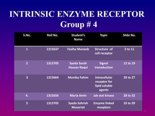
INTRINSIC ENZYME RECEPTOR
Group # 4
S.No. Roll No. Student’s
Name
Topic Slide No.
1 1315637 Fasiha Muneeb Structure of
cell receptor
2 to 11
2 1315705 Syeda Sarah
Hassan Naqvi
Signal
transduction
12 to 19
3 1315664 Muniba Fahim Intracellular
receptor for
lipid soluble
agents
20 to 27
4. 1315656 Maria Amin Jak stat kinase 28 to 32
5 1315705 Syeda Sahrish
Musarrat
Enzyme linked
receptors
33 to 39
2. STRUCTURE OF CELL
RECEPTOR
What is receptor ?
A receptor is a protein molecule usually found inside or on
the surface of a cell that receives chemical signals from
outside the cell.
When such chemical signals bind to a receptor, they cause
some form of cellular/tissue response, e.g. change in the
electrical activity of the cell.
Therefore, a receptor is a protein molecule that recognizes
and responds to endogenous chemical signals,
e.g. the acetylcholine receptor recognized and responds to its
endogenous ligand, acetylcholine.
2
3. However sometimes, the term is also used to
include other proteins that are drug targets,
such as enzymes, transporters and ion
channels.
3
4. Receptor proteins are embedded in either the
cell's plasma membrane (cell surface receptors),
the cytoplasm (cytoplasmic receptors), or in
the nucleus (nuclear receptors).
A molecule that binds to a
receptor is called a ligand,
and can be a peptide
(short protein) or another
small molecule such as a
neurotransmitter, hormone,
pharmaceutical drug, or toxin.
4
5. The endogenously designated molecule for a
particular receptor is referred to as its
endogenous ligand. E.g. the endogenous ligand
for the nicotinic acetylcholine receptor is
acetylcholine but the receptor can also be
activated by nicotine and blocked by curare. Each
receptor is linked to a specific cellular biochemical
pathway.
While numerous receptors are found in most
cells, each receptor will only bind with ligands of a
particular structure, much like how locks will only
accept specifically shaped keys. When a ligand
binds to its corresponding receptor, it activates or
inhibits the receptor's associated biochemical
pathway.
5
6. TYPES OF RECEPTORS
The structures of receptors are very diverse and can
broadly be classified into the following catagories.
1. Type 1: L (ionotropic receptors) :
These receptors are typically the targets of fast
neurotransmitters such as acetylcholine (nicotinic) and
GABA and activation of these receptor results in
changes in ion movement across the membrane.
They have a hetero structure. Each subunit consists of
the extracellular ligand-binding domain and a
transmembrane domain where the transmembrane
domain in turn includes four transmembrane alpha
helixes. The ligand binding cavities are located at the
interface between the subunits.
6
7. 2. Type 2: G protein coupled receptors (metabotropic)
This is the largest family of receptors and includes the
receptors for several hormones and slow transmitters
e.g. dopamine, metabotropic glutamate.
They are composed of seven transmembrane alpha
helices. The loops connecting the alpha helices form
extracellular and intracellular domains.
The binding site for larger peptidic ligands is usually
located in the extracellular domain whereas the
binding site for smaller non-peptidic ligands is often
located between the seven alpha helices and one
extracellular loop.
These receptors are coupled to different intracellular
effector systems via G-proteins.
7
9. 3. Type 3: kinase linked and related receptors :
These receptors are composed of an
extracellular domain containing the ligand
binding site and an intracellular domain, often
with enzymatic function, linked by a single
transmembrane alpha helix. E.g. the insulin
receptor.
4. Type 4: nuclear receptors:
While they are called nuclear receptors, these are
actually located in the cytosol and migrate to the
nucleus after binding with their ligands.
They are composed of a C-terminal ligand binding
region, a core DNA-binding domain (DBD) and
an N-terminal domain that contains
the AF1(activation function 1) region.
9
10. The core region has two zinc fingers that are
responsible for recognizing the DNA sequences
specific to this receptor.
The N-terminal interacts with other cellular
transcription factors in a ligand independent
manner and depending on these interactions it
can modify the binding/activity of the
receptor.
Steroid and thyroid hormone receptors are
examples of such receptors.
10
12. Signal Transduction
• Signal transduction is also known as cell signaling.
• It is the transmission of molecular signals from a
cell's exterior to its interior
• Signals received by cells must be transmitted
effectively into the cell to ensure an appropriate
response.
• This step is initiated by cell-surface receptors.
• Signal transduction occurs when an extracellular
signaling molecule activates a specific receptor
located on the cell surface or inside the cell.
12
14. • In turn, this receptor
triggers a biochemical
chain of events inside the
cell, creating a response.
• Depending on the cell, the
response alters the
cell's metabolism,
shape, gene expression, or
ability to divide.
• The signal can be amplified
at any step. Thus, one
signaling molecule can
cause many responses.
14
15. Products For Signal Transduction
• Calcium Signaling
• Complement
• Cytokine and NF-κB Signaling
• G Proteins (Heterotrimeric)
• G Proteins (Small)
• Gap Channels
• Growth Factor Receptors
• Heat Shock Proteins
• Hedgehog Signaling
• Inositol and cAMP Signaling
• MAPK Signaling
• Nitric Oxide Signaling
• Notch Signaling
• PI 3-Kinase/Akt Signaling
• Post-translational Modifications
• Proteasome
• Transcription Factors
• Translocation, Exocytosis & Endocytosis
• Wnt Signaling
15
16. Signal Transduction Pathways
• Transmission is continued either by a series of biochemical changes
within the cell or by modification of the cell membrane potential by
the movement of ions in or out of the cell.
• Receptors that initiate biochemical changes can do so either
directly via intrinsic enzymatic activities within the receptor or by
activating intracellular messenger molecules.
• Signal transducing receptors are of four general classes:
• Receptors that penetrate the plasma membrane and have intrinsic
enzymatic activity or are enzyme associated (Enzyme-linked
Receptors)
• Receptors that are coupled, inside the cell, to G proteins (7-TM
Receptors)
• Receptors that are found intracellularly and upon ligand binding
directly alter gene transcription (Nuclear Receptors)
• Ligand-gated ion channels
16
18. • The intracellular component of signal transduction is highly
receptor specific, thereby maintaining the specificity of the
incoming signal inside the cell.
• Signal transduction pathways amplify the incoming signal by a
signaling cascade using a network of enzymes.
• These enzymes act on one another in specific ways to ultimately
generate a precise and appropriate physiological response by the
cell.
• Signal transduction involves altering the behavior of proteins in the
cascade, in effect turning them on or off like a switch.
• Adding or removing phosphates is a fundamental mechanism for
altering the shape, and therefore the behavior, of a protein.
• Several small molecules within the cell act as intracellular
messengers (also known as second messengers). These include
cAMP, cGMP, nitric oxide, lipids and Ca2+ ions.
• Activated receptors stimulate second messenger production, which
in turn activate other enzymes and so the cascade continues.
18
20. Intracellular receptor for lipid
soluble agents
Receptors for steroid and thyroid
hormones are located inside
target cells, in the cytoplasm or
nucleus, and function as ligand-
dependent transcription factors.
That is to say, the hormone-
receptor complex binds to
promoter regions of responsive
genes and stimulates or
sometimes inhibits transcription
from those genes.
The mechanism of action of
steroid hormones is to modulate
gene expression in target cells. 20
21. Structure of Intracellular Receptors
• these receptors are composed of a
single polypeptide chain that has,
in the simplest analysis, three
distinct domains:
• The amino-terminus: In most cases,
this region is involved in activating
or stimulating transcription by
interacting with other components
of the transcriptional machinery.
The sequence is highly variable
among different receptors.
• DNA binding domain: Amino acids
in this region are responsible for
binding of the receptor to specific
sequences of DNA.
• The car boxy-terminus or ligand-
binding domain: This is the region
that binds hormone
21
22. Hormone-Receptor Binding and
Interactions with DNA
• Receptor activation is the term used to describe
conformational changes in the receptor induced by
binding hormone. The major consequence of activation
is that the receptor becomes competent to bind DNA.
• Activated receptors bind to "hormone response
elements", which are short specific sequences of DNA
which are located in promoters of hormone-responsive
genes. In most cases, hormone-receptor complexes bind
DNA in pairs, as shown in the figure below.
• Transcription from those genes to which the receptor is
bound is affected. Most commonly, receptor binding
stimulates transcription. The hormone-receptor complex
thus functions as a transcription factor.
22
24. Example
• As a specific
example, consider
glucocorticoids, a type of
steroid hormone that
probably affects the
physiology of all cells in
the body. The image to
the right depicts a pair of
glucocorticoid receptors
(blue and green on the
top) bound to their DNA
hormone response
element (bottom). The
two steroid hormones are
not visible in this
depiction.
24
25. Glucocorticoid Action
1. GR exists in an inactive form in the cytoplasm
complexed with heat shock protein 90 (hsp90).
2. Glucocorticoid (G) diffuses across cell
membrane and enters cytoplasm
3. G binds to GR changes conformation
dissociates from hsp90
4. exposes a nuclear localization signal on GR.
5. G-GR (hormone-receptor complex, HR) enters
nucleus, dimerizes with another HR.
25
26. 6. HR dimmer binds to
enhancer/hormone-
response element
upstream of
hormone activated
7. Binding of HR
dimmer to enhancer
activates
transcription
8. Most contain 2 zinc
fingers (1) controls
DNA binding, (2)
controls
demonization
26
28. JANUS KINASE
JAK Kinase is a family of
intracellular,nonA-receptor tyrosine
kinases that transduced cytokine mediated
signals via JAK-STAT pathway.
These receptors differ in not having any
catalytic domain
.Mammals have four members of this family
jak1, jak2, jak3, and tyrosine kinase 2.
JAK STAT KINASE
BINDING RECEPTOR
28
29. They were initially named as just another
kinase.
Janus kinase possess two near identical
phosphate transferring domain.
One exhibit kinase activity while another
negatively regulates the kinase activity of the
first.
29
30. JAK-STAT SIGNALLING
Cytokine receptor typically consist of two
chains each having an extracellular cytokine
binding domain and an intra-cytoplasmic
domain.
It binds a member of family of protein tyrosine
kinases called as janus kinases or JAK’S.
Cytokine binding to the receptor stabilizes the
dimmer.
30
32. Jaks are brought together that are bound
to cytoplasmic portion of each chain.
Janus kinase phosphorylate the
cytoplasmic tails of cytokine receptor.
STAT binds to phosphorylated cytokines rp
chains and themselves phosphorylated by
JAKS.
STAT dimmerizes and migrate to the
nucleus where they can directly activate
gene transcription.
32
34. •also known as a catalytic receptor
•transmembrane receptor, where the
binding of an extracellular ligand
causes enzymatic activity
on the intracellular side
•integral membrane
protein possessing
both enzymatic catalytic
and receptor functions
•Upon ligand binding a
conformational change is
transmitted which activates
the enzyme, initiating signaling cascades
34
35. Physiology and diseases
• involved in growth, proliferation,
differentiation, or survival
• Because of this, their ligands are
collectively called growth factors.
• The effects of enzyme-linked receptors
typically are slow requiring the
expression of new genes
• Mutations in receptor tyrosine kinases
are responsible for a wide array of
diseases, including cancers,
neurodegeneration, achondroplasia
and atherosclerosis. 35
36. This lecture will focus on:
1. Receptor tyrosine kinases
2. Tyrosine kinase-associated receptors
3. Receptor serine/threonine kinase
36
37. 1. Receptor serine/threonine kinases
There are two types of serine/threonine kinase
receptors, both of which contain an intracellular
kinase domain. They are each dimeric proteins, so
an active receptor complex is made up of four
receptors.
1. Type I receptors
• Inactive unless in complex with type II receptors.
• Do not interact with ligand dimers.
• Contain conserved sequences of serine and
threonine residues near to their kinase domains.
2. Type II receptors
• Constitutively active kinase domains (even in the
absence of the bound ligand).
• Able to phosphorylate and activate the type I
receptor. 37
38. 2. Receptor tyrosine kinases (RTKs)
• RTK ligands, such as fibroblast growth factor (FGF),
epidermal growth factor (EGF), nerve growth factor (NGF)
etc. bind as dimers.
• Ligand binding to RTK monomers results in dimer
formation.
• Receptors possess an intracellular tyrosine kinase domain.
Within the dimer the conformation is changed, locking the
kinase into an active state.
• The kinase of one receptor then phosphorylates a tyrosine
residue contained in the "activation lip"of the second
receptor.
• This forces the activation lip out of the kinase active site,
allowing ATP bind and resulting in enhanced kinase activity.
• This induces phosphorylation at further tyrosine residues.
• Phosphotyrosine is a conserved "docking site" for many
intracellular signal transduction proteins that contain SH2
domains 38
39. 3. Tyrosine-kinase-associated
receptors
• Cytokines are the main ligands that signal through
tyrosine kinase-associated receptors.
• The intracellular side of each receptor is bound to a
cytosolic tyrosine kinase protein.
1. Cytokines bind simultaneously to two receptor
monomers.
2. This brings the two associated kinases closer
together.
3. One kinase phosphorylates the other kinase in an
area called the "activation lip" (similar to RTK
activation).
The activation lip moves out of the active site and
binds ATP therefore enhancing kinase activity.
4. The enhanced kinase phosphorylates more tyrosine
residues on the intracellular portion of the receptor.
5. Phosphotyrosines serve as "docking sites" for SH2
domain-containing proteins 39