Digestion & absorption of carbohydrate
•Télécharger en tant que PPTX, PDF•
95 j'aime•12,501 vues

DIGESTION & ABSORPTION OF CARBOHYDRATE

Recommandé
Contenu connexe
Tendances
Tendances (20)
Similaire à Digestion & absorption of carbohydrate
Similaire à Digestion & absorption of carbohydrate (20)
Plus de sakina hasan
Plus de sakina hasan (12)
Dernier
Dernier (20)
Digestion & absorption of carbohydrate
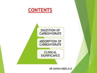
- 1. CONTENTS DIGESTION OF CARBOHYDRATE ABSORPTION OF CARBOHYDRATE CLINICAL SIGNIFICANCE DR SAKINA MBBS,M.D
- 2. Biological Importance Food large molecules small molecules Digestion small molecules Absorption vitamins, minerals, monosaccharides and free amino acids BLOOD
- 3. Carbohydrates present in the diet Polysaccharides Disaccharides Monosaccharides Starch Glycogen Lactose Maltose Sucrose Glucose Fructose Pentose In GIT, all complex carbohydrates are converted to simpler monosaccharide form which is the absorbable form.
- 4. Details of digestion of carbohydrates 2 Types of enzymes are important for the digestion of carbohydrates Amylases Disaccharidases convert polysaccharides to disaccharides Salivary Amylase Pancreatic Amylase Convert disaccharides to monosaccharides which are finally absorbed Maltase Sucrase-Isomaltase Lactase Trehalase
- 5. Digestion in mouth Digestion in stomach Digestion in small intestine
- 6. Digestion in the Mouth Digestion of Carbohydrate starts in the mouth, upon contact with saliva during mastication. Saliva contains a carbohydrate splitting enzyme called salivary amylase , also known as ptylin.
- 7. Action of ptylin (salivary amylase) Location: mouth It is α-amylase and requires Cl− ion for activation with an optimum pH of 6.7 (Range 6.6 to 6.8). The enzyme hydrolyses α-1→ 4 glycosidic linkages deep inside polysaccharide molecules. However, ptylin action stops in the stomach when the pH falls to 3.0.
- 8. Starch, Glycogen and dextrins (Large polysaccharide molecules) α- Amylase Glucose,Maltose and Maltotriose. (Smaller molecules)
- 9. Drawback Shorter duration of food in mouth. Thus it is incomplete digestion of starch or glycogen in the mouth
- 10. Digestion in the Stomach There is no enzyme to break the glycosidic bonds in gastric juice. However, HCl present in the stomach causes hydrolysis of sucrose to fructose and glucose. HCl Sucrose Fructose + Glucose
- 11. Digestion in Duodenum Food bolus reaches the duodenum from the stomach where it meets the pancreatic juice. Pancreatic juice contains a carbohydrate splitting enzyme, pancreatic amylase (amylopsin) similar to salivary amylase.
- 12. Action of pancreatic amylase It is an α- Amylase Optimum pH=7.1 Like ptylin, it requires Cl− ion for its activity. It hydrolyses α-1→ 4 glycosidic linkages situated well inside polysaccharide molecules. Note: Pancreatic amylase, an isoenzyme of salivary amylase, differs only in the optimum pH of action. Both the enzymes require Chloride ions for their actions (Ion activated enzymes).
- 13. Reaction catalyzed by pancreatic amylase Starch/Glycogen Pancreatic Amylase Maltose/ Isomaltose + Dextrins and oligosaccharides
- 14. Digestion in Small Intestine Note: Main digestion takes place in the small intestine by pancreatic amylase Digestion is completed by pancreatic amylase because food stays for a longer time in the intestine.
- 15. What are Disaccharidases? They are present in the brush border epithelium of intestinal mucosal cells where the resultant monosaccharides and others arising from the diet are absorbed. The different disaccharidases are : 1) Maltase, 2) Sucrase-Isomaltase (a bifunctional enzyme catalyzing hydrolysis of sucrose and isomaltose) 3) Lactase
- 16. Reactions catalyzed by Disaccharidases Maltase Maltose Glucose + Glucose Sucrase Isomaltase Sucrose Isomaltose 3Glucose + fructose Lactase Lactose Glucose + Galactose
- 17. Clinical significance of Digestion Lactose intolerance is the inability to digest lactose due to the deficiency of Lactase enzyme. Causes Congenital Acquired during lifetime Primary Secondary
- 18. Congenital Lactose intolerance It is a congenital disorder There is complete absence or deficiency of lactase enzyme. The child develops intolerance to lactose immediately after birth. It is diagnosed in early infancy. Milk feed precipitates symptoms.
- 19. Primary Lactase deficiency Primary lactase deficiency develops over time There is no congenital absence of lactase but the deficiency is precipitated during adulthood. The gene for lactose is normally expressed upto RNA level but it is not translated to form enzyme. It is very common in Asian population. There is intolerance to milk + dairy products.
- 20. Secondary lactase deficiency It may develop in a person with a healthy small intestine during episodes of acute illness. occurs because of mucosal damage or from medications resulting from certain gastrointestinal diseases, common cause of temporary lactose intolerance is gastroenteritis, particularly when the gastroenteritis is caused by rotavirus. Secondary lactase deficiency also results from injury to the small intestine that occurs with celiac disease, Crohn’s disease, or chemotherapy.
- 21. Clinical manifestations In the form of abdominal cramps, distensions, diarrhea, constipation, flatulence upon ingestion of milk or dairy products Biochemical basis Undigested lactose in intestinal lumen is acted upon by bacteria and is converted to CO2 , H2 , 2 carbon compounds and 3 carbon compounds or it may remain undigested.
- 22. CO2 and H2 causes Distensions and flatulence Lactose + 2C + 3C are osmotically active. They withdraw H2O from intestinal mucosal cell and cause osmotic diarrhea or constipation because of undigested bulk. Abdominal distension Flatulence
- 23. Diagnosis Two tests are commonly used: - Hydrogen Breath Test The person drinks a lactose-loaded beverage and then the breath is analyzed at regular intervals to measure the amount of hydrogen. Normally, very little hydrogen is detectable in the breath, but undigested lactose produces high levels of hydrogen. The test takes about 2 to 3 hours.
- 24. Stool Acidity Test The stool acidity test is used for infants and young children to measure the amount of acid in the stool. Undigested lactose creates lactic acid and other short chain fatty acids that can be detected in a stool sample. Glucose may also be present in the stool as a result of undigested lactose. Besides these tests, urine shows- positive test with Benedict’s test, since lactose is a reducing sugar and a small amount of lactose is absorbed in the intestinal cell by pinocytosis and is rapidly eliminated through kidneys in to urine.(Lactosuria) Mucosal biopsy confirms the diagnosis.
- 25. Sucrase-Isomaltase deficiency These 2 enzymes are synthesized on a single polypeptide chain,hence , their deficiencies coexist. Signs and symptoms Same as that of lactose intolerance. Urine does not give +ve test with Benedict’s test because of sucrose(Non reducing sugar). History confirms the diagnosis. Most confirmatory test is mucosal biopsy.
- 26. Absorption of carbohydrates 3 mechanisms Passive diffusion Facilitated diffusion/Carrier mediated Active transport
- 27. Features Passive diffusion Facilitated diffusion Active transport Concentration gradient Down the concentration gradient from high to low. Down the concentration gradient from high to low. Against a concentration gradient from low to high Energy expenditure none none Energy expenditure is in the form of ATP Carrier protein/ transporter Not required required required Speed Slowest mode Fast Fastest mode Note: Glucose is a polar molecule. It cannot pass through lipid bilayer of cell.
- 28. Glucose transporters Glucose transporters Na+ dependent transporter Na+ independent transporter 2 types Also called Also SGLT GLUT called
- 29. Absorption of Monosaccharides The major monosaccharides resulting from carbohydrate digestion are – D-glucose, D-galactose and D-fructose. Absorption is carrier mediated. Pentoses are absorbed by simple diffusion. Monosaccharides are first transported from the lumen to the small intestinal epithelial cells and then into capillaries of portal venous system.
- 30. Absorption of Glucose from the small intestinal lumen by carrier mediated mechanism involving transporter proteins 1) Na+-dependent transporter by secondary active transport and to a less extent by 2) Na+-independent transporter by passive transport into the intestinal epithelial cells
- 31. Absorption of Glucose facilitated transport secondary active transport Intestinal Epithelial Cell Glucose Glucose Glucose GLUT-5 Intestinal Lumen Na+ Na+-dependent transporter (SGLT) Na+ K+ Na+ K+ GLUT-2 Portal Capillary Blood ATP ADP + Pi Na+–K+ ATPase
- 32. Absorption of Glucose Galactose GLUT-5 Na+ Na+-dependent transporter (SGLT) Na+ K+ Na+ K+ GLUT-2 ATP ADP + Pi Na+–K+ ATPase Galactose Fructose Fructose Galactose Fructose
- 33. Factors affecting rate of absorption of Monosaccharides The absorption is faster through intact mucosa. The absorption is decreased if there is some inflammation or injury to the mucosa. Thyroid hormones ↑ the rate of absorption of glucose. Mineralocorticoid,i.e Aldosterone ↑ the rate of absorption.
- 34. Vitamin B6,B12, pantothenic acid, folic acid are required for absorption of glucose. With advancing age, rate of absorption declines.
- 35. Clinical significance In deficiency of SGLT- 1, glucose is left unabsorbed and is excreted in feces. Galactose is also malabsorbed. In deficiency of SGLT- 2, the filtered glucose is not reabsorbed back, it is lost in urine, causing glycosuria.
- 36. THANK YOU