

Recommandé
Contenu connexe
Tendances
Tendances (20)
Similaire à Respiratory system
Similaire à Respiratory system (20)
Plus de Salah Nazar
Plus de Salah Nazar (20)
Dernier
Dernier (20)
Respiratory system
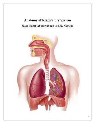
- 1. 1 Anatomy of Respiratory System Salah Nazar Abdulwahhab M.Sc. Nursing
- 2. 2 The Respiratory System The major functions of the respiratory system are: 1. Inhalation and exhalation (breathing). It is supplying oxygen O2 to the cells and removing their gaseous waste product (carbon dioxide) CO2. 2. External respiration exchanges gases between the lungs and the bloodstream 3. Internal respiration exchanges gases between the bloodstream and body tissues 4. Air vibrating to create sound from the vocal cords 5. Olfaction (smelling)
- 3. 3 The Respiratory System Organs 1 Upper respiratory tract (outside thorax) Nose and nasal cavity Paranasal sinuses Pharynx Larynx 2 Lower respiratory tract (within thorax) Trachea Bronchi and their smaller branches Lungs, which contain the terminal air sacs (alveoli) The Nose The nose is the only visible part of the respiratory system jutting external portion is supported by bone and cartilage. Internal nasal cavity is divided by midline (nasal septum) and lined with mucosa. The openings in the nose called the nostrils. The nose located between the roof of the mouth and the cranium. Roof of nasal cavity contains olfactory epithelium (Receptors for sense of smell) The nasal cavity produces mucus; filters, warms, and moistens incoming air; resonance chamber for speech
- 4. 4 Paranasal Sinuses • They are air filled spaces lined by mucous membrane present around and communicate with the nasal cavity. • These are pairs of maxillary, ethmoidal, frontal, sphenoidal sinuses. Same functions of nasal cavity; also lighten skull The Pharynx Passageway connecting nasal cavity to the larynx and oral cavity to esophagus containing of three parts: 1. Nasopharynx cavity 2. Oropharynx cavity 3. Laryngopharynx cavity Containing tonsils (lymphoid tissue masses involved in protection against pathogens.
- 5. 5 The Larynx An air passageway connects pharynx to trachea, has framework of cartilage and connective tissue. Opening (glottis) can be closed by epiglottis to prevents food from entering lower respiratory tract and containing vocal cords responsible of voice production. The Trachea (Windpipe) It is flexible tube running from larynx at the level of C6 and dividing inferiorly into two main bronchi right and left at the lower border of T6. Walls of trachea contain C-shaped rings cartilages that are incomplete posteriorly where connected by tracheal muscles. The trachea is air passageway; cleans, warms, and moistens incoming air The Bronchial tree Consists of right and left main bronchi, which subdivide within the lungs to form bronchi and bronchioles, bronchiolar walls lack cartilage but contain complete layer of smooth muscle. Constriction of this muscle impedes breathing. Considered Air passageways connecting trachea with alveoli; cleans, warms, and moistens incoming air The Alveoli Microscopic chambers at termini of bronchial tree. Walls of simple squamous epithelium are underlain by thin basement membrane. External surfaces are intimately associated with pulmonary capillaries. Alveoli considered the main sites of gas exchange.
- 6. 6 The Lungs The lungs are pyramid-shaped of spongy air-filled organs that are connected to the trachea by the right and left bronchi, the lungs are bordered by the diaphragm. The diaphragm is the flat, dome-shaped muscle located at the base of the lungs and thoracic cavity the lungs are enclosed by the pleurae. Each lung is composed of smaller units called lobes. Fissures separate these lobes from each other. The right lung consists of three lobes: the superior, middle, and inferior lobes. The left lung consists of two lobes: the superior and inferior lobes. Pleura of the Lungs It is Serous membranes, Parietal pleura lines thoracic cavity and visceral pleura covers external lung surfaces. Produce lubricating fluid acts as a lubricant allowing the lungs to slip smoothly as they expand and contract with each breath