
Recommandé
Contenu connexe
Tendances
Tendances (20)
En vedette
En vedette (20)
Similaire à 4to 9 human reproduction for students
Similaire à 4to 9 human reproduction for students (20)
Plus de Andrea Sánchez del Rio
Plus de Andrea Sánchez del Rio (13)
Dernier
Dernier (20)
4to 9 human reproduction for students
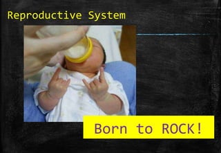
- 1. Reproductive System Born to ROCK!
- 2. 1) Single celled creatures -> divide into two 2) Multi cellular -> produce offspring by breaking away a part of their bodies. •Fission In binary fission the parent organism is replaced by two daughter organisms, because it literally divides in two •Budding Some cells split, resulting in a 'mother' and 'daughter‘ cell. The offspring organism is smaller than the parent. •Spore formation These spores grow into multicellular individuals without a fertilization event •Fragmentation a new organism grows from a fragment of the parent. •Regeneration the process of producing new individuals ASEXUAL SEXUAL
- 3. ▪ Two sexes: each produce reproductive cells called gametes ▪ Male: produces sperms (spermatozoa). Male always produce a large number of them. ▪ Female: ova (many ovum) or eggs. They are produced in a smaller and limited number. *much smaller *in larger quantities to increase the chance of successful fertilization. *Sperms feed on nutrients in the semen fluid * have long tails which helps them swim *has a large number of mitochondria *bigger *it stores nutrients. *one egg at a time. *Eggs can’t move WHY?
- 4. The Reproductive System * We need a Reproductive System so the human race will continue. This system is designed to *produce, nourish, and transport either the egg or sperm. *ensure fertilisation *look after a developing baby until birth. Back to contents
- 5. ▪ the fusion of two gametes, such as a sperm and an egg, that form a zygote which is capable of maturing into an adult individual of that species. ▪ Only one of the approximately 300 million sperm released into a female‘ s vagina during intercourse can fertilize the single female egg cell To produce a new individual, a sperm has to reach an ovum and fuse with it. (the nuclei) They form a zygote. It will grow by cell division to produce an embryo and then a fully formed animal.
- 6. *The sperms swim through the cervix and into the uterus, enter the oviduct. If there is an ovum in the oviduct, one of the sperms may enter *It passes through the thick coating surrounding the egg the zona pellucida ( sugars and proteins) *The tip of the head of the sperm cell contains enzymes which break through the zona pellucida and aid the penetration of the sperm into the egg *It enters the cytoplasm of the ovum male nucleus of the sperm fuses with the female nucleus. •Once the head of the sperm is inside the egg, the tail of the sperm falls off *The perimeter of the egg thickens to prevent another perm fromentering. *The released ovum survives for about 24 hs. Fertilization is able to happen for about 2 or 3 days (middle life od a sperm)
- 7. •The eggs are produced from the female reproductive organs called the ovaries. •3-4 cm long, the ovaries lie on each side of the uterus. •Close to the ova is the opening of the oviduct, the tube down which the ova pass when released from the ovary. (Sometimes called the Fallopian tube). • The oviducts are narrow tubes that open into the uterus, or womb, lower down in the abdomen. • The uterus leads to the outside through a muscular tube, the vagina. • The cervix is a ring of muscle closing the lower end of the uterus where it joins the vagina. • The urethra, from the bladder, opens into the vulva just in front of the vagina.
- 8. hymen
- 9. The ova are present in the ovary from the time of birth. No more are formed during the lifetime. Between the ages of 10 and 14 some of the ova are released, one at a time from alterntate ovaries about every 4 weeks. As each ovum matures, the cells round it divide rapidly and produce a fluid-filled sac, called follicule. When mature, it projects from the surface of the ovary and finally it bursts and releases the ovum into the funnel of the oviduct. The released ovum is enclosed in a jelly-like coat called the zona pellucida and is still surrounded by a layer of follicle cells. For fertilization to take place, the sperm has to get through this layer and penetrate the zona. In humans, usually only one egg is fertilized at a time; two eggs being fertilized produces (non-identical) twins.
- 11. The ovaries release an ovum about every 4 weeks. The oestrogens act on the uterus and cause its lining to become thicker and develop more blood vessels. Two hormones, the follicle-stimulating and the luteinizing, produced by the pituitary gland at the base of the brain promote ovulation. Once the ovum has been released, the follicle which produced it develops into a solid body called the corpus luteum. This produces a hormone called progesterone, which affects the uterus lining in the same way as the oestrogens. If the ovum is fertilized the corpus luteum continues to release progesterone and so keeps the uterus in a state suitable for implantation. If the ovum is not fertilized, the corpus luteum stops producing progesterone. As a result, the thickened lining of the uterus breaks down and loses blood which escapes through the cervix and the vagina (menstrual period).
- 12. Ovarian Cycle 2 Phases 1- Follicular phase (1-14) • ovarian follicles mature, getting ready to release an egg • a rise in Follicle Stimulating Hormone (FSH) • follicle that reaches maturity Graafian follicle •Lining of OVULATION the uterus thickens (14) •OESTROGEN • Ovulation a mature egg is released •levels of ESTRADIOL reach a threshold, estrogen stimulates the production of a large amount of LUTEINIZING HORMONE (LH) 2- Luteal phase (14-28) •FSH and LH remaining parts of the follicle transform into the CORPUS LUTEUM, which produces PROGESTERONE BUT suppress production of the FSH and LH •the corpus luteum atrophies Falling levels of PROGESTERONE trigger
- 13. The Male Reproductive System Back to contents
- 14. Male Reproductive System The purpose of the organs of the Male Reproductive System is to perform the following functions: * To produce, maintain, and transport SPERM(the male reproductive cells) and protective fluid (semen) •To discharge sperm within the female reproductive tract during sex as close to the uterus as possible •To produce and secrete male sex hormones responsible for maintaining the male reproductive system
- 15. • Sperms are produced in the male reproductive organs called testes . • They lie outside the abdominal cavity in a special sac called scrotum, so as to keep a lower temperature for sperm production. • They consist of a mass of sperm-producing tubes. These join to form ducts leading to the epididymis (coiled tube about 6m long on each side of the testis storage of sperms). This leads into a muscular sperm duct .
- 16. The two sperm ducts open into the top of the urethra just after it leaves the bladder. The seminal vesicle is a short coiled tube which branches from each sperm duct just after entering the prostate gland . The urethra passes through the penis . It consists of connective tissue with many blood spaces in it. This is called erectile tissue . 1 testis/ 2 testes
- 17. The function of the scrotum is to produce sperm and keep the testis at a temperature slightly lower than that of the rest of the body as the sperm dies when it is kept too warm. How does the Scrotum work When the whether is cool, the scrotum tightens to bring the testicles closer to the body, and when the weather gets warmer the scrotum loosen to hang the testicles further away from the body. Back to contents
- 18. The lining of the tubules of the testis consists of rapidly dividing cells. After a series of cell divisions, the cells grow long tails and become sperms which pass into the epididymis.
- 19. THE HEAD: has two important features: *The acrosome contains lytic enzymes which are released when the sperm reaches an ovum. These enzymes digest the outer membrane of the egg, allowing penetration of the sperm * The NUCLEUS (genetic material) THE MIDDLE SECTION:immediately behind the head, contains numerous mitochondria . These respire sugars in the semen to generate ATP to provide the energy for movement of the tail. THE TAIL contains microfilaments running the length of the tail .Rhythmic contraction of the filaments causes the tail to wave and move against the fluid environment, providing forward motion. Parts of the sperm
- 20. • Sexual arousal in the male results in an erection. The penis becomes firm and erect as a result of blood flowing into the erectile tissue. • During copulation, the male inserts the penis into the female’s vagina. This causes a reflex in the male which results in the ejaculation of semen. • The epididymis and sperm ducts contract and force sperms out of urethra. • The prostate gland and seminal vesicle add fluid to the sperms. This fluid added to the sperms is called semen.
- 21. Sperm AA Proteins Vitamin C TESTES SEMINAL VESICLE Citric acid Zinc Anti coagulant factors Enzymes Mucus liquid PROSTATE GLAND
- 23. The fertilized ovum divides into 2 cells, then 4, they continue dividing producing a solid ball of cells, an early stage in the development of the embryo. The embryo travels down the oviduct to the uterus, where is implanted to the walls of the uterus. After 8 weeks, when all the organs are formed, the embryo is called a fetus. One of the first organs to form is the heart. Inside the uterus the embryo becomes enclosed in a fluid-filled sac called the amnion sac, which protects it from damage and prevents unequal pressures from acting on it.
- 24. Some cells, instead of forming the organs of the embryo, grow into a disc-like structure called the 1- Provides the oxygen and food needed to keep the embryo alive and growing are obtained from the mother's blood It is attached to the * lining of the uterus *embryo by a tube called the umbilical cord. The blood vessels in the placenta are very close to the blood vessels in the uterus so that oxygen, glucose, amino acids and salts can pass from the mother's blood to the embryo's blood. The carbon dioxide and urea in the embryo's blood escape from the vessels in the placenta and are carried away by the mother's blood in the uterus. 2- It can prevent some harmful substances in the mother's blood from reaching the embryo. 3- Placenta produces hormones(OESTROGENS/PROGESTERONE) that play an important part in maintaining the pregnancy and preparing for birth. There is NO DIRECT COMMUNICATION BETWEEN THE MOTHER’S BLOOD AND THE EMBRYO’S
- 27. It is the way a woman should look after herself during pregnancy, so that the birth will be safe and her baby healthy. She must take more iron and folic acid than usually, so as to prevent anemia. SMOKE Pregnant women who drink or smoke are more likely to have babies with low birth weight or miscarriages ALCOHOL Heavy drinking is suspected of damaging the developing brain of the fetus and babies with low birth weight If a women catches RUBELLA during the first 4 months of pregnancy, there is a danger that the virus may affect the fetus and cause abortion/ still-birth or defects in the eyes/ears or nervous system
- 28. A woman can release two ova when she ovulates. If both are fertilized, they may form twin embryos, each with its own placenta and amnion. Twins formed in this way are called fraternal twins. As they are fertilized by different sperms, they can be a boy and a girl and are not more alike than other brothers and sisters. Another cause of twining is when a single fertilized egg forms two separate embryos. Sometimes this may share a placent and amnion. This type of twins must be the same sex, because only one sperm fertilized the ovum. These ‘one-egg’ twins are sometimes called identical twins.
- 30. 1º MONTH The baby is an embryo consisting of two layers of cells from which all her organs and body parts will develop.
- 31. 2º MONTH The baby is now about the size of a bean and is constantly moving. He has distinct, slightly webbed fingers.
- 32. 3º MONTH By now the baby is about 7.52cm long and weighs nearly 28g. Her tiny, unique fingerprints are now in place.
- 33. 4º MONTH The baby is now about 12.7cm long and weighs 141g. His skeleton is starting to harden from rubbery cartilage to bone.
- 34. 5º MONTH Eyebrows and eyelids are now in place. The baby would now be more than 25.4cm long if you stretched out her legs.
- 35. 6º MONTH The baby weighs more than 600g. His wrinkled skin is starting to smooth out as he puts on baby fat
- 36. 7º MONTH By now, the baby weighs about 1.5kg and is more than 38cm long. She can open and close her eyes and follow a light.
- 37. 8º MONTH The baby now weighs about 2kg. His layers of fat are filling him out, making him rounder, and his lungs are well developed.
- 38. 9º MONTH The average baby is more than 48.26cm long and weighs nearly 3.100kg now, but babies vary widely in size at this stage
- 39. The gestation period is from fertilization to birth, and it takes about 38 weeks. A few weeks before the birth, the fetus has come to lie head downwards in the uterus. When birth starts, the uterus begins to contract rhythmically (labour). These contractions become stronger and more frequent. The opening of the cervix widens enough to let the baby's head pass through and the contractions are assisted by muscular contractions of the abdomen. WATER SAC breaks and fluid escapes through the vagina The muscular contractions of the uterus and abdomen push the baby head-first through the cervix and vagina. The umbilical cord, which still connects the child to the placenta, is tied and cut. The placenta breaks away from the uterus and is pushed out.
- 40. •When the water sac breaks, there’s a sudden fall in • the temperature Stimulates the baby to take the first breath and CRY * When the remains of the umbilical cord falls There is a scar in the abdominal wall NAVEL ▪ If the pregnancy lasted more than 38 weeks or the placenta doesn’t cope with the demands of the fetus The birth is INDUCED (started artificially) by Breaking the membrane of the amniotic sac Inject OXYTOCIN (hormone) Amniocentesis Taking a sample of amniotic fluid to know if the baby has got Down’s syndrome or other conditions/ to find out the sex
- 41. The time has come!!!
- 42. * During pregnancy mammary glands enlarge increase in the number of milk-secreting cells but breasts are stimulated to release milk when the baby starts sucking * Liquid (with more proteins and antibodies) produced during the first days COLOSTRUM * WEANING process by which the baby starts eating solid food FEEDING Mother’s milk Cows’ milk proteins Fats Sugar Vitamins Salts Little iron Antibodies Right temperature + Free from bacteria Emotional / psychological benefits More proteins Sodium phosphorus Less Sugar Vitamins A and C NO Antibodies
- 43. Ova start to be released when the female reaches an age of about 10-14 years. Sex hormones are released into the bloodstream, called oestrogens. They bring the development of secondary sexual characteristics. In girls, there is an increased growth of breasts, widening of hips and growth of hair in pubic regions and armpits. Also an enlargement of the uterus and vagina. In boys, testosterona is released by testes into the bloodstream. Testes and penis are enlarged, voice is deepen, growth of hair in pubic parts, armpits, chest and face. There are also emotional and psychological changes.
- 44. Between 40 and 55, the ovaries cease to release ova or produce hormones. As a consequence, menstrual periods cease, the woman can no longer have children, and sexual desire is gradually reduced. Female infertility Usually caused by a failure to ovulate or a blockage or distortion of the oviducts. The latter can often be corrected by surgery, if not the couple may use ‘in vitro’ fertilization. Failure to produce ova can be treated with fertility drugs. They are similar to hormones and act by increasing the levels of FSH and LH. Their administration is aimed to promote ovulation to coincide with copulation.
- 45. ▪ Caused by an inadequate quantity of sperms in the semen or by sperms which are insufficiently mobile to reach the oviducts. ▪ Artificial Insemination involves injecting semen through a tube into the top of the uterus.
- 46. • The woman may be give fertility drugs to release several mature ova simultaneously. • The ova are collected by laparoscopy and sucked up in a fine tube inserted through the abdominal wall. • Then the ova are mixed with the husband’s seminal fluid and watched under the microscope to see if cell division take place. • One or more of the dividing zygotes are introduced to the woman’s uterus through the cervix by an inserted tube. • Usually one or none zygote develops though, occasionally, there are multiple births.