1. AMER. ZOOL., 22:795-805 (1982)
The Renewing Cell Populations of Ascidians1
THOMAS H. ERMAK
Department of Physiology, University of California,
San Francisco, California 94143
SYNOPSIS. Renewing cell populations are tissues or groups of cells which rapidly prolif-
erate and whose cell division is balanced by cell loss. The rapid proliferation of cells can
be determined in autoradiograms by the uptake of tritiated thymidine into DNA synthe-
sizing cells, and the migration and loss of cells can be followed by taking samples of tissues
at increasing time intervals after exposure to the radioisotope. In ascidians, renewing
populations are the testis, ovary, blood cells, and epithelial lining of the digestive tract.
They are made of subpopulations called compartments: 1) germinal compartments of
Downloaded from http://icb.oxfordjournals.org by Thomas Ermak on April 24, 2010
relatively undifferentiated, dividing stem cells (these cells are labeled at short time inter-
vals), 2) mature compartments of fully differentiated, nondividing cells (these become
labeled with time by the migration of stem cells), and 3) transitional compartments of cells
in intermediate stages. The stomach groove cell population is a model system for cell
differentiation and renewal in the digestive tract. Germinal compartments can be distin-
guished from mature compartments by their morphology (pseudostratified vs. simple
columnar epithelia), by the uptake of tritiated thymidine in autoradiograms, by histo-
chemical staining, by their surface features in the scanning electron microscope, and by
their intracellular organelles when viewed in the transmission electron microscope. Stem
cells differentiate into absorptive cells and zymogen cells. In differentiating, stem cells
increase in size and lose their abundant free ribosomes. Absorptive cells develop longer
microvilli, a long cilium, smooth apical vesicles, and large supranuclear lysosomes. Zym-
ogen cells produce abundant rough endoplasmic reticulum and numerous zymogen gran-
ules. The renewing cell populations of blood (lymph nodule) and digestive tract are im-
portant model systems for studying cell differentiation, morphogenesis, and the phylogeny
of vertebrate hemopoietic, immune, and digestive systems.
INTRODUCTION that the turnover of these renewing cell
From a developmental perspective, a re- populations could truly be established and
newing cell population is an adult tissue or followed. The pioneering work was per-
group of cells which maintains a subpop- formed by Leblond and his associates with
ulation of embryonic-like cells which con- a variety of radioisotopes to label dividing
tinually replace aging differentiated cells cells and follow their fate with time (Le-
during the lifetime of the animal. These blond and Stevens, 1948; Leblond and
renewing populations serve a variety of Walker, 1956). They found that tritium
functions in the body. For example, they gave the best resolution in autoradiograms
may form a protective barrier of cells and that thymidine preferentially labeled
against the external environment, such as replicating DNA. Although thymidine is
in the digestive tract or skin, or they may not normally a precursor of DNA, it can
provide a constant supply of a cellular nonetheless enter the biosynthetic pathway
product, such as the blood cells or ga- and label DNA during both pre-mitotic and
metes. pre-meiotic DNA synthesis (for review, see
Cleaver, 1967). When administered, the
The presence of these embryonic-like tritiated thymidine is available for only a
cells, today called stem cells (Cairnie et ai, short period of time, and only those cells
1976), in rapidly dividing tissues was rec- that are synthesizing DNA at the time of
ognized for years based upon histological injection and immediately afterwards are
observations. However, it was not until the labeled. After DNA synthesis, all cells pre-
use of autoradiography in the late 1940s sumably undergo cell division. The fate of
the DNA synthesizing cells can be followed
1
From the Symposium on The Developmental Biology in autoradiograms by taking samples of the
of the Ascidians presented at the Annual Meeting of tissues at increasing time intervals after the
the American Society of Zoologists, 27-30 December initial exposure period.
1981, at Dallas, Texas.
795
2. 796 THOMAS H . ERMAK
cell
extrusion
Downloaded from http://icb.oxfordjournals.org by Thomas Ermak on April 24, 2010
cell cell absorption
division differentiation and secretion
B
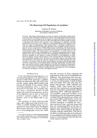
FIG. 1. The compartments of renewing cell populations as shown for a columnar epithelium. A. Germinal
compartment with mitotic figure along the lumenal edge. B. Transitional compartment. C. Mature compart-
ment of fully differentiated cells.
Based upon the uptake of tritiated thy- compartment system, and a third com-
midine and its change in cells with time, partment of transitional cells can be rec-
Leblond classified the cell populations of ognized as cells in the process of matura-
mammals into three categories: static, ex- tion. The stem cell compartment, also called
panding, and renewing cell populations the germinal compartment, is composed of
(Messier and Leblond, 1960). Static pop- relatively undifferentiated, rapidly divid-
ulations do not divide and expanding pop- ing cells that can also be subdivided ac-
ulations have a slow rate of proliferation. cording to their position in the cell cycle.
Renewing populations have a rapid rate of Cells undergoing mitosis (mitotic figures)
cell proliferation and most cell production can be morphologically distinguished from
is used for the replacement of lost cells cells in interphase (Fig. 1A). Interphase
rather than for growth. A large percentage cells undergoing DNA synthesis can be la-
of the cells are labeled with tritiated thy- beled with tritiated thymidine and detect-
midine, and with increasing time intervals ed with autoradiography, as explained
after exposure, the percentage of labeled above. Other cells in interphase, unlabeled
cells drops to zero due to loss of labeled in autoradiograms, are presumed to be in
cells from the tissue. In some tissues, one of two resting stages, one between mi-
migration of groups of labeled cells can ac- tosis and DNA synthesis and the other af-
tually be followed (for example, the epi- ter DNA synthesis and before mitosis.
thelium of the digestive tract or sperma- The mature compartment is composed
togenic cells). The system is thus in a steady of fully differentiated cells, such as absorp-
state with cell production balanced by cell tive or secretory cells of the digestive tract
loss. (Fig. 1C). These cells do not usually take
Renewing populations are made up of up tritiated thymidine (thus, they do not
several compartments based upon mor- usually divide). With time, they eventually
phological as well as kinetic criteria (Fig. become labeled by the migration and dif-
1). Stem cells and mature cells create a two ferentiation of stem cells. Aging mature
3. RENEWING CELL POPULATIONS OF ASCIDIANS 797
TABLE 1. Compartments of asddian renewing cell populations classified according to morphology and cell kinetics.
Cell population Type of cell Type of compartment
Testis Spermalogonia germinal
Spermatocytes dividing transitional
Spermatids transitional
Spermatozoa mature
Ovary Oogonia germinal
Primary oocytes dividing transitional
Oocytes I and II transitional
Oocytes III mature
Follicle cells dividing transitional
Downloaded from http://icb.oxfordjournals.org by Thomas Ermak on April 24, 2010
Test cells mature
Blood cells Hemoblasts germinal
Leukocytes mature
Digestive tract Basophilic cells germinal
Absorptive, zymogen and ciliated cells mature
* Each compartment originates from the preceding compartment except in the ovary, where germinal cells
give rise to both oocytes and follicle cells.
cells are lost from the population by cell the gonads occurs in all animal phyla (but
extrusion, cell death, or cell excretion. not all adult species) and the morphologi-
The time that elapses between a cell en- cal events characterizing cell differentia-
tering a compartment and that cell or its tion are familiar to most biologists. I will,
progeny leaving it is its transit time. I use therefore, only briefly review the kinetics
this term to denote the shortest time for a of renewal in ascidians. On the other hand,
labeled stem cell to migrate through the the renewal of blood cells and digestive
transitional and mature compartments. tract are less common, occurring mainly in
Renewing cell populations have been ex- the vertebrates and a few invertebrates.
tensively investigated in mammals and in- These populations provide important
clude the epidermis, the epithelial lining models for the differentiation of somatic
of the alimentary canal, the hemopoietic cells in the adult and are important sys-
cells of bone marrow, the lymphatic cells tems for studying the phylogeny of verte-
of thymus, spleen, and lymph nodes, the brate hemopoietic, immune (Wright, 1977),
spermatogenic cells, and the uterine and and digestive systems (Ermak, 1981). For
vaginal epithelial cells (Messier and Le- these populations, the kinetics of cell re-
blond, 1960). The list of renewing cell newal will be described as well as the over-
populations in ascidians is almost as im- all aspects of cell differentiation during
pressive: the epithelial lining of the diges- maturation and migration of cells.
tive tract (Ermak, 1975a, c,' 1976a, 1981),
the hemopoietic cells of lymph nodules and GONADS
circulating blood (Ermak, 19756, 1977), the In the gonads, germ cells in both pre-
spermatogenic cells (Ermak, 19766), and meiotic and pre-mitotic DN A synthesis can
the ovarian cells (Ermak, 19766). As in be labeled with tritiated thymidine and de-
mammals, these populations can be sub- tected with autoradiography (Ermak,
divided into germinal, transitional, and 19766). In the testis, each compartment is
mature compartments (Table 1). I have ex- in a direct transit line with the next. Sper-
amined the kinetics of cell renewal in a sin- matogonia (stem cells) form the basal layer
gle, solitary species, Styela clava (Ermak, around the circumference of the follicle.
1975a, b, c, 1976a, b, 1977), and compared Spermatocytes and spermatids form con-
them to those in a variety of other ascidian tinuous layers within the basal layer and
species (Ermak, 1977, 1981). Renewal of spermatozoa fill the lumen. In Styela, the
4. 798 THOMAS H. ERMAK
transit time from DNA synthesis in the pri- geal or atrial epithelium and along blood
mary spermatocyte to the appearance of channels.
labeled spermatozoa is about 10 days (Er- In Styela, blood cells are renewed within
mak, 19766). A one-hour exposure to tri- several weeks (Ermak, 19756). A one-hour
tiated thymidine labels many spermato- exposure to tritiated thymidine labels he-
gonia and spermatocytes, and after 10 days moblasts in the centers of nodules and in
spermatids and spermatozoa become la- circulating blood. By 20 days after expo-
beled. With even longer times (20-30 days), sure, cells in the peripheral parts of a nod-
labeled spermatozoa appear in the sperm ule become labeled whereas cells in the in-
ducts. terior are no longer labeled. By 60 days, a
Downloaded from http://icb.oxfordjournals.org by Thomas Ermak on April 24, 2010
In the ovary, the germinal epithelium nodule as well as most circulating blood
gives rise to oocytes in sequential stages cells are no longer labeled.
(usually noted as I, II, and III) as well as Although some differentiated blood cells
follicle cells surrounding the oocytes can apparently divide, most labeled cells
(Tucker, 1942; Kessel and Kemp, 1962). are probably hemoblasts. These cells are
Test cells are presumably derived from also sensitive to X rays (Freeman, 1964,
follicle cells. DNA synthesizing cells are 1970), a characteristic shared by stem cells
oogonia and preleptotene primary oo- of vertebrates (Patt and Quastler, 1963).
cytes. The smallest oocytes lie within the Most fully differentiated blood cells prob-
ovarian wall, and larger oocytes become ably do not divide. Vacuolated cells in Stye-
progressively displaced from the germinal la are not labeled at early time intervals
epithelium as they grow larger, become less (Ermak, 19756). In Perophora annectens,
basophilic, and acquire accessory cells. Al- compartment cells and phagocytes but not
though not followed for more than 60 days, other fully differentiated cells are report-
the total time period for oogenesis is prob- ed to be labeled with tritiated thymidine
ably on the order of several months (Er- (Freeman, 1970).
mak, 19766). At one hour to 20 days after The lymph nodule of Styela is a model
exposure, cells in the germinal epithelium system for the differentiation of blood cells
as well as follicle cells around the oocytes (Ermak, 1977). Hemoblasts are relatively
are labeled. By 60 days, a few stage I oo- undifferentiated, spherical cells which have
cytes and test cells are labeled. Test cells a large nucleus containing one or more nu-
are presumed to originate from follicle cells cleoli and little chromatin. The cytoplasm
whereas oocytes are presumed to originate is filled with numerous polyribosomes, a
from gonial cells in the ovarian wall as few cisternae of rough endoplastic reticu-
originally proposed by Tucker (1942). lum, and an occasional small, dense gran-
ule. A pair of centrioles (probably involved
BLOOD CELLS in centromere formation during mitosis) is
The hemopoietic tissue of ascidians is sometimes observed near the Golgi com-
organized into clusters of stationary cells plex.
called lymph nodules and circulating cells Differentiating leukocytes around the
(George, 1939; Millar, 1953). Lymph nod- central hemoblasts lose their prominent
ules occur in the pharyngeal wall, around nucleus as the amount of chromatin in-
the digestive tract, and in the body wall of creases (Ermak, 1977). Large electron
advanced species (Ermak, 1977). In prim- dense granules usually appear in the cy-
itive ascidians, hemopoietic tissue appears toplasm before the nuclear changes are
diffuse whereas in advanced species, it is completed. As one proceeds away from the
organized into distinct nodules. In Styela, center of a nodule, maturing blood cells
a nodule is composed of a few clusters of increase in size and their dense granules
dividing hemoblasts (the stem cells) sur- become larger and more numerous. Cell
rounded by non-dividing, maturing blood differentiation is also marked by the loss
cells (Ermak, 1977). Lymph nodules lie of polyribosomes and the development of
within connective tissue below the pharyn- elongate mitochondria, a larger Golgi
5. RENEWING CELL POPULATIONS OF ASCIDIANS 799
complex, and long cisternae of rough en- the underlying connective tissue through
doplasmic reticulum. the transition zone and up the sides of the
folds into the non-proliferating mature
DIGESTIVE TRACT zone of absorptive and zymogen cells. The
Most of the epithelia lining the digestive mature zone forms a simple, columnar ep-
tract are renewing cell populations. In Styeki ithelium and represents about 80% of the
they are the dorsal tubercle, dorsal lamina, height of the fold. Migrating labeled cells
branchial bars, zone 1 of the endostyle, reach the top of the fold in about 16 days
stigmata, esophagus, stomach, and intes- (Ermak, 1976a). Aging mature cells are
tine (Ermak, 1975c). In the postbranchial presumably extruded into the gut lumen
digestive tract of most other ascidians, re- at so-called "extrusion zones" at the top of
Downloaded from http://icb.oxfordjournals.org by Thomas Ermak on April 24, 2010
newing populations are also a characteris- the fold (at the junction with the crest pop-
tic feature of the esophageal and stomach ulation of ciliated mucous cells). Migration
epithelia, but expanding populations rates in other regions of the digestive tract
sometimes cover all or part of the intestine range from 10 days for the stigmata to 5
(Ermak, 1981). The renewing populations weeks in the intestine (Ermak, 1975c).
are adapted to different degrees of body Germinal regions can be distinguished
size, organization, and evolutionary ad- from mature regions at the light micro-
vancement, and with an increase in body scopic level with the use of histochemical
size, the digestive organs undergo exten- stains. With routine hematoxylin and eo-
sive folding and multiplication of pairs of sin, the stem cell is characterized by baso-
germinal and mature compartments (cell philic cytoplasm, and with toluidine blue
renewal units). Colonial ascidians gener- on epon sections, only the lysosomal re-
ally have a single cell renewal unit per cell gions of mature cells are stained. The ma-
population in their small organs whereas ture region also gives a strong positive re-
solitary ascidians have highly folded gut action for acid phosphatase3 which is absent
epithelia with multiplication of cell renew- from the cytoplasm of stem cells (Fig. 3).
al units (Ermak, 1981). The groove pop- The brush border at the base of the groove
ulation in the stomach of Styela provides a usually shows only a weak alkaline phos-
model system for cell renewal and differ- phatase reaction in comparison to the in-
entiation in the digestive tract. This epi- tense reaction on the sides of the folds (Er-
thelium covers the majority of each stom- mak, 1975a). Finally, the cytoplasm of the
ach fold as shown in the scanning electron germinal region takes up large amounts of
micrograph2 of the stomach wall (Fig. 2). tritiated leucine (Ermak, 1975a), a reflec-
Germinal zones occur at the base of each tion of active protein synthesis in these cells.
fold and form a pseudostratified columnar Germinal regions can also be distin-
epithelium (Ermak, 1975c). A one-hour guished from mature regions in surface
exposure to tritiated thymidine labels about view with the scanning electron micro-
10% of the groove height (Ermak, 1976a). scope (Fig. 4). Stem cells appear free of
Stem cell nuclei migrate to the cell apex surface organelles marking differentia-
and undergo mitosis there. Mitotic figures tion, namely cilia or long microvilli. In-
are labeled within several hours after ex- stead the germinal compartment is marked
posure (Ermak, 1975c). With time, stem by smooth, short microvilli. At the top of
cells migrate as a band of labeled cells over the fold, the "extrusion zone" can be iden-
- For scanning electron microscopy, stomach tissue
3
was fixed in 2% glutaraldehycle in 74% sea water for To test for the presence of acid phosphatase, tis-
24 hr (Holland and Jespersen, 1973; Nemanic and sues were prepared according to the standard cou-
Pitelka, 1971) and dried in a Freon critical point dryer. pling azo dye technique (Pease, 1968) using frozen
Specimens were coated with about 15 nm of gold- sections incubated at room temperature with sodium
palladium alloy and examined with a Stereoscan S4 a-naphthyl phosphate, poly vinyl pyrrolidone, and fast
scanning electron microscope. garnet CBC salt in 0.1 M acetate buffer at pH 5.0.
6. 800 THOMAS H. ERMAK
Downloaded from http://icb.oxfordjournals.org by Thomas Ermak on April 24, 2010
FIG. 2. Stomach folds of Styela clava as viewed with the scanning electron microscope. The stomach wall has
been cut across with a razor blade and viewed from within. On the cut surface of the folds, the demarcation
between the inner columnar epithelium and the underlying connective tissue and blood spaces is distinct.
Crest cell populations are conspicuous on the top of each fold, x 100.
FIG. 3. Section through the base of a stomach groove stained for acid phosphatase. Germinal cells are
relatively unreactive. Mature cells (probably lysosomal regions of absorptive cells) give a strong positive re-
action. X125.
Fit;. 4. The base of a stomach groove showing the surface features ot the germinal (g) and mature (in)
regions. Germinal /ones with short microvilli appear "naked" in comparison with mature regions with nu-
merous long cilia and longer microvilli. x 1,000.
7. RENEWING CELL POPULATIONS OF ASCIDIANS 801
tified as the junction between the groove
population and the ciliated crest cell pop-
ulation (Fig. 5).
The differentiation of stem cells into ab-
sorptive cells and zymogen cells (Fig. 6) has
been followed by transmission electron
microscopy4 in the stomach groove popu-
lation of S. clava (Ermak, 1975a; Thorn-
dyke, 1977) and on the stomach folds of
Ciona intestinalis (Thomas, 1970). These
mature cells appear to be characteristic of
Downloaded from http://icb.oxfordjournals.org by Thomas Ermak on April 24, 2010
the stomach and digestive diverticulum of
other ascidians as well (Degail and Levi,
1964; Burighel and Milanesi, 1973).
Stem cells (Fig. 6A) are smaller in size,
have a higher nucleus-to-cytoplasm ratio,
and are relatively undifferentiated in
structure (Fig. 7). Their most chracteristic
features, like those of the hemoblast in a
lymph nodule, are a nucleus containing a
prominent nucleolus and little chromatin
(Thorndyke, 1977) and numerous free ri-
bosomes in the cytoplasm (Fig. 8). The api-
cal ends of the cells bear several microvilli
which are shorter (about 3 /xm) and less
numerous than those on the absorptive cells
and several small, dense granules. Below A BC
the cell apex, mitotic figures are occasion- FIG. 6. Schematic diagrams of the stem cell (A), ab-
ally observed (Fig. 7). There are no cilia sorptive cell (B), and zymogen cell (C) in the stomach
extending into the stomach lumen, but a groove cell population of Styela clava. c, cilium; dc,
pair of diplosomal centrioles is located just diplosomal centrioles; go, Golgi apparatus; ly, lyso-
below the microvilli. These organelles are some; mb, multivesicular body; rer, rough endoplas-
mic reticulum; sg, secretory granule of stem cell; v,
presumably involved in centromere for- apical vesicles; z, zymogen granule.
mation during mitosis and probably also
give rise to the cilia of absorptive cells.
During differentiation into absorptive
cells (Fig. 6B) and zymogen cells (Fig. 6C), ules and numerous free robisomes. The
stem cells lose their dense secretory gran- nucleus of the absorptive cell loses its
prominent nucleolus, but it is retained in
the zymogen cell.
4
For transmission electron microscopy, stomach Absorptive cells (Fig. 6B) predominate
tissue was fixed in 3% glutaraldehyde in 0.1 M phos- in the mature region and are characterized
phate buffer (pH 7.3) with 0.70 M sucrose for 1.5 hr by long microvilli (about 8 /xm long), a long
followed by postfixation in ice cold 1% OsO4 in the
same sucrose-buffered medium for 1 hr. Samples were cilium, apical smooth vesicles, elements of
embedded in Epon 812 and examined in a JEM-100B rough endoplasmic reticulum, and a stack
electron microscope. of large supranuclear lysosomes (Figs. 9 —
FIG. 5. Junction (asterisk) of a groove population (gp) with a crest population (cp) at the top of a stomach
fold. Cell extrusion presumably occurs at this point. Crest population also shows differentiation between
"naked" germinal (g) regions and ciliated mature (m) regions, x 1,000.
8. 802 THOMAS H. ERMAK
iV/'<
•? /
Downloaded from http://icb.oxfordjournals.org by Thomas Ermak on April 24, 2010
•a*
8
FIG. 7. Transverse section through portions of the germinal (g) and transitional (tr) regions of the stomach
groove population of Styela clava. Stem cells contain small, dense granules. Differentiating zvmogen cells
contain larger granules ot light electron density. The germinal zone also contains mitotic figures (mi). x4,5(>0.
FK.. 8. Detail of secretion granules and cvtoplasm of stem cells and transitional zvmogen cells from Figure
7, The cytoplasm contains numerous polyribosomes. x 15,000.
9. RENEWING CELL POPULATIONS OF ASCIDIANS 803
''•' '•'•xXiyi. . * *'• .^m. ^BLJ^ . '^ .-.••'/?".- ' ^~ ' x">-:
Downloaded from http://icb.oxfordjournals.org by Thomas Ermak on April 24, 2010
FIG. 9. Apical portion of transitional absorptive cells showing intermediate stage of apical vesicle (v) accu-
mulation and short, blunt cilia (c). The latter appear to be true intermediates rather than tangentially sectioned
long cilia, x 9,000.
11). Transitional cells have smaller lyso- ments of rough endoplasmic reticulum
somes, an intermediate number of apical (Fig. 11).
vesicles and profiles of short, blunt cilia
suggestive of ciliary growth (Fig. 9; see also CONCLUSIONS
Thorndyke, 1977). The lysosomes mea- The renewing cell populations of ascid-
sure about 2-4.5 /u.m in diameter and are ians are characterized by a rapid rate of
filled with membrane lamellae and clumps proliferation, with cell production bal-
of light and dark granular material (Fig. anced by cell loss; thus, the system is in a
11). Although digestion in ascidians has steady state. These populations are un-
been described as extracellular (van Weel, common among invertebrates, particularly
1940; Morton, 1960), this organelle might renewing blood cells and digestive epithe-
be an important site of intracellular diges- lia. Considering the affinities of ascidians
tion (Ermak, 1975a; Burighel, 1979). to vertebrates (Berrill, 1955), it is possible
Differentiating zymogen cells are en- that renewing cell populations are char-
countered just outside of the zone of stem acteristic of chordates as a whole. These
cells prior to the development of lysosomes populations provide excellent model sys-
in the absorptive cells (Figs. 7, 8). Mature tems for the study of cell differentiation
zymogen cells (Fig. 6C) contain a large, ve- and have been useful in examining the
sicular Golgi complex, numerous zymogen origins of cell renewal (Ermak, 1981) and
granules (1—2 yjm in diameter), both in the the phylogeny of the vertebrate immune
cell apex (Fig. 10) and the vicinity of the system (Ermak, 1977; Wright, 1977). The
Golgi complex, and abundant tubular ele- role of these populations in morphogene-
10. 804 THOMAS H . ERMAK
.L.
f
Downloaded from http://icb.oxfordjournals.org by Thomas Ermak on April 24, 2010
i ;
*
*» •
,'i m
"•• * * •*-%'„'
.- /
:
•l~ft l i
.<» ^v*'
FIG. 10. Apical portion of a mature zymogen cell (center) and two absorptive cells (left and right) containing
numerous apical vesicles (v). x 12,000.
Fit.. I I . Supranuclear region of an absorptive cell (center) showing a single, large lysnsome (Iy) and a
zymogen cell (left) showing abundant rough endoplasmic retitulum (iei). x 18,000.
11. RENEWING CELL POPULATIONS OF ASCIDIANS 805
sis, regeneration, and regression (of colo- ation patterns of the digestive tract of ascidians.
nial ascidians) is virtually unknown and de- J. Exp. Zool. 217:325-339.
serving of further exploration. Freeman, G. 1964. The role of blood cells in the
process oF asexual reproduction in the tunicate
Perophora viridis. J. Exp. Zool. 156:157—184.
ACKNOWLEDGMENTS Freeman, G. 1970. The reticuloendothelial system
Part of this work (scanning electron mi- of tunicates. J. Reticuloendothelial Soc. 7:183-
194.
croscopy, enzyme histochuinistry, and fix- George, W. C. 1939. A comparative study of the
ation of tissues for transmission electron blood of the tunicates. Quart. J. Microscop. Sci.
Downloaded from http://icb.oxfordjournals.org by Thomas Ermak on April 24, 2010
microscopy) was conducted at the Scripps 81:391^128.
Institution of Oceanography, La Jolla, Cal- Holland, N. D. and A. Jespersen. 1973. The fine
ifornia 92037. structure of the fertilization membrane of the
feather star Comanthus japonica (Echinodermata:
Crinoidea). Tiss. Cell 5:209-214.
REFERENCES
Kessel, R. G. and N. E. Kemp. 1962. An electron
Berrill, N. J. 1955. The origin of vertebrates. Oxford microscope study on the oocyte, test-cell, and fol-
University Press, London. licular envelope of the tunicate, Molgula munhat-
Burighel, P. 1979. Peroxidase absorption in the as- tensis. J. Ultrastruct. Res. 6:57-76.
cidian gut. J. Exp. Zool. 207:131-142. Leblond, C. P. and C. E. Stevens. 1948. The con-
Burighel, P. and C. Milanesi. 1973. Fine structure stant renewal of the intestinal epithelium in the
of the gastric epithelium of the ascidian Botryllus albino rat. Anal. Rec. 100:357-378.
schlosseri. Vacuolated and zymogenic cells. Z. Zell- Leblond, C. P. and B. E. Walker. 1956. Renewal of
Forsch. 145:541-555. cell populations. Physiol. Rev. 36:255-276.
Cairnie, A. B., P. K. Lala, and D. G. Osmond, (eds.) Messier, B. and C. P. Leblond. 1960. Cell prolif-
1976. Stem cells of renewing cell populations. Aca- eration and migration as revealed by radioautog-
demic Press, New York. raphy after injection of thymidine-H3 into male
Cleaver, J. E. 1967. Thymidine metabolism and cell ki- rats and mice. Am. J. Anat. 106:247-265.
netics. John Wiley and Sons, Inc., New York. Millar, R. H. 1953. Ciona. L.M.B.C. Memoirs XXXV.
Degail, L. and C. Levi. 1964. Etude au microscope University Press, Liverpool.
electronique de la glande digestive des Pyuridae Morton, J. E. 1960. The functions of the gut in
(Ascidies). Cah. Biol. Mar. 5:41 1-422. ciliary feeders. Biol. Rev. 35:92-140.
Ermak, T. H. 1975a. Cell proliferation in the asci- Nemanic, M. K. and D. R. Pitelka. 1971. A scanning
dian Styela clava: An autoradiographic and elec- electron microscope study ot the lactating mam-
tron microscopic investigation emphasizing cell mary gland. J. Cell Biol. 48:410-415.
renewal in the digestive tract of this and fourteen Patt, H. M.and H. Quastler. 1963. Radiation effects
other species of ascidians. Ph.D. Diss., Univ. Cal- on cell renewal and related systems. Physiol. Rev.
if., San Diego. 43:357-396.
Ermak, T. H. 19756. An autoradiographic dem- Pease, A. G. E. 1968. Histochemistry, theoretical and
onstration of blood cell renewal in Styela clava applied. Little, Brown, and Co., Boston.
(Urochordata: Ascidiacea). Experientia 31:837- Thomas, N. W. 1970. Morphology of cell types from
838. the gastric epithelium of Ciona intestinalis. J. Mar.
Ermak, T. H. 1975c. Cell proliferation in the diges- Biol. Ass. U.K. 50:737-746.
tive tract oF Styela clava (Urochordata: Ascidi- Thorndyke, M. C. 1977. Observations on the gas-
acea) as revealed by autoradiography with triti- tric epithelium of ascidians with special reference
ated thymidine. J. Exp. Zool. 194:449^166. to Styela clava. Cell Tiss. Res. 184:539-550.
Ermak, T. H. 1976a. Cell migration kinetics in the Tucker, G. H. 1942. The histology of the gonads
stomach of Styela clava (Urochordata: Ascidi- and development of the egg envelopes of an as-
acea). J. Exp. Zool. 197:339-346. cidian (Styelaplicata Lesueur). J. Morphol. 70:81-
Ermak, I. H. 19766. Renewal of the gonads in Stye- 113.
la clava (Urochordata:Ascidiacea) as revealed by Weel, P. B. van. 1940. Beitrage zur Ernahrungs-
autoradiography with tritiated thymidine. Tiss. biologie der Ascidien. Pub. Sta. Zool. Napoli 18:
Cell 8:471-478. 50-79.
Ermak, I. H. 1977. The hematogenic tissues of tu- Wright, R. K. 1977. Phylogenetic origin of the ver-
nicates. In R. K. Wright and E. L. Cooper (eds.), tebrate lymphocyte and lymphoid tissue. In R. K.
The phytogeny of thymus and bone marrow-bursa cells, Wright and E. L. Cooper (eds.), The phylogeny of
pp. 45-56. Elsevier/North Holland, Amsterdam. thymus and bone marrow-bursa cells, pp. 57-70. El-
Ermak, T. H. 1981. A comparison of cell prolifer- sevier/North Holland, Amsterdam.