
Recommandé
Contenu connexe
Tendances
Tendances (20)
Similaire à Blood supply to head and neck
Similaire à Blood supply to head and neck (20)
Dernier
Dernier (20)
Blood supply to head and neck
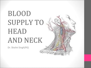
- 1. BLOOD SUPPLY TO HEAD AND NECK Dr. Shalini Singh(PG)
- 2. OBJECTIVES • General Principles of Arterial Supply • AORTA • Common Carotid Arteries • External Carotid Artery • Internal Carotid Artery • Subclavian Artery BLOODSUPPLYTOHEAD ANDNECK 2
- 4. GENERAL PRINCIPLES OF ARTERIAL SUPPLY • Arteries carry blood away from the heart. • All arteries, carry oxygenated blood • except the pulmonary and umbilical arteries, which carry deoxygenated blood to the lungs (postnatal) and to the placenta (prenatal) respectively • The flow of blood depends on the pumping action of the heart. • There are no valves in the arteries. • The branches of arteries supplying adjacent areas normally • anastomose with one another freely providing backup routes for blood to flow if one link is blocked. 4 BLOODSUPPLYTOHEAD ANDNECK
- 5. AORTA • It is the largest artery in the body. • Originates from the left ventricle. • It is divided into 3 parts. • It carries oxygenated blood to all parts of the body. 5 BLOODSUPPLYTOHEAD ANDNECK
- 6. ARCH OF AORTA Branches of Arch of Aorta 1. Left Subclavian artery. 2. Left Common Carotid artery. 3. Brachiocephalic trunk. -Right subclavian artery. -Right common carotid artery. 6 BLOODSUPPLYTOHEAD ANDNECK
- 7. COMMON CAROTID ARTERY – The right common carotid artery arises from the brachiocephalic artery behind the sternoclavicular joint. -- The left artery arises directly from the arch of aorta behind the manubrium sternum. 7 BLOODSUPPLYTOHEAD ANDNECK
- 8. COMMON CAROTID ARTERY – In the neck, each CCA extends upwards & laterally with in the carotid sheath to the level of upper border of lamina of thyroid cartilage. -- The bifurcation takes place in carotid triangle opposite the disc between c3 & c4 vertebra. 8 BLOODSUPPLYTOHEAD ANDNECK
- 9. BRANCHES OF COMMON CAROTID ARTERY External Carotid Artery Internal Carotid Artery 9 BLOODSUPPLYTOHEAD ANDNECK
- 10. EXTERNAL CAROTID ARTERY It lies anterior to ICA and is the chief arterial supply to structures in front of neck and face. Under cover of anterior border of sternocleidomastoid 10 BLOODSUPPLYTOHEAD ANDNECK
- 11. Course At the origin- Artery lies in the carotid triangle, antero- medial to ICA. Begins lateral to the upper border of the thyroid cartilage, at level with the disc b/w c3 & c4 . A little curved & with a gentle spiral, it first ascends slightly forward & then backwards & a little laterally to pass b/w mastoid tip & mandibular angle and lies lateral to the ICA. 11 BLOODSUPPLYTOHEAD ANDNECK
- 12. Terminates in the substance of the parotid gland behind the neck of mandible by dividing into: Superficial temporal artery Maxillary artery BLOODSUPPLYTOHEAD ANDNECK 12
- 13. Relations 13 BLOODSUPPLYTOHEAD ANDNECK • Superficial- In carotid triangle • Skin, superficial fascia • Loop b/w the facial nr, cervical branch & transverse cutaneous nr. of neck. • Deep fascia & ant. margin of sternocleidomastoid.
- 14. Crossed by- Hypoglossal nr & its vena comitans. Lingual, facial, sup. Thyroid vein. After leaving triangle- Crossed by- • Posterior belly of digastric & stylohyoid • Posteromedial surface of parotid gland. Lying medial to- facial nr., superficial temporal & maxillary veins. 14 BLOODSUPPLYTOHEAD ANDNECK
- 15. • Medial- • Pharyngeal wall • Sup. Larngeal nr. • Asc. Pharngeal art. • ICA separated from ECA by • Styloid process • Styloglossus & stylopharyngeus • Glossopharyngeal nr. • Pharyngeal br. Of vagus nr. • Part of parotid gland BLOODSUPPLYTOHEAD ANDNECK 15
- 16. Branches • Anterior : • Superior thyroid • Lingual • Facial • Posterior: • Occipital • Posterior auricular • Medial: • Ascending pharyngeal • Terminal: • Maxillary • Superficial temporal BLOODSUPPLYTOHEAD ANDNECK 16
- 17. SUPERIOR THYROID ARTERY COURSE: arises from the front of ECA below the tip of greater cornu of hyoid bone. Dividing into terminal branches at the apex of the thyroid lobe i.e ant. & post. BLOODSUPPLYTOHEAD ANDNECK 17
- 18. Relations • From origin- under sternocleidomastoid muscle descends forward in triangle. • Along lateral border • thyrohyoid • omohyoid • sternothyroid • sternohyoid BLOODSUPPLYTOHEAD ANDNECK 18
- 19. • Medial to artery- • constrictor pharyngis • external laryngeal nr. 19 BLOODSUPPLYTOHEAD ANDNECK
- 20. Branches • Infrahyoid artery- runs along the lower border of the hyoid deep to thyrohyoid anastomose with its fellow. • Sternocleidomastoid artery- descends laterally along carotid sheath. • superior laryngeal artery- accompanying the internal laryngeal nr. deep to thyrohyoid. Supply- larynx. Anastomose with its fellow & inf. larngeal br. of inf. thyroid art. BLOODSUPPLYTOHEAD ANDNECK 20
- 21. Branches • Cricothyroid artery- crosses high on cricothyroid ligament anastomose with its fellow. • Glandular branches- • Anterior- along the medial side of the upper pole of the lateral lobe, supplying mainly ant. surface by crossing above isthmus to anastomose with its fellow. • Posterior- descending on post. border. Supplying the medial & lateral surfaces & anastomosing with the inf. thyroid art. BLOODSUPPLYTOHEAD ANDNECK 21
- 22. LINGUAL ARTERY Introduction: Principal artery of tongue Arises anteromedially from ECA opposite the tip of greater cornu of hyoid bone b/w thyroid & facial art. Divided into 3 parts by hyoglossus muscle. BLOODSUPPLYTOHEAD ANDNECK 22
- 23. FIRST PART – In carotid triangle, extends from origin to the posterior border of hyoglossus. • Rests on the middle constrictor, crossed by hypoglossal nerve. SECOND PART – Deep to hyoglossus, runs horizontally forward along the upper border of hyoid bone between hyoglossus laterally and middle constrictor, stylohyoid ligament medially. accompanied with lingual vein. Relations:- Superficial- hyoglossus muscle tendon of digastric, stylohyoid lower pat of submandibular gland posterior part of mylohyoid. Medially- middle pharyngeal constrictor stylohyoid ligament BLOODSUPPLYTOHEAD ANDNECK 23
- 24. THIRD PART – [ arteria profunda linguae],ascends along the anterior Border of hyoglossus, then horizontally forward on the undersurface of tongue on each side of frenum linguae. In vertical course, lies b/t the genioglossus medially & inferior constrictor of tongue laterally. Horizontal part is accompanied by lingual nerve. BLOODSUPPLYTOHEAD ANDNECK 24
- 25. Branches • Suprahyoid artery- small, runs along hyoid’s upper border to anastomose contralateral art. • Dorsal lingual artery- medial to hyoglossus. Supply:- • mucous mem. Of tongue • palatoglossal arch • Tonsil, soft palate & epiglottis • Sublingual artery- arise from anterior margin of hyoglossus goes forward b/w genioglossus & mylohyoid to sublingual gland. • Supply- • sublingual gland • mylohyoid • buccal and gingival mucous mem. BLOODSUPPLYTOHEAD ANDNECK 25
- 26. Facial Artery Arises anteriorly from the ECA just above the tip of greater cornu of hyoid bone. Tortuous course— on neck-- allows free movements of pharynx during deglutition on face -- free movements of mandible , lips, & cheek during mastication & facial expressions, escapes traction & pressure during movements. BLOODSUPPLYTOHEAD ANDNECK 26
- 27. • Course: Runs upwards on superior constrictor of pharynx deep to the, posterior belly of digastric with stylohyoid & to the ramus of mandible Grooves the posterior border of submandibular gland Makes S-bend [2 loops] 1st winding down over submandibular reaching the surface of the mandible it curves round its inf. border, ant. to masseter to enter the face. Ascends forward across the mandible and buccinator to traverse a cleft in the modiolus near the buccal angle. Ascends side of nose & ends at the medial palpebral commisure. BLOODSUPPLYTOHEAD ANDNECK 27
- 28. Branches • Cervical: • Ascending palatine artery • Tonsilar artery • Submental artery • Glandular branches • Facial: • Inferior labial artery • Superior labial artery • Lateral nasal branch to nasalis muscle • Angular artery- terminal branch 28 BLOODSUPPLYTOHEAD ANDNECK
- 29. Cervical Branches • Ascending palatine artery- arise near facial origin, ascending b/w the styloglossus & stylopharyngeus to side of pharnx. Supply- • pharynx • soft palate • tonsil • auditory tube • Tonsilar artery- supply tonsil • Glandular branch- supply submandibular gland & lymph nodes. • Submental artery- largest cervical br. Runs forward along lower border of mandible( over the mylohyoid mus.). It supplies muscles of the region including those of chin & lower lip. BLOODSUPPLYTOHEAD ANDNECK 29
- 30. • Inferior labial artery- arises near the buccal angle, pass up & forward under the depressor anguli oris, b/w the orbicularis oris and mucous mem. • Supply- inf. labial glands, mucous mem. & muscles. • Superior labial- more tortuous course along sup. labial margin. • Lateral nasal artery- ascends the side of the nose. Supply- nasal ala & dorsum. • Angular artery- terminal part. BLOODSUPPLYTOHEAD ANDNECK 30
- 31. Occipital Artery • Arises in carotid triangle from posterior aspect of ECA 2 cm from its origin. • Passes backward, upward along & under cover of post. Belly of diagastric , crossing superficial to contents of carotid sheath, hypoglossal & accessory nerve. • Appears in the sub occipital region , rests on the rectus capitis ,obliqus capitis superior &semispinalis capitis, crosses the apex of post. triangle of neck, finally piercing trapezius and sternocleidomastoid. • Ascends tortuously in the dense superficial fascia of the scalp and divides into many branches. BLOODSUPPLYTOHEAD ANDNECK 31
- 32. Branches • Sternomastoid branch – two in no. upper & lower, supply sternomastoid m. • Mastoid branch – enters cranial cavity through mastoid foramen, supplies mastoid air cells in the dura. Sometimes absent. • Meningeal branch – enters the skull through jugular foramen & condylar canal, supplies dura & bone of posterior cranial fossa. BLOODSUPPLYTOHEAD ANDNECK 32
- 33. • Muscular branch- supply adj. muscles. Digastric, stylohyoid, splenius, longissimus capitis. • Occasional auricular branch supplies cranial surface of auricle. • Descending branch- superficial --anastamoses with sup.br. of transverse cervical art.; deep br.anastamoses with vertebral & deep cervical art.(costocervical trunk) • Occipital br. – supply the scalp upto vertex. BLOODSUPPLYTOHEAD ANDNECK 33
- 34. Ascending Pharyngeal Artery • Smallest, posteriorly near the ECA. • Ascends to base of skull between wall of pharynx & ICA. • Relations- crossed by styloglossus, stylopharyngeus. • Supply- • Sympathetic trunk • Hypoglossal • Glossopharyngeal • Vagus nr. • Cervical lymph nodes BLOODSUPPLYTOHEAD ANDNECK 34
- 35. Branches • Pharyngeal br. – supply:- • Constrictors & stylopharyngeus • Soft palate • Tonsil • part of auditory tube. • Inferior tympanic branch – supply:- • medial wall of tympanic cavity • Tympanic br. of glossopharyngeal nr. • Meningeal br. –supply:- • dura mater & adj. bones. BLOODSUPPLYTOHEAD ANDNECK 35
- 36. Posterior Auricular Artery • Branches posteriorly from external carotid just above the digastic & stylohyoid. • Ascends b/w the parotid gland & styloid process to the groove b/w the auricular cartilage & mastoid process. • Dividing into auricular & occipital branches. BLOODSUPPLYTOHEAD ANDNECK 36
- 37. Branches • Stylomastoid- enter stylomastoid foramen. • Supply- facial nr., tympanic cavity, mastoid antrum, air cells & semicircular canals. • Auricular branch- supply lateral aspect. • Occipital branch- supply occipital belly of the occipitofrontalis & scalp above and behind the ear. BLOODSUPPLYTOHEAD ANDNECK 37
- 38. Superficial temporal artery • Smaller terminal br. of ECA. • Begins in the parotid gland behind the mandible neck, crosses the post. root of the zygomatic process of the temporal bone. • About 5cm above this divides into ant. & post. branches. • Relations— • Zygoma-covered by auricularis ant. • Parotid gland- temporal & zygomatic br. of facial nr. cross it. • Scalp-accompanied by occipital vein & post. to it lies the auriculotemporal nr. BLOODSUPPLYTOHEAD ANDNECK 38
- 39. Branches • Transverse facial artery- arise within the substance of parotid gland. • Supply- parotid gland & duct, masseter & skin. • Anterior auricular branch • supply to lobule & ant part of auricle, external acoustic meatus. • Zygomatico-orbital artery– runs forward along upper border of zygomatic arch up to lateral angle of the eye. • Supply orbicularis oculi. • Middle temporal artery • temporalis BLOODSUPPLYTOHEAD ANDNECK 39
- 40. • Frontal branch(ant.)- runs upward & forward in the part of the scalp overlying temporal & frontal bone. • Supply musscles, skin & pericranium. • Parietal branch(post.)- runs backward in the scalp overlying the temporal & parietal bones. BLOODSUPPLYTOHEAD ANDNECK 40
- 41. Maxillary Artery • Origin– larger terminal branch of external carotid, arises behind and below the mandibular neck, in substance of parotid gland • Course – • Mandibular part • Pterygoid part • Pterygopalatine part BLOODSUPPLYTOHEAD ANDNECK 41
- 42. Mandibular part ( first part) Passes between the mandibular neck and the sphenomandibular ligament, below auriculotemporal nerve Branches: ◦ Deep auricular artery ◦ Anterior tympanic branch ◦ Middle meningeal artery Frontal & Parietal ◦ Accessory meningeal artery ◦ Inferior alveolar artery BLOODSUPPLYTOHEAD ANDNECK 42
- 45. Pterygoid part (Second part) • Ascends obliquely forwards medial to temporalis and superficial to lower head of lateral pterygoid • Branches: • Deep temporal branches • Pterygoid branches • Massetric artery • Buccal artery BLOODSUPPLYTOHEAD ANDNECK 45
- 46. Pterygopalatine part Passes between the heads of lateral pterygoid, through pterygomaxillary fissure into the pterygopalatine fossa Branches: ◦ PSA Artery ◦ Infraorbital ◦ Greater palatine ◦ Pharyngeal branch ◦ Artery of pterygoid canal ◦ Sphenopalatine artery BLOODSUPPLYTOHEAD ANDNECK 46
- 47. Collateral Circulation In occlusion of CCA -- anastamoses between branches of SCA & ECA. Achieved through : 1] Br. Of Right & left ECAs., 2] between left & right ICA via circle of willis. 3] superior thyroid A. with inferior thyroid A. 4] descending branch of occipital A. with deep cervical & asc. Branch of transverse cervical A. 5] vertebral A. may take over entire supply of carotids with in skull. BLOODSUPPLYTOHEAD ANDNECK 47
- 48. Circle of Willis Circulus arteriosus – polygonal Anterior cerebral arteries through anterior communicating arteries Basilar artery Posterior cerebral arteries each joins the ipsilateral internal carotid artery by a posterior communicating artery BLOODSUPPLYTOHEAD ANDNECK 48
- 49. Internal Carotid Artery Has no branches in the neck and enters the cranial cavity. Supplies structures inside skull. Arises from the common carotid at the level of the superior border of the thyroid cartilage It is embedded in the carotid sheath with internal jugular vein and vagus nerve. It Supplies: ◦ Brain ◦ Nose ◦ Scalp ◦ Eye BLOODSUPPLYTOHEAD ANDNECK 49
- 50. Course Vertically upwards – neck Horizontally forwards and medially- petrous carotid canal Upwards – foramen lacerum Horizontally forwards – cavernous sinus Vertically upwards medial- anterior clinoid process Backwards and upwards – to its terminal branches BLOODSUPPLYTOHEAD ANDNECK 50
- 51. Internal carotid artery • Divided into- • Cervical • Petrous • Cavernous • Cerebral BLOODSUPPLYTOHEAD ANDNECK 51
- 52. BRANCHES OF ICA : From petrous part – carotico-tympanic branches. branches to pterygoid canal. From cavernous part – inferior hypophysial artery. meningeal branch. From cerebral part – superior hypophyseal artery. opthalmic artery. posterior communicating artery. anterior choriod artery. anterior cerebral artery. middle cerebral artery. 52 BLOODSUPPLYTOHEAD ANDNECK
- 53. Cervical part Relations Posteriorly -sup cervical ganglion,sup laryngeal nerve Medially - ascending pharyngeal artery Anterolaterally - sternocleidomastoid muscle Inferiorly-digastric, hypoglossal nerve At the level of digastric - stylohyoid muscle, posterior branches of ECA Above the digastric - styloid process,deeper part of parotid gland Internal carotid artery BLOODSUPPLYTOHEAD ANDNECK 53
- 54. Petrous part Relations Surounded by venous and sympathetic plexuses Posterolaterally-middle ear and cochlea Anterolaterally- auditory tube and tensor tympani Superiorly- trigeminal ganglion Internal carotid artery BLOODSUPPLYTOHEAD ANDNECK 54
- 55. ICA Cavernous part ◦ Ascends to the posterior clinoid process ◦ Emerges through the dorsal roof of the cavernous sinus Branches ◦ Cavernous branches ◦ Hypophyseal branches ◦ Meningeal branches BLOODSUPPLYTOHEAD ANDNECK 55
- 56. ICA Cerebral part Lies at base of the brain. Divides into Anterior and Middle cerebral arteries. Gives off 5 branches: ◦ Ophthalmic artery ◦ Anterior cerebral artery ◦ Middle cerebral artery ◦ Posterior communicating artery ◦ Anterior choroid artery BLOODSUPPLYTOHEAD ANDNECK 56
- 57. ICA Ophthalmic artery Artery enters the orbit through optic canal. Terminates near the medial angle of the eye, dividing into supratrochlear and dorsal nasal branches BLOODSUPPLYTOHEAD ANDNECK 57
- 58. Ophthalmic artery Branches Central artery of retina Lacrimal branch Muscular branch Ciliary arteries Supraorbital artery Posterior ethmoidal artery Anterior ethmoidal artery Meningeal artery Medial palpebral artery Supratrochlear artery BLOODSUPPLYTOHEAD ANDNECK 58
- 59. The Subclavian System of Arteries ORIGIN – • Right subclavian art. Arises from the brachiocephalic trunk. • Left subclavian art. arises from the arch of aorta. BLOODSUPPLYTOHEAD ANDNECK 59
- 60. Branches of Subclavian • vertebral, • internal thoracic • thyrocervical trunk. • costo cervical trunk. • Dorsal scapular artery. BLOODSUPPLYTOHEAD ANDNECK 60
- 61. Course: Cervical part -- curved course with upward convexity. extends from the sternoclavicular joint to the outer border of first rib, enters through the apex of axilla & continued as axillary artery. Each art. Arches over the cervical pleura n apex of the lung, subdivided into 3 parts by scalenus anterior muscle 1st part -- upto medial border of muscle, 2nd part--- behind the muscle, 3rd---- lateral border of muscle to the outer border of 1st rib. BLOODSUPPLYTOHEAD ANDNECK 61
- 63. Vertebral Artery Origin-- from the upper surface of the first part of SC A.passes through-- foramina transversaria of upper six cervical vertebrae, winds backward around the lateral mass of atlas,enters the cranial cavity through foramen magnum, and at the lower border of pons. unites with similar artery of opposite side forms-- the basilar artery. BLOODSUPPLYTOHEAD ANDNECK 63
- 64. Branches Cervical branches – ◦ spinal branches – enter the vertebral canal through intervertebral foramina ; supplies spinal cord,meninges, vertebra. ◦ muscular branches – from 3rd part ; supply sub-occipital muscles. B] cranial branches – ◦ meningeal branches ◦ posterior spinal artery ◦ ant. Spinal artery., ◦ post. Inferior cerebellar artery, ◦ medullary arteries. BLOODSUPPLYTOHEAD ANDNECK 64
- 65. Parts First part:- extends from the origin of the artery to the transverse process of c6. Runs upwards and backwards in the triangular space b/w scalenus anterior and longus colli muscles called vertebral triangle Second part– runs through the foramina transverseria of upper C6.it course is vertical upto the axis vertebrae BLOODSUPPLYTOHEAD ANDNECK 65
- 66. Third part :Lies in the sub-occipital triangle emerging from foramen tranversarium of atlas. Enters the vertebral canal by passing deep to the lower arched margin of the posterior atlanto- occipital membrane . Fourth part :Pierces the dura & arachnoid maters,& passes upward & medially through the foramen magnum in front of first tooth of ligamentum denticulum. At lower border of pons ,it unites with the fellow of opp. Side to form basilar art. BLOODSUPPLYTOHEAD ANDNECK 66
- 67. Internal Thoracic Artery • Arises from the inferior surface of 1st part of SCA, opposite the origin of thyrocervical trunk.,2cm above the sternal end of clavicle. • BRANCHES --- • Pericardico-phrenic artery. • Mediastinal branches. • Pericardial branches • Sternal branches • Ant. Inter-costal artery. • Perforating artery. • Musculo-phrenic artery. • Superior epigastric artery. BLOODSUPPLYTOHEAD ANDNECK 67
- 68. Thyro Cervical Trunk • Arises from the upper surface of 1st part of SCA, just distal to the origin of vertebral art. • 3 branches : inferior thyroid art. asc. Cervical art. inf laryngeal art. tracheal, oesophageal, laryngeal br. Transverse cervical art. suprascapular art. BLOODSUPPLYTOHEAD ANDNECK 68
- 69. Costo-Cervical Trunk •Arises from the back of 1st part of SCA on left side2nd part of same art. On rt. Side. Branches – • deep cervical artery • superior intercostal art. BLOODSUPPLYTOHEAD ANDNECK 69
- 70. Dorsal Scapular Artery Arises from 3rd part of SCA. Passes laterally b/w upper & middle or middle & lower trunks of bracheal plexus. supply the rhomboids & enters in formation of scapular anastamoses. BLOODSUPPLYTOHEAD ANDNECK 70
- 71. APPLIED ANATOMY CAROTID PULSE : CCA may be compressed against the carotid tubercle of transverse process of C6 vertebra ( carotid tubercle of chassaignac ) about 4cm above the sternoclavicular joint. • Patency of carotid system can be investigated by angiography by injecting a contrast medium into CCA.
- 72. APPLIED ANATOMY • LIGATION OF ECA : Done at 2 points Artery exposed at its origin & ligature above superior thyroid artery upper part of neck, superficial & deep structures of neck Ligation higher up, behind the angle of lower jaw- maxillary artery injuries • UNILATERAL LIGATION – will not stop hemorrhage
- 73. A] LIGATION OF ECA IN CAROTID TRIANGLE:- • Skin incision-- at the level of angle of mandible behind anterior border of sternocleidomastoid muscle ,continued downward to the level of cricoid cartilage. -- Platysma, superficial sheath of sternomastoid incised, muscle exposed & retracted ,deep layer of sternomastoid head is visible & IJV through it. -- Fascia in front of vein is cut to expose the arteries.
- 74. LIGATION IN RETROMANDIBULAR FOSSA Skin incision--- at line starting at the tip of mastoid process , circling the mandibular angle, continuing forward below the mandible one inch. Passing scalpel through skin & posterior fibers of platysma , the retromandibular vein or EJV is located, tied & cut. Branches of great auricular nerve cut -- permit mobilization of cervical lobe of parotid gland.
- 75. Attachment of parotid capsule to the anterior border of sternomastoid severed with scalpel. Parotid gland retracted , post. Belly of digastric ,stylohyoid muscle is visible. Above this stylomandibular ligament can be palpated if lower jaw of the patient is pulled forward. This movement--- widens the entrance into retromandibular fossa , tenses the stylomandibular ligament. Pulsations of ECA are felt , isolated & tied.
- 76. sublingual artery -- injury occurs in premolar & molar region, when sharp instrument or rotating disks slips off a lower molar & injure the floor of mouth.
- 77. • Applied anatomy In surgical removal of tongue , first part of artery is ligatured before it gives any branches to the tongue or tonsil. • sublingual artery -- injury occurs in premolar & molar region, when sharp instrument or rotating disks slips off a lower molar & injure the floor of mouth.
- 78. LIGATION OF LINGUAL ARTERY : • Incision – circling the lower pole of submandibular gland. • Posterior part – towards tip of mastoid ; anterior part – towards chin. • Skin, platysma, deep fascia incised, submandibular gland exposed , lifted,tendon of diagastric visible. • Free border of mylohyoid muscle ascertained, hypoglossal nerve identified. • Digastric tendon pulled downwards –enlarges the digastric triangle, hyoglossus muscle visible. • Muscle divided bluntly, in the gap of its vertical fibers lingual artery found & ligated.
- 79. • • VARIATIONS : May arise in common with lingual artery constituting “linguo-facial trunk”. Occasionly ends by forming submental artery& not infreqently extends only as high as the angle of mouth or nose. Deficiency is compensated by enlargement of one of neighbouring arteries. • 3] facial artery – can be injured –during operative procedures on lower premolars & molars, if instrument enters the cheek at inferior vestibular fornix., also while attempt to open a buccal abscess.
- 80. LIGATION OF FACIAL ARTERY.• Exposed --at the point crossing the lower border of mandible • Using contracted masseter as a landmark, pulse of facial artery felt at point situated anterior to the attachment of masseter. • Artery is accompanied by facial vein & crossed superficially by marginal mandibular branch of facial nerve. • Taking this into consideration, incision -- at least half inch below the border of mandible & parallel to it. • Skin, platysma, deep fascia are cut , soft tissues retracted, pulse of facial artery felt. • Artery-- isolated, tied & cut.
- 81. • POSTERIOR SUPERIOR ALVEOLAR ARTERY- APPLIED SURGICAL ANATOMY site of hematoma during PSA block. - prevented by aspirating before giving LA in the site. • GREATER PALATINE AND ANTERIOR PALATINE ARTERY. case of abscess from palatal root of first molar,incision should be made in a antero-posterior direction ,then transversly. Incision– made near free margin of gingiva. Edge of knife directed outward, upward.
- 82. Superficial temporal artery • Origin: smaller of the two terminal branches, begins in the parotid gland behind mandible’s neck • Course: crosses the posterior root of zygomatic process of temporal bone, divides into anterior and posterior branches
- 83. APPLIED ANATOMY • Control of temporal haemorrhage • Anastomose freely; partially detached with scalp also heal with reasonable hope even if one vessel is intact • Placement of incisions in craniotomy • In reduction of zygomatic arch fractures – Gilli’s approach
- 85. Thank You