SlideShare une entreprise Scribd logo
1  sur  23
ULCER
An ulcer is a break in the continuity of
the covering epithelium either skin or mucous
membrane .
PARTS OF AN ULCER
1 : Margin
2 : Edge
3 : Floor
4 : Base
1 : Margin
It may be regular or irregular . It may be
rounded or oval .
2 : Edge
Edge is one which connects floor of the
ulcer to the margin .
3 : Floor
Floor is a deepened part and may contain
the discharge , granulation tissue , or slough .
4 : Base
Base is the one on which ulcer lies . It may
be bone or soft tissue .
DIFFERENT TYPES OF EDGES
A : SLOPING EDGE
It is seen in a healing ulcer its inner
part is red because of healthy granulation
tissue and outer part is white due to scar .
B : UNDRMINED EDGE
It is seen in cases of Tuberculous
ulcer .
C : PUNCHED OUT EDGES
It is seen in granulomatous
( syphilitic ) ulcer and bed sores .
D : RAISED AND BEADED EDGES
( Pearly white )
It is seen in rodent ulcer .( BCC ).
E : EVERTED EDGE ( Rolled out edge )
It is seen in carcinomatous ulcer
due to spillage of the proliferating malignant
tissues over the normal skin .
CLASSIFICATION OF ULCER ( CLINICAL )
1 : SPREADING ULCER
In this edge is inflamed and
edematous .
2 : HEALING ULCER
sloping edge with healthy , pink and
red tissue .
3 : CALLOUS ULCER
Floor contains pale unhealthy
granulation tissue with indurated edge . This
ulcer is for months and years because of
callous attitude of the patient .
CLASSIFICATION OF ULCER
( PATHOLOGICAL)
1 : SPECIFIC ULCER
- Tuberculous ulcer
- Syphilitic ulcer
- Actinomycosis
2 : MALIGNANT ULCER
- Carcinomatous ulcer
- Rodent ulcer
- Melanotic ulcer
3 : NON SPECIFIC ULCER
- Traumatic ulcer
- Arterial ulcer
- Venous ulcer or
- Gravitational ulcer
- Trophic ulcer / pressure sore
- Diabetic ulcer
WAGNER’S GRADING OF AN ULCER
GRADE : 0
Preulcerative lesion / healed ulcer
GRADE : 1
Superficial ulcer
GRADE : 2
Ulcer deeper to subcutaneous tissue ,
exposing soft tissues or bone .
GRADE : 3
Abscess formation / osteomylitis
GRADE : 4
Gangrene of part of tissue / limb / foot
GRADE : 5
Gangrene of entire one area / foot
INVESTIGATIONS OF AN ULCER
1 : STUDY OF A DICHARGE
- Culture and sensitivity
- AFB study and cytology
2 : WDGE BIOPSY
- Biopsy is always taken from
edge because edge contains multiplying cells .
- At least 2 biopsies are taken .
3 : X-RAY OF THE PART to look for
- Periostitis / osteomylitis
4 : FNAC of the lymph node
TREATMENT OF AN ULCER
Cause should be found and treated .
Debridement of an ulcer .
All dead , devitalized necrotic tissue is
removed and dressing is applied like :
- Liquid paraffin dressing
- Cotton dressing
INTRODUCTION
TO
SURGICAL INFECTIONS
SURGICAL INFECTION
Surgical infection is a major surgical
problem in surgical practice and here are the
protective mechanisms like phagocytes ,
antibodies , leucocytes and complement
system . They have an important role in
protecting the infection .
SEPSIS
clinical evidence of infection .
SEPSIS SYNDROME
clinical evidence of infection plus evidence
of altered organ perfusion.
SEPTIC SHOCK
Septic syndrome plus evidence of decreased
blood pressure unresponsive to fluid therapy .
CLINICAL INDICATORS OF INFECTION
CHANGES IN CORE TEMPERATURE
- Fever > 37. 8 C
- hypothermia < 36 C
Unexlained hypotension
oliguria
confusion
POSSIBLE FOCI OF INFECTION
ABDOMINAL EXAMINATION
Bowel
inflammatory bowel dissease ,
perforation , abscess
hepatobiliary
cholecystitis , cholangitis
genitiurinary
uti
RESPIRATORY EXAMINATION
Pneumonia
C V S
endocarditis
skin
surgical wound infection
cns
meningitis , enchephalitis
SEPTIC SCREENING
BLOOD TEST
New microsoft office power point presentation

Contenu connexe

Tendances (20)

Examination of Swelling
Examination of SwellingExamination of Swelling
Examination of Swelling
 
Leg Ulcers - Clinical diagnosis
Leg Ulcers - Clinical diagnosisLeg Ulcers - Clinical diagnosis
Leg Ulcers - Clinical diagnosis
 
Solitary oral ulcers and systemic diseases
Solitary oral ulcers and systemic diseasesSolitary oral ulcers and systemic diseases
Solitary oral ulcers and systemic diseases
 
Ulcer
UlcerUlcer
Ulcer
 
Ulcer by Dr.Prashant patil
Ulcer by Dr.Prashant patilUlcer by Dr.Prashant patil
Ulcer by Dr.Prashant patil
 
Ulcer by Dr. AmrithaAnilkumar
Ulcer by Dr. AmrithaAnilkumarUlcer by Dr. AmrithaAnilkumar
Ulcer by Dr. AmrithaAnilkumar
 
Sinus and fistula
Sinus and fistulaSinus and fistula
Sinus and fistula
 
Swelling - Examination
Swelling  - ExaminationSwelling  - Examination
Swelling - Examination
 
Haemangioma
HaemangiomaHaemangioma
Haemangioma
 
Ulcer (2)
Ulcer (2)Ulcer (2)
Ulcer (2)
 
Clinical examination of swelling
Clinical examination of swellingClinical examination of swelling
Clinical examination of swelling
 
EXAMINATION OF SWELLING
EXAMINATION OF SWELLING EXAMINATION OF SWELLING
EXAMINATION OF SWELLING
 
Hemangioma
HemangiomaHemangioma
Hemangioma
 
Discuss keloid and hypertrophic scars
Discuss keloid and hypertrophic scarsDiscuss keloid and hypertrophic scars
Discuss keloid and hypertrophic scars
 
Basal cell carcinoma
Basal cell carcinomaBasal cell carcinoma
Basal cell carcinoma
 
Bullous pemphigoid
Bullous pemphigoidBullous pemphigoid
Bullous pemphigoid
 
Clinical examination of swelling
Clinical examination of swelling Clinical examination of swelling
Clinical examination of swelling
 
Ulcer
UlcerUlcer
Ulcer
 
palpation of ulcer
palpation of ulcerpalpation of ulcer
palpation of ulcer
 
Ulcers & skin infections
Ulcers & skin infectionsUlcers & skin infections
Ulcers & skin infections
 

En vedette

En vedette (9)

Ulcers & wounds
Ulcers & woundsUlcers & wounds
Ulcers & wounds
 
Clinical surgery(History & Physical)
Clinical surgery(History & Physical)Clinical surgery(History & Physical)
Clinical surgery(History & Physical)
 
Ulcers
UlcersUlcers
Ulcers
 
Evaluation Of Oral Ulcerations
Evaluation Of Oral UlcerationsEvaluation Of Oral Ulcerations
Evaluation Of Oral Ulcerations
 
Wound management
Wound managementWound management
Wound management
 
Skin ulcers
Skin ulcersSkin ulcers
Skin ulcers
 
Benign n pre malignant diseases of cx
Benign n pre malignant diseases of cxBenign n pre malignant diseases of cx
Benign n pre malignant diseases of cx
 
Stomas
StomasStomas
Stomas
 
Intestinal stomas
Intestinal stomasIntestinal stomas
Intestinal stomas
 

Similaire à New microsoft office power point presentation

Tumor,ulcer,cyst,sinus,fistula.pptx
Tumor,ulcer,cyst,sinus,fistula.pptxTumor,ulcer,cyst,sinus,fistula.pptx
Tumor,ulcer,cyst,sinus,fistula.pptxDrYashSharma
 
clinicalexaminationofulcers-180118174404.pdf
clinicalexaminationofulcers-180118174404.pdfclinicalexaminationofulcers-180118174404.pdf
clinicalexaminationofulcers-180118174404.pdfNituKumari826353
 
Ulcer sinus fistula
Ulcer sinus fistulaUlcer sinus fistula
Ulcer sinus fistuladrsunilnegi
 
ulcer-190501174026.pdf
ulcer-190501174026.pdfulcer-190501174026.pdf
ulcer-190501174026.pdfSayanRDey
 
Ulcerative, Vesicular and Bullous Lesions.pptx
Ulcerative, Vesicular and Bullous Lesions.pptxUlcerative, Vesicular and Bullous Lesions.pptx
Ulcerative, Vesicular and Bullous Lesions.pptxManuelKituzi
 
webinar6_pu_woundassesst (1).pdf
webinar6_pu_woundassesst (1).pdfwebinar6_pu_woundassesst (1).pdf
webinar6_pu_woundassesst (1).pdfPreetiChouhan6
 
6-Neck swelling.pptx
6-Neck swelling.pptx6-Neck swelling.pptx
6-Neck swelling.pptxAhmedAbd66
 
clinical examination.pdf
clinical examination.pdfclinical examination.pdf
clinical examination.pdfssusercab735
 
Cervical mases DDx and Radio-imaging by B.H.A.A Malik
Cervical mases DDx and Radio-imaging by  B.H.A.A MalikCervical mases DDx and Radio-imaging by  B.H.A.A Malik
Cervical mases DDx and Radio-imaging by B.H.A.A Malikbushra a malik
 
methods of clinical examination
methods of clinical examinationmethods of clinical examination
methods of clinical examinationEnas Elgendy
 
pressure-ulcers.pdffhfhfhfhfhhuhfhfhkhfhfhfuhriuhuh
pressure-ulcers.pdffhfhfhfhfhhuhfhfhkhfhfhfuhriuhuhpressure-ulcers.pdffhfhfhfhfhhuhfhfhkhfhfhfuhriuhuh
pressure-ulcers.pdffhfhfhfhfhhuhfhfhkhfhfhfuhriuhuhoishia398
 
introduction to basic knowledge onulcer.pptx
introduction to basic knowledge onulcer.pptxintroduction to basic knowledge onulcer.pptx
introduction to basic knowledge onulcer.pptxAndrewsKudjordji
 
Primary Oral Ulceration.pptx
Primary Oral Ulceration.pptxPrimary Oral Ulceration.pptx
Primary Oral Ulceration.pptxMoaatezMohammed
 

Similaire à New microsoft office power point presentation (20)

ULCER.pptx
ULCER.pptxULCER.pptx
ULCER.pptx
 
Tumor,ulcer,cyst,sinus,fistula.pptx
Tumor,ulcer,cyst,sinus,fistula.pptxTumor,ulcer,cyst,sinus,fistula.pptx
Tumor,ulcer,cyst,sinus,fistula.pptx
 
clinicalexaminationofulcers-180118174404.pdf
clinicalexaminationofulcers-180118174404.pdfclinicalexaminationofulcers-180118174404.pdf
clinicalexaminationofulcers-180118174404.pdf
 
Leg ulcers (chronic
Leg ulcers (chronicLeg ulcers (chronic
Leg ulcers (chronic
 
Examination of ulcer
Examination of ulcerExamination of ulcer
Examination of ulcer
 
Ulcer sinus fistula
Ulcer sinus fistulaUlcer sinus fistula
Ulcer sinus fistula
 
ulcer-190501174026.pdf
ulcer-190501174026.pdfulcer-190501174026.pdf
ulcer-190501174026.pdf
 
ulcer-190501174026.pptx
ulcer-190501174026.pptxulcer-190501174026.pptx
ulcer-190501174026.pptx
 
Ulcer.pptx
Ulcer.pptxUlcer.pptx
Ulcer.pptx
 
ulcer ug class.pptx
ulcer ug class.pptxulcer ug class.pptx
ulcer ug class.pptx
 
Ulcerative, Vesicular and Bullous Lesions.pptx
Ulcerative, Vesicular and Bullous Lesions.pptxUlcerative, Vesicular and Bullous Lesions.pptx
Ulcerative, Vesicular and Bullous Lesions.pptx
 
SKIN ULCERS.pptx
SKIN ULCERS.pptxSKIN ULCERS.pptx
SKIN ULCERS.pptx
 
webinar6_pu_woundassesst (1).pdf
webinar6_pu_woundassesst (1).pdfwebinar6_pu_woundassesst (1).pdf
webinar6_pu_woundassesst (1).pdf
 
6-Neck swelling.pptx
6-Neck swelling.pptx6-Neck swelling.pptx
6-Neck swelling.pptx
 
clinical examination.pdf
clinical examination.pdfclinical examination.pdf
clinical examination.pdf
 
Cervical mases DDx and Radio-imaging by B.H.A.A Malik
Cervical mases DDx and Radio-imaging by  B.H.A.A MalikCervical mases DDx and Radio-imaging by  B.H.A.A Malik
Cervical mases DDx and Radio-imaging by B.H.A.A Malik
 
methods of clinical examination
methods of clinical examinationmethods of clinical examination
methods of clinical examination
 
pressure-ulcers.pdffhfhfhfhfhhuhfhfhkhfhfhfuhriuhuh
pressure-ulcers.pdffhfhfhfhfhhuhfhfhkhfhfhfuhriuhuhpressure-ulcers.pdffhfhfhfhfhhuhfhfhkhfhfhfuhriuhuh
pressure-ulcers.pdffhfhfhfhfhhuhfhfhkhfhfhfuhriuhuh
 
introduction to basic knowledge onulcer.pptx
introduction to basic knowledge onulcer.pptxintroduction to basic knowledge onulcer.pptx
introduction to basic knowledge onulcer.pptx
 
Primary Oral Ulceration.pptx
Primary Oral Ulceration.pptxPrimary Oral Ulceration.pptx
Primary Oral Ulceration.pptx
 

New microsoft office power point presentation

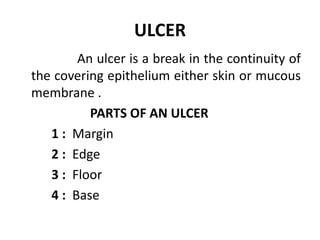
  • 1. ULCER An ulcer is a break in the continuity of the covering epithelium either skin or mucous membrane . PARTS OF AN ULCER 1 : Margin 2 : Edge 3 : Floor 4 : Base
  • 2. 1 : Margin It may be regular or irregular . It may be rounded or oval . 2 : Edge Edge is one which connects floor of the ulcer to the margin . 3 : Floor Floor is a deepened part and may contain the discharge , granulation tissue , or slough . 4 : Base Base is the one on which ulcer lies . It may be bone or soft tissue .
  • 3. DIFFERENT TYPES OF EDGES A : SLOPING EDGE It is seen in a healing ulcer its inner part is red because of healthy granulation tissue and outer part is white due to scar . B : UNDRMINED EDGE It is seen in cases of Tuberculous ulcer .
  • 4. C : PUNCHED OUT EDGES It is seen in granulomatous ( syphilitic ) ulcer and bed sores . D : RAISED AND BEADED EDGES ( Pearly white ) It is seen in rodent ulcer .( BCC ). E : EVERTED EDGE ( Rolled out edge ) It is seen in carcinomatous ulcer due to spillage of the proliferating malignant tissues over the normal skin .
  • 5.
  • 6. CLASSIFICATION OF ULCER ( CLINICAL ) 1 : SPREADING ULCER In this edge is inflamed and edematous . 2 : HEALING ULCER sloping edge with healthy , pink and red tissue . 3 : CALLOUS ULCER Floor contains pale unhealthy granulation tissue with indurated edge . This ulcer is for months and years because of callous attitude of the patient .
  • 7. CLASSIFICATION OF ULCER ( PATHOLOGICAL) 1 : SPECIFIC ULCER - Tuberculous ulcer - Syphilitic ulcer - Actinomycosis 2 : MALIGNANT ULCER - Carcinomatous ulcer - Rodent ulcer - Melanotic ulcer 3 : NON SPECIFIC ULCER - Traumatic ulcer - Arterial ulcer
  • 8. - Venous ulcer or - Gravitational ulcer - Trophic ulcer / pressure sore - Diabetic ulcer
  • 9. WAGNER’S GRADING OF AN ULCER GRADE : 0 Preulcerative lesion / healed ulcer GRADE : 1 Superficial ulcer GRADE : 2 Ulcer deeper to subcutaneous tissue , exposing soft tissues or bone . GRADE : 3 Abscess formation / osteomylitis GRADE : 4 Gangrene of part of tissue / limb / foot GRADE : 5 Gangrene of entire one area / foot
  • 10. INVESTIGATIONS OF AN ULCER 1 : STUDY OF A DICHARGE - Culture and sensitivity - AFB study and cytology 2 : WDGE BIOPSY - Biopsy is always taken from edge because edge contains multiplying cells . - At least 2 biopsies are taken . 3 : X-RAY OF THE PART to look for - Periostitis / osteomylitis 4 : FNAC of the lymph node
  • 11. TREATMENT OF AN ULCER Cause should be found and treated . Debridement of an ulcer . All dead , devitalized necrotic tissue is removed and dressing is applied like : - Liquid paraffin dressing - Cotton dressing
  • 13. SURGICAL INFECTION Surgical infection is a major surgical problem in surgical practice and here are the protective mechanisms like phagocytes , antibodies , leucocytes and complement system . They have an important role in protecting the infection .
  • 14. SEPSIS clinical evidence of infection . SEPSIS SYNDROME clinical evidence of infection plus evidence of altered organ perfusion. SEPTIC SHOCK Septic syndrome plus evidence of decreased blood pressure unresponsive to fluid therapy .
  • 15. CLINICAL INDICATORS OF INFECTION CHANGES IN CORE TEMPERATURE - Fever > 37. 8 C - hypothermia < 36 C Unexlained hypotension oliguria confusion
  • 16. POSSIBLE FOCI OF INFECTION ABDOMINAL EXAMINATION Bowel inflammatory bowel dissease , perforation , abscess hepatobiliary cholecystitis , cholangitis genitiurinary uti RESPIRATORY EXAMINATION Pneumonia
  • 17. C V S endocarditis skin surgical wound infection cns meningitis , enchephalitis