2. 98 TURKSTRA ET AL.
and scheduled for elective noncardiac surgery requiring placed in the vallecula. The large size ATQ was used for
general anesthesia with tracheal intubation. Exclusion men and the small size was used for women. Protraction
criteria were gastroesophageal reflux disease, body mass of the mandible was performed if necessary, but it was
index greater than 35 kg/m2, possibility of pregnancy, minimized in an attempt to limit displacement of the
previous neck surgery, unstable C-spine, or known dif- C-spine via connecting structures. With both tech-
ficult airway. Preoperative clinical assessment of the niques, the operator attempted to minimize neck move-
patients included height, weight, physical status, Mal- ment, accepting the first view10 that offered a reasonable
lampati score, dentition, tongue size, thyromental dis- opportunity to adequately position the ETT at the glottic
tance, and neck mobility. opening. Intubation completed the study; the rigid
While awake, patients were placed on the operating board was removed, and anesthesia and surgery contin-
room table with a rigid board beneath them to simulate ued in the usual fashion.
field spinal precautions or the table on which trauma All laryngoscopies were performed by one operator
patients are placed in the emergency department. The (Dr. Turkstra) to minimize interoperator variability. Be-
patient’s head rested on a pillow in a position judged by fore this study, this operator had performed more than
the patient to be neutral. After verification that the pa- 50 intubations with the ATQ and more than 3,000 with
tient was properly centered, the fluoroscopy unit and the Macintosh blade. The fluoroscopy video monitor was
operating room table remained fixed for the remainder not visible to the operator during the study.
of the study. Standard monitors were placed. After
breathing 100% oxygen for 3 min, anesthesia was in- Study Data and Data Analyses
duced with fentanyl (2– 4 g/kg IV) and propofol (2–3 Similar to previous work, fluoroscopy of the C-spine
mg/kg IV). Upon loss of eyelid reflex, paralysis was during laryngoscopy and intubation was recorded at four
induced with rocuronium (0.6 – 0.8 mg/kg IV). frames per second by a digital video fluoroscopy unit
Manual in-line stabilization was provided by an assis- (Series 9800 mobile C-Arm with vascular package and
tant with Advanced Trauma and Life Support1 certifica- 1 k 1 k video monitor; GE Medical Systems, Salt Lake
tion. Care was taken to avoid obscuring the radiographic City, UT) for review by the radiologist (Dr. Pelz) to assess
landmarks during the fluoroscopy. Study personnel used cervical vertebrae movement.4,7 The fluoroscopic video
radiation-resistant surgical gloves and eyewear as well as was analyzed using Centricity software (GE Centricity
upper and lower lead aprons with thyroid protection; Picture Archiving and Communication System, Version
patients were shielded with lead aprons for areas not 3.0.4; GE Medical Systems) to determine the duration of
under investigation. laryngoscopy. Duration was defined as the time from
After positioning and induction, a sealed opaque enve- when the ATQ or Macintosh blade passed the central
lope containing a computer-generated random assign- incisors to the time when the ETT was positioned just
ment was opened, assigning patients to the following past the vocal cords. If the laryngoscopy sequence took
groups: group 1, Macintosh laryngoscopy first, followed longer than 120 s, it would be deemed a failure.
by ATQ laryngoscopy; group 2, ATQ laryngoscopy first, Using the radiology software, the orientation in the
followed by Macintosh laryngoscopy. All patients under- sagittal plane of the occiput and C1 through C5-C7 can
went laryngoscopy using both techniques. The order of be determined at any frame (point in time) in the fluo-
laryngoscopy was randomized to prevent a consistent roscopic video, with a precision of 0.1 degree. The
bias in favor of one group. absolute rotation of the occiput or vertebrae in global
After stabilization was completed, the operator venti- coordinates was not the focus, but rather the motion of
lated the patient with sevoflurane in 100% oxygen via each relative to adjacent vertebrae. (i.e., Trendelenburg
bag and mask until 90 s had elapsed from the administra- rotation of the operating room table would result in
tion of rocuronium. To minimize neck extension, a low global “extension” of all components, but no flexion or
threshold was adopted for use of an oral airway. Patients extension of the vertebrae relative to one another).
then underwent laryngoscopy using both techniques se- Motion segments were defined by two vertebrae, sim-
quentially; intubation was completed as part of the second ilar to previous work,4,7,11 and denoted M0-1 for Occi-
laryngoscopy. Intubation was not fully completed during put-C1, M1-2 for C1-C2, and M2-5 for C2-C5 (fig. 1).
the first laryngoscopy, as required by our ethics board to Motion segment M5-T comprised C5 through whatever
prevent potential trauma of multiple intubations; the distal vertebrae remained stationary on the backboard. The
end of the ETT was advanced just beyond the vocal cords relative angle between the two bones of each motion
and withdrawn. C-spine movement was recorded with segment at any point in time was denoted A0-1TIME,
continuous fluoroscopy during both laryngoscopies and A1-2TIME, A2-5TIME, andA5-TTIME.
intubation; the entire time interval was recorded to capture The reference for the occiput was defined by a line
the maximal extent of C-spine movement. between the base of the sella and the opisthion (fig. 1).
The ATQ was used according to the instructions pro- The C1 reference was a line between the lower cortical
vided by the manufacturer, with the distal tip of the ATQ margin of the anterior arch of C1 and the lower cortical
Anesthesiology, V 111, No 1, Jul 2009
3. C-SPINE MOTION: AIRTRAQ VS. MACINTOSH LARYNGOSCOPE 99
Statistical Analyses
Using data from the control group of a previous study4
and estimation that a 30% reduction in C-spine move-
ment would be clinically relevant, the two-tailed sample
size was calculated to be 11 patients for each group
( 0.05, 0.20). Twenty-four patients were re-
cruited to allow for patient dropout and/or potential
failure of the fluoroscopic equipment or recording device.
Statistical analysis was conducted using SAS version 9.1
(SAS Institute Inc., Cary, NC). For the study design, all
patients received both the Macintosh and the ATQ device
in random order. As a crossover study, a repeated measures
analysis of variance with one within-subject factor (device)
and one between-subject factor12 (the sequence in which
the devices were used) was used to analyze the C-spine
movement data. The between-subject factor (sequence)
tested for carry-over, i.e., whether the use of one device
was affected by previous use of the other device.12
Because of the skewed nature of the duration of laryn-
goscopy data, the median provides an estimate of the cen-
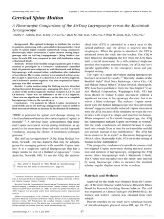
Fig. 1. Vertebral reference lines. The reference for the occiput ter of the distribution, and the interquartile range (IQR)
was defined by a line between the base of the sella and the provides a measure of variation.13 The IQR is the difference
opisthion (Line A). The C1 reference was a line between the
lower cortical margin of the anterior arch of C1 and the lower
between the upper quartile (Q3) and the lower quartile
cortical margin of the C1 spinous process (Line B). The C2 (Q1). As a result of the crossover trial design, the duration
reference was a line between the anterior, inferior margin of of laryngoscopy data has also been compared using the
the C2 body and the lower cortical margin of the C2 spinous
process (Line C). The C5 reference line was a tangent along the
repeated measures analysis of variance.12 Kaplan-Meier
superior end-plate of the C5 vertebral body (Line D). The Occi- curves have been used to graphically illustrate the distribu-
put-C1 segment is defined by the angle between Lines A and B. tion of laryngoscopy duration data for the two treatments.
The C1-C2 segment is defined by the angle between Lines B and
C. The C2-C5 segment is defined by the angle between Lines C
and D. The C5-Thoracic segment is defined by the angle be-
tween Line D and the global reference.
Results
margin of the C1 spinous process. The C2 reference was a Twenty-nine patients were invited to participate in the
line between the anterior, inferior margin of the C2 body study between January and May of 2008. Of the 29 patients,
and the lower cortical margin of the C2 spinous process. 5 declined; 24 patients were enrolled and gave written
The C5 reference line was a tangent along the superior consent. No patients were lost to follow-up; all patients
endplate of the C5 vertebral body. Stationary vertebrae of were analyzed in the group to which they were assigned.
Segment M5-T remained fixed relative to the fluoros- The patient characteristics are summarized in table 1.
copy unit. When necessary, other anatomic landmarks Patients underwent ATQ use and direct Macintosh
were used by the radiologist, remaining consistent for laryngoscopy in random order, and C-spine motion was
a given subject. This was acceptable because the study
compared the change in the angle of the motion Table 1. Patient Characteristics
segments, so any consistent landmarks would suffice.
Group 1, Group 2,
The first frame of each fluoroscopic sequence pro- Macintosh, then AirTraq, then
vided the baseline angles for the motion segments. View- Characteristic AirTraq, n 13 Macintosh, n 11
ing the sequence in real time at various speeds and on a Age, years 48 18 49 15
frame-by-frame basis, the varying angle of each motion Height, cm 168 10 171 10
segment was analyzed to determine the maximum change Weight, kg 76 15 80 23
BMI, kg . m 2 27 3 27 8
in angle from the baseline values. Extension was arbitrarily Gender, male/female 7/6 3/8
defined as positive, and flexion as negative.4,8,11 The dura- Mallampati Score, 1/2 5/8 9/2
tion and maximal change for each ATQ laryngoscopy was TMD (3 fingers) 13 (100%) 11 (100%)
compared to those with direct Macintosh laryngoscopy at Any upper dentition 11 (85%) 9 (82%)
ASA classification, 1/2/3 2/6/5 2/5/4
each motion segment, using a repeated measures analysis
of variance (ANOVA). Blinding of the radiologist was not Mean standard deviation.
feasible, so the fluoroscopic videos were presented to the ASA American Society of Anesthesiologists Physical Status; BMI body
radiologist in random order. mass index; TMD thyromental distance.
Anesthesiology, V 111, No 1, Jul 2009
4. 100 TURKSTRA ET AL.
Fig. 3. Percentage of laryngoscopy completed versus time with
Fig. 2. Mean segmental cervical spine movement with AirTraq AirTraq and Macintosh laryngoscopes; P 0.32.
laryngoscope® versus Macintosh use. * P 0.008, 0.009, and
0.005, respectively, Oc-C1, C2-C5, and C5-Th. Oc Occiput, Th
thoracic vertebra. direct Macintosh laryngoscopy. Thus, the ATQ may be a
useful tool for experienced users to intubate patients
compared. Segmental C-spine movement using the ATQ with an “uncleared” C-spine, particularly if the C-spine
was 6 5 degrees, 3 3 degrees, 1 4 degrees, and injury is suspected in the Occiput-C1, C2-C5, or C5-
–3 4 degrees, at the Occiput-C1, C1-C2, C2-C5, and Thoracic areas of the C-spine. This study agrees in gen-
C5-Thoracic motion segments, respectively, versus 12 6 eral with previous work,8 although that study8 did not
degrees, 4 4 degrees, 4 5 degrees, and –7 6 find a difference at the Occiput-C1 level as well. The sig-
degrees using the Macintosh blade. Figure 2 shows the nificant methodology and measurement technique differ-
distribution of C-spine movement during laryngoscopy ences prohibit direct comparison, especially the examina-
with the two techniques. C-spine motion was 53%, 95%, tion of the entire time interval versus one point in time and
and 60% less during laryngoscopy with ATQ compared the lack of in-line stabilization in the previous study.
to the Macintosh blade at the Occiput-C1, C2-C5, and There was no significant difference in the duration of
C5-Thoracic motion segments, respectively (all P laryngoscopy between the ATQ and Macintosh laryngo-
0.01). The trend towards 33% reduced movement at the scopes. There was a trend toward faster intubation using
C1-C2 segment was not statistically significant (P the ATQ; however, even if a larger study were to find a
0.26). Two-way ANOVA demonstrated no evidence of statistically significant finding, the clinical significance of a
device carryover between the crossover sample sets. 4 s difference would be questionable. This suggests that
There were no laryngoscopy failures using the ATQ or there is no inherent “time penalty” associated with using
Macintosh blade. The median time required for ATQ the ATQ to minimize C-spine movement, similar to the
laryngoscopy was 8.8 s (IQR 6.7–10.6 s) compared to lighted stylet.4 It is important to note that this duration data
12.4 s (IQR 10.2–14.5 s) for the Macintosh, but this include only equipment use and not equipment set-up
result was not statistically significant (P 0.32). To
time, which might favor Macintosh laryngoscopy in time-
illustrate the distribution of the laryngoscopy duration
critical situations because the ATQ may require 30–60 s to
data, figure 3 shows a Kaplan-Meier plot illustrating the
warm the lens and prevent fogging. These data are similar to
percentage of laryngoscopy completed versus time.
a previous study that found a statistically significant difference
of 7 s favoring the ATQ with a similar sample size.9
During direct Macintosh laryngoscopy, the ETT was
Discussion
positioned at the glottic opening while attempting to
The principal finding of this study is that, in healthy minimize C-spine movement, which resulted in Cormack
individuals with in-line stabilization, there is less C-spine and Lehane10 Grade 1, 2, and 3 views 20%, 70%, and 10%
motion with the ATQ in comparison to the Macintosh of the time, respectively. During ATQ use, the optical
laryngoscope at the Occiput-C1, C2-C5, and C5-Thoracic view resulted in a Grade 1 view for 90% of laryngoscopy
motion segments, when the ETT is placed by an experi- attempts (P 0.0001). The remaining two patients had
enced operator. On average, C-spine movement was a Grade 2 view during ATQ use and a Grade 1 and a
reduced 66% at these segments by using the ATQ versus Grade 2 view during Macintosh Laryngoscopy. This im-
Anesthesiology, V 111, No 1, Jul 2009
5. C-SPINE MOTION: AIRTRAQ VS. MACINTOSH LARYNGOSCOPE 101
proved view might be valuable in situations of suspected the prehospital setting, where patients with uncleared
trauma to the larynx or vocal cords. C-spines could be expected more frequently. In the
The reduction in C-spine movement with the ATQ is prehospital setting, the AirTraq has a number of benefits.
similar to that observed in a previous study of the lighted It is self-contained, it requires no maintenance, and it can
stylet4 at segments Occiput-C1, C2-C5, and C5-Th. The be used with minimal setup delay. As a single-use prod-
ATQ did not reduce C-spine movement at segment uct, the potential for infectious disease transmission is
C1-C2, unlike the lighted stylet, which did reduce C-spine minimized. Also the cost to equip multiple prehospital
movement at segment C1-C2. units would be much lower than other video laryngoscopy
Although the operator was technically more experienced devices. However, the per-use cost of this disposable prod-
with the Macintosh blade by approximately two orders of uct and potential restocking costs (shelf-life is rated at 3 yr)
magnitude, the ATQ performed better in terms of minimiz- should not be ignored by potential purchasers.
ing C-spine movement. It is likely that the results still In conclusion, average C-spine motion was reduced
compare reasonably experienced use of both devices; no 66% using the AirTraq laryngoscope® as compared to
learning effect was observed in the study data for ATQ use. the Macintosh blade in the setting of in-line stabilization
To facilitate laryngoscopy, the ideal initial position for (at the Occiput-C1, C2-C5, and C5-Thoracic segments).
the patient’s head and neck in the setting of possible There was no difference in time to intubation between
C-spine injury has not been standardized. 2,4,11,14 As a the two techniques. When used by an experienced op-
result, patients in different studies may begin with dif- erator, the AirTraq laryngoscope® may be beneficial to
ferent initial extension and/or flexion of the C-spine. reduce C-spine movement during tracheal intubation.
Accordingly, the amount of extension or flexion ob-
served during laryngoscopy will likely be different. For The AirTraq® laryngoscopes for this trial were provided by King Medical
Systems, Newark, Delaware, for the trial. King Medical Systems had no input with
this reason, patients in this study were randomized after respect to study design or data analysis and provided no financial support.
positioning to prevent a potential “initial position” bias
from influencing the results.
Each patient underwent laryngoscopy twice, but intu- References
bation only once. With the first laryngoscopy, the ETT 1. American College of Surgeons Committee on Trauma: Initial Assessment
was not fully inserted into the trachea to avoid the and Management, Airway and Ventilatory Management, Spine and Spinal Cord
Trauma, Advanced Trauma Life Support for Doctors. ATLS Student Course Man-
potential trauma of multiple intubations, as required by ual. 7 edition. 2004, pp 1–202
our institution’s ethics review board. With initial ATQ 2. Crosby E: Airway management after upper cervical spine injury: What have
we learned? Can.J Anaesth 2002; 49:733–44
and Macintosh use, visualizing the ETT tip just past the 3. Crosby ET: Airway management in adults after cervical spine trauma. ANES-
cords defined successful placement; identical criteria THESIOLOGY 2006; 104:1293–318
4. Turkstra TP, Craen RA, Pelz DM, Gelb AW: Cervical spine motion: A
were used for the second laryngoscopy, all of which pro- fluoroscopic comparison during intubation with lighted stylet, GlideScope, and
ceeded to successful intubation. In addition, the fluoro- Macintosh laryngoscope. Anesth Analg 2005; 101:910–5
5. Watts AD, Gelb AW, Bach DB, Pelz DM: Comparison of the Bullard and
scopic images were reviewed with “soft tissue windows” Macintosh laryngoscopes for endotracheal intubation of patients with a potential
(contrast and brightness settings); which confirmed the cervical spine injury. ANESTHESIOLOGY 1997; 87:1335–42
6. Rudolph C, Schneider JP, Wallenborn J, Schaffranietz L: Movement of the
position of the ETT tip at the glottis for each laryngoscopy. upper cervical spine during laryngoscopy: A comparison of the Bonfils intubation
Cricoid force15 was not used during this study. There fibrescope and the Macintosh laryngoscope. Anaesthesia 2005; 60:668–72
7. Turkstra TP, Pelz DM, Shaikh AA, Craen RA: Cervical spine motion: A
is still some controversy in regard to actual benefit in fluoroscopic comparison of Shikani Optical Stylet versus Macintosh laryngo-
patients with C-spine injury,1,15 and it is the authors’ scope. Can J Anaesth 2007; 54:441–7
8. Hirabayashi Y, Fujita A, Seo N, Sugimoto H: A comparison of cervical spine
experience that few anesthesiologists would apply cricoid movement during laryngoscopy using the Airtraq or Macintosh laryngoscopes.
force in the setting of an unstable C-spine. The application Anaesthesia 2008; 63:635–40
9. Maharaj CH, Buckley E, Harte BH, Laffey JG: Endotracheal intubation in
of cricoid force was also avoided because it would have patients with cervical spine immobilization: A comparison of Macintosh and
involved additional hand X-ray exposure and could poten- AirTraq laryngoscopes. ANESTHESIOLOGY 2007; 107:53–9
10. Cormack RS, Lehane J: Difficult tracheal intubation in obstetrics. Anaes-
tially obscure areas of interest on fluoroscopy. thesia 1984; 39:1105–11
A limitation of this study is that healthy patients were 11. Sawin PD, Todd MM, Traynelis VC, Farrell SB, Nader A, Sato Y, Clausen JD,
Goel VK: Cervical spine motion with direct laryngoscopy and orotracheal intu-
examined as a model for C-spine injured patients. It is bation. An in vivo cinefluoroscopic study of subjects without cervical abnormal-
likely, however, that an intubating technique that re- ity. ANESTHESIOLOGY 1996; 85:26–36
12. Jones B, Kenward M: Design and Analysis of Cross-Over Trials, 2nd edition.
duces C-spine motion in healthy patients may also result Boca Raton, Chapman & Hall/CRC, 2003, pp 37-42
in reduced vertebral movement in the setting of an 13. Pocock SJ: Clinical Trials - A Practical Approach. Chichester, John Wiley &
Sons Ltd., 1983, pp 110 –22
unstable C-spine; the technique may involve less force 14. Lennarson PJ, Smith D, Todd MM, Carras D, Sawin PD, Brayton J, Sato Y,
being applied to the cervical structures. Indirect video Traynelis VC: Segmental cervical spine motion during orotracheal intubation of
the intact and injured spine with and without external stabilization. J. Neurosurg
laryngoscopy has been shown to employ less force than 2000; 92:201–6
direct Macintosh laryngoscopy.16 15. Brimacombe JR, Berry AM: Cricoid pressure. Can J Anaesth 1997; 44:414–25
16. Lee RA, van Zundert AA, Maassen RL, Willems RJ, Beeke LP, Schaaper JN,
Although this study examined elective patients in the van Dobbelsteen J, Wieringa PA: Forces applied to the maxillary incisors during
operating room, the data are perhaps more relevant to video-assisted intubation. Anesth Analg 2009; 108:187–91
Anesthesiology, V 111, No 1, Jul 2009