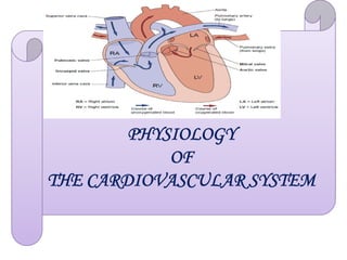
2. Learning Objectives
At the end of teaching/learning activities, students
are expected to:
Identify functional structure of the cardiovascular system
Characterize histology of the heart muscle cells
Characterize histology of the heart muscle cells
Describe pumping function of the heart chambers
Explain cardiac output and its regulation
Explain blood pressure and its regulation
Describe capillary fluid exchange
2
3.
4. Components of the CVS
1. The Heart: central pump
2. Systemic arteries:
Designed to carry blood under high pressure out to the
tissue beds
3. Arterioles & pre capillary sphincters:
Act as control valves to regulate local flow
4
4. Capillaries: one cell layer thick
Exchange between tissue (cells) & blood
5. Venules:
Collect blood from capillaries
6. Systemic veins:
Return blood to heart/dynamic storage
5. Division of the Circulation
• In the CVS, blood passes through two (double)
circulations:
1. Systemic circulation
2. Pulmonary circulation
• Systemic circulation:
Starts in the LV→ Aorta → Systemic arteries →Systemic
5
capillaries →Veins →SVC & IVC →ends in RA
• Pulmonary circulation:
Starts in the RV→Pulmonary trunk →Pulmonary arteries →
Pulmonary capillaries →Pulmonary veins →ends in the LA.
-LA=left atrium -LV=left ventricle,
-SVC=superior vena cava -IVC=inferior vena cava
8. General Functions of the CVS
1. Convective (mass movement of fluid caused by pressure
gradient) transport of O2, nutrients, water, hormones,
electrolytes, and drugs
2. Rapid washout of metabolic wastes
3. Control function relating to distribution of hormones to
tissues and secretion of some hormones like ANP
tissues and secretion of some hormones like ANP
4. Contribution to regulation of temperature and blood flow
5. Vital role in reproduction-hydraulic mechanism for penile
erection
6. Contribution to defense mechanisms by delivering
antibodies, platelets and leucocytes to affected areas of
the body.
8
9. The Heart
• Heart is the hollow, muscular organ that plays a central
pumping role
• Vertically divided into left & right sides by a structure
called septum
• Composed of 4-chambers:
– 2 atria and 2 ventricles
9
– 2 atria and 2 ventricles
• Has 4 valves:
– 2 AV valves (TCV &BCV) and 2 SLV (PV &AV)
• Size: Approximately equivalent to clenched fist
• Weight: 280 to 320 grams in average adults
• Located in the midiastenum
11. Histology of the heart
• Surfaces/Layers: The
heart is composed of
three layers:
1. Endocardium: -
innermost layer;
epithelial tissue that lines
the entire circulatory
system
11
system
2 - Myocardium - thickest
layer; consists of cardiac
muscle
3 - Epicardium - thin,
external membrane
around the heart
12. Histology of the heart…
The Pericardium
Is a fibrous closed sac investing the entire heart and
cardiac portion of great vessels.
contains a small amount of fluid for lubrication -
facilitating the continuous movement of the enclosed
heart.
Contains:
Contains:
1. Visceral layer (epicardium) immediately lining
the outer surface of the heart and
2. Parietal layer forming protective outer lining of
the pericardial sac.
The sac contains a small amount of fluid for
lubrication - facilitating the continuous movement
of the enclosed heart.
12
14. Histology of the heart…
Chambers of the Heart
The Atria: Two thin-walled overlying muscular
sheaths, serving as reservoirs and pumps
The Ventricles: Thicker-walled portion of the heart
that pumps blood from the low-pressure venous
system into the higher pressure arterial system.
Cardiac Valves
Thin flaps of flexible, endothelium-covered fibrous
tissue firmly attached to fibrous rings at the base of
the heart
Responsible for the unidirectional flow of blood
through the heart.
14
15. Types of Cardiac valves
1. The Mitral and Tricuspid (atrio-ventricular-AV)
valves
Thin-walled and located b/n the atria and the
ventricles.
Mitral (two cusps) valve lies b/n left atrium and
left ventricle
15
left ventricle
Tricuspid (three cusps) valve lies b/n Rt atrium
and Rt ventricle
16. …cont’d
The semilunar valves
Constitute the aortic and pulmonary valves locate at
the exits of the right and left ventricles.
a. Aortic valve: is three-cusped and allows blood to
flow into the aortic tree and through their cusps to
the left and right main coronary arteries
b. Pulmonary valve: allows blood to flow into the
pulmonary artery.
16
pulmonary artery.
• SL valves open when pressure in the ventricles is
greater than pressure in the arteries (i.e., during
ventricular systole) and close when pressure in the
pulmonary trunk and aorta is greater than pressure in
the ventricles (i.e., during ventricular diastole).
18. Pathway of Blood Through the Heart and Lungs
• Right atrium tricuspid valve right ventricle
• Right ventricle pulmonary semilunar valve
pulmonary arteries lungs
• Lungs pulmonary veins left atrium
• Left atrium bicuspid valve left ventricle
18
• Left atrium bicuspid valve left ventricle
• Left ventricle aortic semilunar valve aorta
• Aorta systemic circulation
19. Cardiac muscle (myocardium)
It is composed of 3 types of cardiac muscles
1. The atrial muscle
2. The ventricular muscle
3. The specialized excitatory and conductive muscle fibres
(Autorhythmic Cells)
• Atrial and ventricular muscles are contractile components
19
• Atrial and ventricular muscles are contractile components
(Contain too many actin & myosin)
• The atrial and the ventricular muscles are separated by the
fibrous skeleton of the heart
Function: -form cardiac valves
-serve as a means of attachment and insertion of
cardiac muscles
20. Pacemaker tissue of the Heart
• The SA node in the right atrium is the primary pacemaker
of the heart.
• The AV node located b/n right atrium and right ventricle
is the secondary pacemaker.
• Specialized Conductive Tissue of the Heart
• Consists of Purkinje fibers ramifying over the sub-
• Consists of Purkinje fibers ramifying over the sub-
endocardial surfaces of both ventricles.
• Purkinje cells are broad cells (70-80 µm in diameter)
compared with ventricular myocardial cells (10-15 um in
diameter).
20
21. Specialized excitatory and conductive system of the heart
It is comprised of the following components:
1. Sino-atrial node (SA-node): in which the normal (80-120
x/min) rhythmical self-excitatory impulse is generated
2. Internodal pathways: conduct impulse from the SA-node
to the Atrioventricular node (AV-node)
3. AV-node: in which impulse from the atria delayed to be
21
conducted to the ventricle. Site of nodal delay. Rhythm=40-
60 x/min
4. Atrioventricular bundle (bundle of His): which conducts
impulse from the atria to the ventricle.
Rhythm=20-40 x/min
5. Purkinje fibers: conduct cardiac impulse to the ventricles:
Rhythm=20-40 x/min
23. Velocity of conduction of AP in cardiac muscles
Structures Conduction velocity (m/s)
SA-node 0.05
Internodal fibers 1.0
Atrial muscle 0.3
23
Atrial muscle 0.3
AV-node 0.05
AV-bundle 1.0
Purkinje fibres 4.0
Ventricular muscles 1.0
24. Blood supply to the Heart
• Types of coronary vessels:
– two main coronary arteries that supply the
myocardium arise from sinuses behind two of the
cusps of the aortic valve.
a. Right Coronary Artery (RCA) supplies: Right atrium
and Posterior ventricles
b. Left Coronary Artery divides into:
i. Circumflex Artery (CA) supplying Atrium and L.
Ventricle
ii. Anterior descending artery (LDA) supplying
Right and L. ventricles
24
25. …cont’d
Coronary flow variations:
– The RCA has a greater flow in 50% of individuals;
– The left has greater flow in 20% and flow is equal in
30%.
• The heart receives arterial blood from the coronary artery,
• The heart receives arterial blood from the coronary artery,
which is the branch of ascending aorta.
• Resting coronary blood flow = 250 ml/min, 5% CO
• Coronary arterial diseases leads to Angina pectoris and
myocardial infarction (MI)
25
26. Venous Drainage
• The major venous drainage
system of the human heart
consists of four
intercommunicating parts
which generally open into the
right atrium.
• The major cardiac veins
26
• The major cardiac veins
include
o the great vein,
o the left marginal vein,
o the posterior vein of the
left ventricle and
o the middle and small
cardiac veins.
27. Coronary venous drainage
Venules
Small veins
Great cardiac vein Middle cardiac vein
27
Great cardiac vein Middle cardiac vein
From the anterior part From the posterior part
of the heart of the heart
Coronary sinus
R Atrium
28. Innervation of the Heart
• Heart has dual autonomic
innervation from both SNS and
PNS with afferent and efferent
components.
• The sympathetic nerve supply
to the heart is controlled by the
medullary vasoconstrictor/
cardio accelerator center
• Preganglionic sympathetic
28
• Preganglionic sympathetic
fibers arise from the lateral
horn of the upper 5-thoracic
spinal segments
• Postganglionic sympathetic
fibers arise from the cervical
and thoracic ganglia and
proceed to supply atria,
ventricles and nodal areas
29. Innervation of the heart cont’d…
• The parasympathetic nerve supply to the heart is controlled by the
vasodilator/ cardio inhibitor center.
• Preganglionic parasympathetic fibers arise from cardio inhibitory
center in the medulla and proceed as vagal fibers to relay in
terminal ganglia in the wall of the atria
• Short postganglionic fibers arise from terminal ganglia and supply
the atria, SA-node and the AV-node
29
the atria, SA-node and the AV-node
• The right vagus has a strong influence on SA node, while the
left vagus has dominant effect on AV node
• Ventricles are not supplied by vagus nerve
Afferent cardiac nerves include
– Pain receptors which are visceral afferent fibers
– Stretch receptors transmitted through
– Chemo receptors Sympathetic/vagus
30. Electrical Activity of Cardiac Cells
• Cardiac Excitability: varies considerably depending on
whether the action potentials are fast responses (from muscle
of atrium and ventricle) or slow responses (from SA Node,
AV node).
AV node).
• Trans-membrane Potentials: occurring across cell
membranes include:
• Resting Membrane Potential (RMP)
• Action Potential (AP)
30
31.
32. Ionic Basis of Cardiac Action Potential
• Various phases of cardiac AP are associated with changes in
the permeability of the cell membrane to, mainly, Na, K, and
Ca ions.
• During AP, the influx of sodium into the cardiac cell occurs via
two channels:
two channels:
1. A fast channel that accounts for the early influx of Na+
(atria, ventricles, and)
2. A slow channel that permits Ca++ and some sodium to
move down its concentration and electrical gradients into
the cell (SA node and AV node).
32
33. Electrophysiology of the heart
Phases and ionic basis of
myocardial action potential
It has the following phases
Phase-0: Rapid depolarization
Caused by rapid Na-influx
Phase-1: Early brief
repolarization
Caused by Cl- influx
↑PCl
33
Caused by Cl- influx
Phase-2: The plateau (prolonged
depolarization)
Caused by Ca2+influx
Phase-3: Repolarization
Caused by K+ efflux
Phase-4: complete repolarization
RMP re-established
Caused by Na+-K+-ATPase
RMP = -90 mv
34. Electrical Activity of the Pacemaker
• The pacemaker is composed of small myocytes with only
scanty myofibrils and an electrically unstable resting
membrane potential (pre-potential, about-60 to -70mV).
• Following firing of an AP, membrane potential decreases
gradually from a basal value of – 60 or-70 mV (maximum
diastolic potential) to a critical firing level of-40 to-45
mV.
mV.
• A wave of depolarization spreads across the two atria
passing from cell to cell at a rate of about 1m/s and
initiating atrial systole.
34
35. Electrical Activity …cont’d
• The pre-potential is primarily due to a slow decrease
in K+ permeability and reduced K+ efflux.
• As K+ efflux decreases, membrane begins to
depolarize forming the first part of the pre-potential.
• Pre-potential is altered by sympathetic and
parasympathetic stimulation or other factors like
drugs
35
36.
37.
38. Important terms
• Ventricular volumes: The volume of blood in the
ventricles
• Ventricular end diastolic volume (VEDV):
The volume of blood in the ventricle at the end of
ventricular diastole (relaxation phase)
EDV = 120-140
38
EDV = 120-140
• Ventricular end systolic volume (VESV):
The volume of blood that remains in the ventricle at the
end of ventricular systole (contraction phase).
VESV = 50-60 ml
39. Important terms…cont’d
• Stroke volume (SV): the volume of blood ejected
from the ventricle during ventricular systole.
SV = VEDV – VESV, 70 – 80 ml
• Cardiac output: the volume of blood ejected from the
heart per minute.
CO = SV x HR =6 L/min
39
CO = SV x HR =6 L/min
• Ejection fraction: the blood proportion that enters the
ventricles during diastole to the amount ejected.
EF = SV/VEDV, 60% - 70%
40. Heart Rate
• HR is the number of cardiac cycles per minute
• Normal HR: 60 to 100 beats/minute
– < 60 beats/minute, bradycardia
– > 100 beats/minute, tachycardia
How to count HR?
• Counting arterial pulsation, heart sound and ECG
40
• Counting arterial pulsation, heart sound and ECG
cycles
41. Heart Rate…
HR varies with the following factors
1. Age: higher in new born infants (120 b/min)
2. Sex: higher in females (85 b/min)
3. Time of the day: ↓morning, ↑evening
3. Time of the day: ↓morning, ↑evening
4. Resting and sleep: decreased
5. Physical training: low in athletes (45-60 b/min)
6. Body position: ↑standing, ↓supine positions
42. ECG…cont’d
ECG Conventions
1. 1mV input→10mm deflection
2. Paper speed 25mm/sec.
3. Recording points wrist, ankle, skin on chest
4. Right leg ground(earth)
4. Right leg ground(earth)
• ECG: Fluctuations of potential that represent the
algebraic sum of the action potentials of myocardial
fibres recorded extra-cellularly during the cardiac
cycle.
42
46. ECG Recordings
• There are 3 types of recording:
1. Bipolar Limb Leads
2. Augmented (Unipolar) Limb Leads
3. Precordial (Chest) Leads
A. Bipolar Limb Leads
• Record voltage b/n two electrodes (leads) placed on the wrists
and legs.
46
and legs.
• These leads include:
1. Lead I= LA-RA: electric potential d/ce b/n Lft arm & Rt arm
2. Lead II=LL-RA: electric potential d/ce b/n Lft leg & Rt arm
3. Lead III= LL-LA: electric potential d/ce b/n Lft arm & Lft leg.
• Each lead shows waves of depolarization and repolarization: P-
wave, QRS complex,T-wave and occasionally U-wave.
47.
48. Bipolar Limb L…cont’d
• Einthoven triangle:
– Drawn around the heart.
– The triangle shows that the two arms and the left leg form
apices of a triangle surrounding the heart.
– The two arms with electrodes connect electrically with the
fluid around the heart and the left leg with another electrode
also connects with the fluid. (Fig. below)
also connects with the fluid. (Fig. below)
• Einthoven’s law:
– If electrical potentials of any two of the three leads are given,
the 3rd one can be determined.
– The relation, Lead II = Lead I+ Lead III, can be used to
estimate electric potential of each of the leads.
48
51. ECG Recordings…cont’d
B. Unipolar (Augmented) Limb Leads
• Voltage is recorded b/n a single “exploratory electrode”
placed on the body and an electrode that is built into
the electrocardiograph and maintained at zero potential
(ground).
• In this system, two of the limbs are connected through
51
• In this system, two of the limbs are connected through
electrical resistances to the negative (indifferent)
terminal of the electrocardiograph and the 3rd limb is
connected to the positive terminal.
• When positive terminal is placed on right arm, the lead
is designated as aVR; when on the left arm aVL, and
when on left leg, aVF (Fig. below).
52.
53.
54. ECG Recordings…cont’d
C. Precordial (Chest) Limb Leads
• Unipolar leads labeled V1
through V6.
• One electrode is connected to
the positive terminal of the
Electrocardiograph and the
negative electrode (indifferent
54
negative electrode (indifferent
electrode) is connected
through equal electrical
resistances to the right arm,
left arm, and left leg.
• Electrodes are placed on the
chest.
55. …cont’d
• Heart surfaces are close to the chest wall and each chest
lead records mainly the electrical potential of the cardiac
muscle immediately beneath the electrode,
– i.e., relatively minute abnormalities in the ventricles
can cause marked changes in the ECG.
• QRS in
– V1, V2, are negative because the chest electrodes are
nearer the base of the heart (direction of
electronegativity).
– V3 is in between.
– QRS of leads v4-v6 are positive because they are
nearer the apex (direction of electro positivity).
55
58. Application of ECG
• By analyzing electric potential fluctuations, the
physician can get some insight into:
– Anatomical orientation of the heart,
– Heart rate determination
– Relative size of heart chambers,
– A variety of disturbances of rhythm and conduction
– A variety of disturbances of rhythm and conduction
arrhythmia and conduction block
– Extent, location and progress of ischaemic damage
(myocardial infarction)
– Hypertrophy
– Pericarditis and myocarditis
58
70. Cardiac cycle
Activities in the heart in a single beat
Contraction (systole) and relaxation (diastole) of cardiac
chambers
A single cardiac cycle comprised of
- Atrial systole and atrial diastole
-Ventricular diastole + ventricular systole
70
-Ventricular diastole + ventricular systole
75 cycles completed per minute
Duration of each cycle = 0.8 second
-Ventricular diastole = 0.5 second
-Ventricular systole = 0.3 second
71. Means of exploring activities accomplished in
a cardiac cycle
1. Auscultation of heart sounds, phonograph
2. ECG tracing
3. Measuring aortic pressure
4. Measuring atrial pressure
5. Measuring ventricular pressure and volumes
5. Measuring ventricular pressure and volumes
72. Phases of the Cardiac Cycle
• Includes the following events:
– Electrical events of summated ECG voltage changes-P
wave, QRS complex,T wave and U wave in some cases
– Mechanical events of myocardial diastole and systole with
opening and closing of cardiac valves, pressure and
volume changes, heart sounds, atrial (jugular) a,c,v, waves.
72
volume changes, heart sounds, atrial (jugular) a,c,v, waves.
• Phases:
I. Atrial and Ventricular diastole
II. Phase of systole
73. Phases of …cont’d
A. Phase of Diastole (Atrial and ventricular diastole):
Atrial pressure rises before the tricuspid valve opens during
diastole. This is mirrored by v-wave
Atria and ventricles are relaxed.
As pressure falls below that in the atrium, it leads to opening of
AV valves and closing of semi-lunar valves
73
AV valves and closing of semi-lunar valves
Blood flows into the heart filling both atrial and ventricular
chambers throughout diastole.
Rapid filling, proto-diastole occurs for 0.04 sec.
When pulse rate is slow and diastole is long, inflow is slowed in
the latter part of diastole→ a period of diastasis.
74. Phases of …cont’d
The 3rd heart sound begins with end of rapid inflow and ends
with beginning of diastasis.
Proto-diastole + diastasis =70% of inflow
About 70% of the ventricular filling occurs passively.
The remaining 30% of filling occurs by active atrial systole
P-wave formation begins with diastasis and ends with atrial
74
P-wave formation begins with diastasis and ends with atrial
systole.
The beginning of the 1st heart sound and the end of QRS
complex coincide with isovolumetric or isometric contraction
(0.02-0.06 sec).
2nd heart sound occurs with closure of semilunar valves and
during isometric relaxation (0.09-0.12 sec)
75. Figure Events of the cardiac cycle for left ventricular function (left atrial pressure, left
ventricular pressure, aortic pressure, ventricular volume, the electrocardiogram, and the
phonocardiogram
75
76. Phases of …cont’d
B. Phase of Systole
a. Atrial systole (contraction):
Propels an additional blood of about 30% of EDV into
ventricles
Preceded by P-wave
EDV =120 mL
76
EDV =120 mL
Forms a-wave, formed by atrial systole. This is one of the
three atrial pressure waves (a,c,v) transmitted to thoracic
and neck veins (Fig.below)
– ↑ ventricular volume
– ↑ventricular pressure (atrial kick)
78. Phases of …cont’d
b. Ventricular Systole
– During this period:
– Isovolumetric contraction (0.02-0.06 sec) occurs
– i.e, there will be:
No change in ventricular volume or length of
ventricular muscle fibers
78
ventricular muscle fibers
Rise of BP in left ventricle from 4 to 80 mmHg
Closed AV valves
Closed SL valves
Aortic and pulmonary pressures are at their lowest
levels
79. Phases of …cont’d
• c-wave as transmitted manifestation of the rise in atrial
pressure is produced by the bulging of tricuspid valve into
atria during iso-volumetric contraction (ventricular systole).
• c-wave is synchronous with pulse wave in carotid artery, and
hence the use of the letter “c”,
• During isovolumetric contraction part of the first heart sound
develops, QRS complex develops fully and Ventricular
pressure
79
pressure
rises from 4mmHg to 80 mmHg
• At the end of isovolumetric contraction:
– SL valves open
– Ventricular pressure rises from 80 mmHg to 120 mmHg
– Rapid ejection period (0.10 sec) occurs…70% of SV
– Slow (reduced) ejection (0.08 sec) follows,,,30% of SV
– Isovolumetric relaxation follows
80.
81. …cont’d
• SV= 80 ml, i.e. ejection fraction (80/120), EF=67%,
ESV=120-80ml =40 ml
• Ventricular volume goes down to ESV
• Most of the T-wave occurs during the ejection
period
• Pressure in ventricle falls slightly below atrial pressure
• Valves and aorta being distensible, undergo recoil and
produce a secondary pressure wave in aortic curve with a
notch between systolic and diastolic pressure curves.
• This is referred to as dicrotic notch - a small oscillation
on the falling phase of the pulse wave (Fig. below)
formed when the aortic valve snaps shut following full
emptying of blood.
81
82. 120 mm Hg
Systolic pressure
Aortic pressure
120 mm Hg
Systolic pressure
80 mm Hg
Dicrotic notch (incisura)
Diastolic pressure
80 mm Hg
Dicrotic notch (incisura)
Diastolic pressure
84. Heart Sounds
Heart sounds (lubb-dubb) are associated with closing of
heart valves
4-separate audible heart sounds (S1, S2, S3 and S4)
Means of identification
1. Auscultation: direct/immediate auscultation
Stethoscope mediated auscultation
2. Phonocardiographic based recording
84
2. Phonocardiographic based recording
S4 S1
S2
S3
S1: is always audible, has a LUBB-sound
S2: is always audible, has DUBB-sound
Continuous heart sound: Lubb-Dubb, Lubb-Dubb
Duration of Lubb is shorter than that of Dubb
S3: is audible in children and in adults during exercise
S4: is audible very rarely
85. S1: First heart sound
– Slightly prolonged “lub”sound
• Caused by sudden closure of AV-valves.
• Timing: occurs at the beginning of ventricular systole
– Associated with isometric contraction of ventricles
– Intensity directly proportional to rate of IVP rise
Heart Sounds (cont’d)
85
– Intensity directly proportional to rate of IVP rise
– Best heard over cardiac apex
– Asynchronous closure of tricuspid and mitral valves in
health →splitting of S1
86. S2: Second heart sound
– Shorter, high pitched “ dub” sound
– Caused by sudden closure of semi-lunar valves and
vibration of the valves and large arteries
– Occurs at the beginning of ventricular diastole,
– Less than 0.05 sec duration
– Associated with isometric relaxation of ventricles...minor
Heart Sounds (cont’d)
86
– Associated with isometric relaxation of ventricles...minor
contribution to sound
– Intensity directly proportional to rate of IVP fall
– Asynchronous closure of aortic and pulmonic valves
during inspiration....
– Increased filling of RV→delay in closure of pulmonary
valve →splitting of 2nd heart sound.
– BBB→Split 2nd heart sound
87. S3: Third heart sound
• Usually inaudible
• Detected by phonocardiograph
• 0.04 esc duration
• Develops 0.10 to 0.20 sec after 2nd heart sound
Heart Sounds (cont’d)
87
• Develops 0.10 to 0.20 sec after 2 heart sound
• Caused by rapid filling of the ventricles with blood
during ventricular diastole
• Audible in children and in adults during exercise
88. S4: Fourth heart sound
– Very rarely audible
– Detected by phonocardiograph
– 0.04 sec duration atrial systole→blood flow on
ventricular walls →vibrations
Heart Sounds (cont’d)
88
ventricular walls →vibrations
– Caused by rapid ventricular filling during atrial systole
– Occurs during atrial systole
89. Abnormal Heart sounds
• Heart murmurs: Abnormal sounds heard in various
parts of the vascular system.
a. Systolic murmurs:
• AV-valve insufficiency
• Semilunar valve stenosis
• High CO (physiological)
89
• High CO (physiological)
b. Diastolic murmurs
• Semilunar valve insufficiency
• Av-valve stenosis
• Valvular insufficiency causes regurgitation
90. Graphic presentation of phonocardiograms of murmurs under various
conditions is as shown below.
91. Cardiac Output (CO)
Cardiac Output:
– It is the amount of blood pumped by each ventricle in
one minute
– It is the product of heart rate (HR) and stroke volume
(SV)
oHR is the number of heart beats per minute
91
oHR is the number of heart beats per minute
oSV is the amount of blood pumped out by ventricle
with each beat
oSV= ejection fraction x EDV
oEjection fraction =2/3 of blood contained and
ejected by the heart
92. …cont’d
Cardiac reserve:
– It is the difference between maximal exercise and
resting CO
oCO = 5-6 liters per minute at rest
oCO = 20-40 liters per minute at maximum exercise
92
E.g. 35L-5 L =30L
Cardiac Index:
– It is the amount of blood pumped of the left ventricle
each minute
CI = Cardiac output÷ Body surface area
E.g. 5.5 L/min ÷1.7 =3.23 L/m2
93. Effect of Venous Return (VR) on C.O
Venous return is:
1. volume of blood flowing from veins into RA/min
2. 5.5L/min at rest; about 35L/min in well trained athletes
during strenuous exercise
3. influenced by RAP like C.O as shown on cardiac
function curves (Fig..)
93
function curves (Fig..)
– C.O = VR at the “operating point” of the cardiac
function curves (Fig..).
– Any factor influencing C.O or VR shifts the “operating
point” of C.O/VR intersection to a new position.
97. Factors affecting Venous Return
1. Mean Systemic Filling Pressure (MSFP)
– Pressure measured every where in the systemic
circulation 1 min after blood flow has been stopped by
clamping large blood vessels near the heart.
– It is 7 mmHg and indicates the degree of filling of the
systemic circulation
97
– It is the driving force for VR.
2. Right Atrial Pressure(RAP)
– Mean pressure in the right atrium (Central venous
pressure) =2 mmHg
– Pressure gradient= MSFP-RAP
– RAPVR
98. Factors affecting ....cont’d
3. Resistance to venous return (RVR)
– Resistance blood meets(1mmHg/L/min) during its flow
from arterial side to RA
– Occurs mainly at arterioles
– VR= MSFP-RAP/RVR=MAP-RAP/TPR
4. Sympathetic stimulation
98
4. Sympathetic stimulation
– VR by inducing vasoconstriction, improved cardiac
pumping power and arteriolar dilatation
5. Blood volume
– Blood volume VR by MSFP and vice versa.
6. Respiratory movements
– VR increases with inspiration and decreases with
expiration
99. Factors affecting ....cont’d
7. Arteriolar dilatation RVR and VR
8. Capillary dilatation vascular capacity, MSFP and
VR
9. Skeletal muscle contraction
– Squeezes veins b/n muscles MSFP VR
99
10. Gravity
– Standing motionless for some time pooling of
blood in lower extremities MSFP VR
C.O hypotension brain ischaemia syncope.
100. Regulation of Pumping Activity of the Heart
• There are two types of regulatory mechanisms:
1. The Intrinsic Auto-regulatory Mechanisms (The Frank
Starling Mechanism)
“Within physiological limits, the contractile force of the
heart is proportional to the amount of the blood enters
the heart”.
2. The Extrinsic Regulatory Mechanisms
Sympathetic, parasympathetic stimulation
Catecholamine
Electrolytes (Ca2+, K+)
Drugs: Cardiac glycosides, xanthenes
Hormones: AD, Glucagon, T3/T4
100
101. Frank Starling Law of the Heart
• Frank-Starling mechanism means the greater the heart
muscle is stretched during filling →↑force of contraction
→↑blood pumped to the aorta.
• Pre-load or degree of stretch of cardiac muscle cells before
they contract is the critical factor controlling stroke volume
– It depends on end diastolic volume
– It depends on end diastolic volume
• Slow heart beat and exercise → ↑VR to the heart →
increasing EDV that in turn, ↑SV
• Blood loss and extremely rapid heartbeat → decreases
ventricular filling time and decreasing EDV → that
decreases SV
101
104. …cont’d
• A few seconds of heterometric autoregulation(pre-
load)→↑ metabolic state of heart→ more forceful
contraction and 30-60 sec. after, pressure elevation
occurs; the heart expels more blood than is brought into it
• EDV & ESV persists for 2-3 minutes.
• This increase in contractile force without a change in
EDV is referred to as Homeometric (after-load)
EDV is referred to as Homeometric (after-load)
regulation, a condition mediated by a change in metabolic
state or contractility of the heart.
NB:
– Heterometric autoregulation is related to filling
pressure of the heart
– while homeometric autoregulation is related to
maximum pressure (contractile force).
104
105.
106. …cont’d
• A sudden increase in venous return (VR) in heart size.
• For example, a change from erect to supine position leads to
shift of blood from lower extremities to thorax CVP(4-
12cm H2O) in heart size.
• The heart may be:
Hyper-effective (capacity more than normal) due to
sympathetic stimulation or hypertrophy.
Hypo-effective and pumping capacity is lower than normal
Hypo-effective and pumping capacity is lower than normal
due to:
• Sympathetic denervation
• Parasympathetic stimulation
• Atrial flutter or fibrillation
• Myocarditis
• Cardiac hypoxia
106
109. • Arteries and veins resemble each other in that their walls
contain three coats.
• However, the vessels adapt to their different circulatory tasks
by differing in the structure of these coats.
• The inner coat (vascular endothelium, tunica intima) consists
of a single layer of endothelial cells applied to a thin
connective tissue layer, the basement membrane.
The Structure of Arteries and Veins
connective tissue layer, the basement membrane.
• The middle coat (tunica media) contains primarily smooth
muscle and elastic tissue fibers.
• The outer coat (tunica adventitia, adventitious coat) embeds
the vessel in its surroundings and consists mainly of
connective tissue.
• In addition, the arteries have an elastic, fenestrated membrane.
109
110. • Arteries are distinguished by an especially well developed
muscle coat, which contains a varying amount of elastic fiber
according to its site (predominantly elastic and
predominantly muscular arteries).
• This layer is the driving force of the blood vessels by
dilating (vasodilatation) and constricting
(vasoconstriction) the diameter of the blood vessels, it
….cont’d
(vasoconstriction) the diameter of the blood vessels, it
regulates blood flow and blood pressure.
• The arteries near the heart contain a high proportion of elastic
fibers and this creates an elastic recoil.
• In the smallest blood vessels, the capillaries, the coats are
reduced to one, the tunica intima; this facilitates the exchange
of fluids and gases.
110
112. The lymphatic system runs parallel to the venous side of the
circulation
It begins near the capillaries as “blind” lymphatic capillaries that
reabsorb fluid that has not been taken up from the tissues by the
blood vessels (lymphatic fluid).
Small and large lymph vessels then return the lymph to the venous
blood.
Lymph Vessels
blood.
The wall of the lymph vessels consists of an endothelium, and a
thin layer of rhythmically contracting smooth muscle cells.
Similarly to the situation in the veins, numerous valves further the
transport of the lymph.
The course of the lymph vessels is interrupted by lymph node
stations, which represent a kind of biological filter and fulfill
important functions in immune defense.
112
113. Functional Classification of Blood Vessels
1. Elastic vessels (Windkassel vessels):
Example: Aorta, big arteries
Pressure storing components
High ability of recoiling
2. Resistance vessels (Stop Cock Vessels)
Example: small arteries and arterioles
113
Example: small arteries and arterioles
High muscular component
Develop high resistance
Regulate blood flow
114. Blood vessels: Classification…cont’d
3. Exchange vessels:
Example: capillaries
Made up of endothelium and basement membrane.
Thin enough for exchange
3 types: continuous, discontinuous and fenestrated
114
3 types: continuous, discontinuous and fenestrated
capillaries
4. Capacitance vessels (big to small veins)
Very high capacity of distension
Can accommodate large volume of blood (65% of
blood volume)
115. • AORTA branches peripheral vascular bed1000 fold
increase in cross-sectional area at capillaries
• Large amount of elastinexpansion and recoil
pressure storage.
• A velocity of 40 cm/sec at aorta <2mm/sec at
capillaries,
i.e. V=Q/A.
Arteries
i.e. V=Q/A.
• Beyond capillaries, the situation is reversed and blood
flow accelerates as total cross-sectional area of veins
decreases.
115
118. •The overall strategy of the cardiovascular system is to :
1. provide all organs with a constant perfusion pressure
and
2. allow each individual organ to regulate its blood flow
in accordance with the local needs of the tissue.
• ABP includes pressure in the aorta and other large
arteries.
Arterial Blood Pressure (BP)
118
arteries.
NB:
• BP is conventionally written as systolic (peak, ejection
pressure)/ diastolic (minimum, filling pressure) blood
pressure.
Normal SBP: 90 – 130 mm Hg (120 mm Hg)
DBP: 60 – 90 mm Hg (80 mm Hg)
PP: SBP – DBP
MAP: Pd + 1/3(Ps - Pd)
120. 1. Invasive (direct) measurement:
– A cannula is inserted into a large artery and pressure measured
directly with a manometer or a strain gauge connected to a
recorder.
– The record is made as shown below.
Measurement of ABP
120
121. 2. Indirect routine measurement
– This method uses Riva Rocci cuff attached to a
sphygmomanometer
a.Auscultatory method: BP(S/D) is read using Korotkoff’s
sound.
– BP reading in:
• Children: muffling of Korotkoff’s sound for DB
...cont’d
• Children: muffling of Korotkoff’s sound for DB
• Adults: complete cessation of Korotkoff’s sound for DB
b.Palpation method: used to estimate systolic BP
– Approximation of mean BP
121
122.
123. Recommended cuff sizes for estimating BP
1. Under one yr of age =2.5cm
2. 1-3 yrs =5-6cm
3. 4-8 yrs- 9-10cm
4. Average adults =12.5cm
5. Obese adults =14cm
...cont’d
• Venous pressure measurement: use a hypodermic needle
to be inserted into a vein and connect it to a sensitive
aneroid manometer.
123
124. 1. Posture: In the upright position any vessel below heart level
has increased BP,
– e.g. BP in dorsalis pedis =200 mmHg
– Gravitational effect on BP=0.77 mmHg/cm
2. Age: Newborn: 70/50, infants: 90/60, children: 100/70
– SBP increases by 1mmHg/yr, secondary to
General Factors that affect arterial BP
atherosclerosis.
– DBP increases by 0.4 mmHg/yr secondary to increase
in TPR.
3. Sex: ♀sex 40-50 yrs low BP,♀ 50 yrs high BP.
– This Bp could be higher in women than in men.
– probably due to hormonal changes during menopause.
124
125. ...cont’d
4. Body weight: both SBP and DBP are directly related to
body wt.
ABP is higher by 10-15 mm Hg in obese persons.
5. Race and socioeconomic status: Both SBP & DBP are
higher in blacks than those in whites at all ages and for
both sexes.
both sexes.
This difference is attributable to genetic factors and/
or economic status.
126. 6. Exercise: SBP and DBP increase with exercise.
SBP is secondary to contractility;
DBP may decrease initially secondary to vasodilatation
in skeletal muscle vasculature but heart rate limits
runoff timeDBP.
Exercise increases ABP by 40 – 50 mm Hg
...cont’d
7. Emotion: increases ABP by 10– 30 mm Hg
8. Deep sleep: decreases ABP by 20 mm Hg
9. Time of the day: ↓BP in the morning, ↑BP in the evening
10. Thermal stress decreases ABP
126
127. Regulation of ABP
A. Short-term controlling mechanisms
CNS ischemic response
Baroreceptor reflex
Chemoreceptor reflex
Atrial stretch reflex
Stress relaxation and reverse stress relaxation
127
Stress relaxation and reverse stress relaxation
Capillary fluid shift
Hormonal mechanisms: AD, NAD, Vasopressin (ADH)
B. Long-term controlling mechanisms
The renal mechanism
The Renin-angiotensin-aldoterone system
129. Baroreceptor Reflex
↓ ABP
b) Aor c Sinus→ Vagus nerve
a) Caro d Sinus→ Hearings nerve →
GPN
Medullary CVC
129
Stimulation of the SNS
On Heart
• ↑HR
• ↑Force of contrac on
• ↑CO
On Blood Vessels
• ↑TPR
↑ABP
130. RENIN-ANGIOTENSIN-ALDOSTERONE MECHANISM
ABP (Kidney JG-cells)
produce Renin
Angiotensinogen (Renin substrate)
(Liver)
Angiotensin-I
Angiotensin-II
ACE (Lung)
Stimulates the
Thirst centre Vasoconstriction
Venoconstriction
ADH
secretion
Kidney
Aldosterone Secretion
Sodium & water retention
ECF/Blood Volume
ABP
131. Blood vessel functions: overview
Strong and elastic arteries
Arterioles control blood
flow and pressure
131
Capillaries: thin and with
large area for diffusional
exchange
Veins: compliant, large, low resistance
veins have valves & assure blood return to the heart
132. CAPILLARIES
• Pressure inside is 15 to 35 mmHg
• 5% of the blood is in capillaries
• Necessary for exchange of gases, nutrients, and wastes
• Flow is slow and continuous
• Blood flow controlled by pre-capillary sphincter muscles via
132
• Blood flow controlled by pre-capillary sphincter muscles via
metabolic regulation
• No smooth muscle, no innervations
• Largest total cross sectional area
• Based on the continuity of their filtration barriers, capillaries
have been labelled as continuous, fenestrated and
discontinuous
134. Capillaries Exchange
• There are two forces that determine the direction of flow of
fluid through the capillaries
– The capillary colloid osmotic pressure (25 mm Hg)
– The capillary hydrostatic pressure (32mmHg)
• Fluid that is leak out of the capillaries is taken up by the
lymphatic vessels to prevent edema
134
lymphatic vessels to prevent edema
• Net outflow into ECF
– Net filtration – net absorption = net outflow
– About 2 L/day collected by lymph vessels
138. • Lowest
velocity
• Largest total
cross sectional
Capillary Blood Flow-Velocity
138
cross sectional
area
• Hydrostatic
pressure drops
slightly
Figure. The velocity of flow depends on the total
cross-sectional area
139. Blood flow
• Blood flow (BF) is the amount of blood that moves to a
particular organ in a given time
• Total blood flow is equal to CO; 5-6 L/min
• BF is determined by 2 factors
1. Pressure difference b/n 2 ends of the vessel
i.e., the force that pushes blood through vessels
139
i.e., the force that pushes blood through vessels
2. Resistance of flow, hindrance to flow through vessels
Where Q = Blood flow
Q = ΔP/R ΔP = Change in pressure
R = Resistance
• Ohms Law: states that BF is directly proportional to the ΔP
but inversely proportional to resistance (R).
140. Vascular resistance
• Poiseuille's Law: Vascular resistance to BF is directly
proportional to the length of the vessel and viscosity of blood, but
inversely proportional to the 4th power of radius of the vessel.
Where, R = Resistance
l = Length
= Viscosity
= Circle constant
(3.14)
R = 8l/r4
Pr4
8l
Q =
140
(3.14)
r = Radius
Factors affecting viscosity of blood
• Velocity of BF: ↑Velocity = ↓Resistance
• Hematocrit: polycythemia = ↑Viscosity = ↑PR
Anemia = ↓Viscosity = ↓PR
• Diameter of blood vessels
• Plasma protein concentration
Q=Blood flow
PR=Peripheral resistance
141. Resistance to blood flow
• It is the hindrance to blood flow
• Has no direct means of measurement, but can be calculated
from values of BF and P in vessels
• If the P is 1 mm Hg and BF 1 ml/sec, then vascular
resistance is said to be 1 peripheral resistance unit (PRU)
Total peripheral resistance and total pulmonary resistance
141
• The rate of BF through the circulatory system at rest is 100
ml/sec or 5 L/min.
• The P between systemic arteries and veins is 100 mm Hg
• Resistance of the entire systemic circulation, called TPR is
100/100 = 1 PRU.
• TPR ranges between 0.2 and 4 PRU during vascular dilation
and constriction
142. TOTAL PERIPHERAL RESISTANCE
TPR = Aortic Pressure - RAP
FLOW
TPR =
100 - 0 mmHg
83.3 ml/sec (5 L/min)
= 1.2 PRUs
SYSTEMIC CIRCULATION: RAP=Right atrial
pressure
142
PULMONARY CIRCULATION:
Pul. R. =Pul. Art. P. - LAP
FLOW
Pul. R. = 15 - 5 mmHg
83.3 ml/sec
= 0.12 PRUs
143. • Water and solutes accumulate in IF compartment oedema,
• It is pitting if firm pressure on swollen area leaves an
impression-occurring in dependent parts- extracellular
oedema.
• If the oedema is non-pitting, it is cellular.
• Oedema in general occurs when the increase in tissue fluid
volume is 30% or more of the basal volume or when
Oedema
volume is 30% or more of the basal volume or when
filtration is >reabsorption + lymphatic drainage
Oedema may be evident from:
• Puffy face (eyes) in the morning
• Puffy feet
• Sacral oedema when patient is lying in bed.
143
144. • Expanded IF volume
• Total body sodium
Possible causes of pitting oedema
1. Increased hydrostatic pressure in capillaries secondary to
heart failure
Generalized Oedema (Anasarca):
heart failure
2. Decreased osmotic pressure due to low plasma proteins
secondary to conditions like nephrotic syndrome
3. Increased capillary permeability
4. Lymphatic obstruction (non-pitting edema). . Localized
144
145. Cellular or non-pitting oedema:
• may be caused by:
1. Depression of cellular metabolism→↓Na+-K+
pump→↓Na+ influx into cell →Water following
Na+→Cellular oedema
2. Inflammation→↑membrane permeability to sodium and
other ions that diffuse into cell with water→oedema.
…cont’d
145
other ions that diffuse into cell with water→oedema.
Accumulation of water inside cells→Water intoxication.
Water intoxication: occurs
• when excessive volumes of water are absorbed too
quickly leading to nausea, vomiting and shock--
conditions caused by undue drop in plasma osmolality
before adequate inhibition of ADH secretion occurs.
146. Other related problems
• Depletion of ECF volume: low plasma vol. low BP,
rapid pulse; low IF vol. poor skin turgor, dry tongue,
sunken eyes.
• Depletion of ICF Volume: Brain goes mad with thirst
…cont’d
• Depletion of ICF Volume: Brain goes mad with thirst
due to intense stimulation of hypothalamic
osmoreceptors
146
147. Characteristics of the Venous System
Veins:
Constitute the low pressure system and extend b/n smallest
venules (>10mm Hg) and largest terminal veins (venae cavae
= 0 mmHg).
Are wide-bored and thin-walled
Are volume storer (high capacitance) of the circulation
Are low pressure vessels
Are low pressure vessels
Have great distensibility
Have 5-6 times greater distensibility and 2-3 times greater
capacity than the arteries.
Have one-way valve to break up columns of blood and to
prevent excessive venous distension.
Faulty valves may lead to development of varicose veins.
Use sympathetic supply to facilitate circulatory homeostasis.
148. Venous Pressure
– 12-18 mmHg in venules
– 5.5 mmHg in large veins
– Mean pressure in ante-cubital vein is 7.1mmHg
– At entrance of large veins into RA (CVP), the average
pressure is 4.6 mmHg
…cont’d
pressure is 4.6 mmHg
– Like ABP, peripheral venous BP is affected by gravity;
it is increased by 0.77mmHg for each cm below RA
and decreased by a similar amount for each cm above
RA (Fig..).
148
150. Pressures, either central (CVP, low) or peripheral (relatively
high), can be estimated by inserting a catheter into the
thoracic great veins.
The pressure in peripheral veins is greater than central venous
pressure by an amount proportional to the distance away from
Measurement of venous pressure
pressure by an amount proportional to the distance away from
the right side of the heart.
CVP can be estimated simply by noting the height to which
the external jugular veins are distended when the subject lies
with the head slightly above the heart.
150
151. Measurement of venous pressure
The vertical distance b/n RA and the place the vein
collapses (pressure=0) is the venous pressure in mm of
blood.
CVP is decreased during negative pressure breathing and
shock. (inspiration)
It is increased by +ve pressure breathing(expiratipon),
straining, expansion of blood volume and heart failure.
Normal venous pulsations are weak and usually do not
appear at levels higher than the sternal angle.
152. To make them visible in the neck veins, the subject has to
recline on his back with the head supported on pillows
(Fig..below). In this position, pulsations in internal jugular
vein can be seen and examined.
Maximum jugular pulsations are observed when the trunk is
CENTRAL VENOUS PRESSURE (CVP)
Maximum jugular pulsations are observed when the trunk is
inclined by less than 30o.
The vertical distance b/n the right atrium (sternal angle) and
the place the vein collapses (the place where the pressure in
it is zero) is the venous pressure in mm of blood.