
Recommandé
Recommandé
Contenu connexe
Similaire à General embryology for students reading part I (2).pptx
Similaire à General embryology for students reading part I (2).pptx (20)
Dernier
Dernier (20)
General embryology for students reading part I (2).pptx
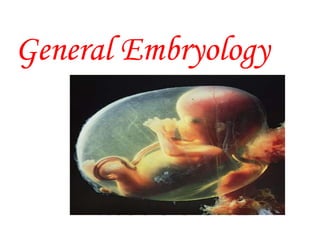
- 2. Introduction to Human Embryology Embryology :- the study of embryos, generally prenatal dev’t. The study of the origin and development of an organism. Developmental anatomy is the field of embryology concerned with the changes that cells, tissues, organs, and the body as a whole undergo from a germ cell of each parent to the resulting adult. Prenatal development is more rapid than postnatal development and results in more striking changes.
- 3. Human development is a continuous process that begins when an oocyte (ovum) from a female is fertilized by a sperm (spermatozoon) from a male. Cell division, cell migration, programmed cell death, differentiation, growth, and cell rearrangement transform the fertilized oocyte, a highly specialized, totipotent cell, a zygote, into a multicellular human being.
- 4. ✽ Development ☞ Growth (↑ in mass of tissues) ☞ Differentiation (↑ in complexity) ✽ Development does not stop at birth. Important changes, in addition to growth, occur after birth. ✽ Divided into: ☞ Prenatal period ►pre & embryonic period ► fetal period ☞ Postnatal period (after birth) ✹ neonatal period and Infancy ✹ childhood ✹ adolescence ✹ adulthood
- 5. Illuminates gross anatomy and explains how normal and abnormal relations develop. bridges the gap between prenatal development &obstetrics and pediatrics. Develops knowledge concerning the beginnings of human life and the changes occurring during prenatal development. helps to understand the causes of variations in human structure. SIGNIFICANCE OF EMBRYOLOGY
- 7. Internal genitalia( uterus, uterine tube ,ovaries and vagina ). External genitalia ( clitoris, labia majora ,labia minora ,vestibule ,mons pubis) Female reproductive structures Includes :-
- 8. Uterus Hollow thick walled, pear shaped muscular organ. Size ; 7-8 cm length , 2-3 cm thick in non pregnant Parts:- body, cervix &isthmus Wall :- perimetrium myometrium & endometrium
- 9. Layers of Endometrium :- 4-5 mm thick During secretory phase of menstrual cycle three layer of endometrium can be identified microscopically . functional layer- shade during menstruation thin compact layer:-densely packed CT thick spongy layer:-edematous CT. Basal layer:- is not sloughed off during menstruation
- 10. The uterine tube
- 11. Female reproductive cycle (sexual cycles Begin at puberty ,prepare reproductive organ for pregnancy. It involves hypothalamus , pituitary gland ,ovaries, uterus, uterine tubes, vagina, and mammary glands Hypothalamus--- GnRH--- pituitary --- LH & FSH. FSH :-dev’t of ovarian follicle & estrogen production LH:-ovulation & progesterone production. Ovarian Cycle :-cyclic change on the ovary, by LH & FSH -development of follicle -ovulation -dev’t of corpus luteum
- 12. 1. Follicular dev’t It is characterized by :- Growth & differentiation of primary oocyte. Proliferation of follicular cells Formation of zona pellucida/helps to prevent Poly spermy and premature implantation Development of the theca folliculi 2. Ovulation -FSH & LH- ovarian follicle , sudden growth burst ,swelling and stigma. -LH surge - follicular rupture & secondary oocyte expel , meiosis I end ,meiosis II started.
- 14. The principle mechanism leading to expulsion of oocyte is: A.Raised intrafollicular pressure B.Contraction of smooth muscles in theca externa C. Enzymatic/collagenaze digestion of follicular wall D. Increased LH production from pituitary gland 3. Corpus luteum Glandular structure Formed by follicular cell & theca folliculi Produce some estrogen & mainly progesterone Type :- of pregnancy :- of menstruation
- 15. Corpus luteum of pregnancy:- formed if ovum is fertilized It enlarge and increase its hormone production human Chorionic Gonadotropin prevent its degeneration Functionality - upto 20weeks of pregnancy Corpus luteum of menstruation formed if the ovum is not fertilized Degenerate 10-12 days after ovulation Subsequently changed to corpus albicans
- 16. 1 Oocyte 2 Pellucid zone 3 Stratum granulosum 4 Theca interna 5 Theca externa 6 Antral follicle 7 Cumulus oophorus (Granulosa cells, together with the oocyte) 8 Basal lamina between theca and stratum granulosum
- 17. Menstrual ( endometrial )cycle A cyclic changes in the endometrium Produced by estrogen and progesterone Time during w/c – oocyte matures, ovulated, and enters the uterine tube Phases of the Menstrual Cycle
- 18. Phases of the Menstrual Cycle 1. Menstrual phase -80ml of blood flows -functional layer sloughed off 2. Proliferative phase/estrogenic/follicular -thickness of functional layer -by increased estrogen -follicles proliferate 3. Secretary phase/luteal/progestetional -progestrone stimulates uterus to secrete glycogen -spiral arteries grow,enlarge -if ova is not fertilizzed there will be intermitent contraction of the uterus. The ova come to uterus. 4. Ischemic phase -corpus luteum degenerate -spiral arteries bacame constricted -marked shrinkage of endometrium.
- 19. • Includes scrotum,testes, spermatic ducts, sex glands and penus. • Work together to produce sperm and semen as well as to deliver it out of body and into the vagina. Male reproductive system
- 20. THE BEGINNING OF HUMAN DEVELOPMENT Gametogenesis & anatomy of female reproductive trac Conversion of primordial Germ Cells into Male and Female Gametes. spermatogenesis and oogenesis The sequence of gametogenesis is the same, but the timing of events during meiosis differs in the two sexes. Involve mitosis , meiosis and cytodifferentiation. prepares these sex cells for fertilization.
- 21. Gametes are derived from primordial germ cells (PGCs) that are formed in the epiblast during 2nd week & that move to the wall of yalk sac . During 4rth week these cells begin to migrate toward the developing gonads where they arrive by 5th week. mitotic division Increase their number during their migration. When they arrive in the gonad ,in preparation for fertilization ,germ cells undergo gametogenesis which includes meiosis to decrease chromosome no.by meiosis &cytodifferentiation to complete their maturation. Phases of Gametogenesis:-
- 22. Meiosis Special type of cell division that takes place in germ cells only in the gonads to generate male and female gametes. Reduce chromosome number by half to haploid. Require two cell divisions meiosis I and meiosis II Meiosis I- Prophase I :- chromosomes condense Homologous chromosomes lie side by side forming tetrad or bivalent. Crossing over ,physical exchange of b/n chromosome segments of nonsister chromatid occurs. sister chromatids of homolog.chrom. attach to spindle fibers by the kinotochores. Metaphase I:-homologous pairs align at the equator randomly
- 23. Anaphase I :-the homologous chr.separate & move towards opposite poles. Telophase I:-chromosome decondense &nuclear envelope reforms. cytokinesis separate the cytoplasm &the two daughter cells form by cleavage furrow.
- 27. Meiosis II Prophase II:- no DNA replication -chromosome condense -spindle fibers form. Metaphase II :-chromosome align at the equator. Anaphase II & telophase II:-single chromatid chromosome is distributed to each daughter cell. Cytokinesis:-cell divide & produce 4 Spermatids in Male -1 definitive oocyte & 3 polar bodies in Female - all contain 23 single chromatid chromosomes.
- 28. Importance of meiosis Provide constancy of chromosome number by reducing it to haploid. Allow random assortment of maternal and paternal chromosome between gametes. Recombination of genetic material by crossing over.
- 29. Spermatogenesis • Primordial germ cell:- migrate from wall of yolk sac to the gonads at 4th wks development • Begins :- 13-16 yrs - old age • Spermatogonia (type A & B)transferred to spermatozoa • Spermatogonia -mitotic division -primary spermatocytes • primary spermatocytes (largest germ cell)-meiosis I - two secondary spermatocytes • secondary spermatocytes-meiosis II -four haploid spermatids • Spermatids -spermiogenesis -mature sperm
- 30. Condensation of nucleus Shading of cytoplasm/unneccessary materials will be discarded Acrosome formation Formation of neck and tail Many genes &molecular factors are implicated in the Process of spermatogenesis. Spermiogenesis:-
- 31. 1 Axonemal structure, first flagellar primordium 2 Golgi complex 3 Acrosomal vesicle 4 Pair of centrioles (distal and proximal) 5 Mitochondrion 6 Nucleus 7 Flagellar primordium 8 Microtubules 9 Sperm cells tail 10 Acrosomal cap
- 32. Spermatogenesis
- 33. The entire spermatogenesis takes about 60-64 days Regulation of spermatogenesis is by LH & FSH.
- 34. • Mature sperms Free-swimming, actively motile cells consists of a head, neck and a tail The head of the sperm forms most of the bulk of the sperm and contains the haploid nucleus which is covered by th Acrosome(saccular organelle ). • Acrosome contain several enzymes that facilitate penetration of the zona pellucida and corona radiata of the follicular cellsduring fertilization The tail of the sperm consists : middle piece, principal and end piece The middle piece contains mitochondria
- 35. Oogenesis • Oogonia transform to mature oocyte. • It begins in utero (3rd month) completed with sexual maturity. Prenatal maturation of oocyte Oogonia -mitotic division -primary oocyte primary oocyte surrounded by follicular epithelia i.e. primordial follicle During puberty ,As primary oocyte enlarge follicular epithelial cell become cuboidal then columnar forming primary follicle
- 36. primary oocyte begin the first meiotic division but completion of prophase arrest until puberty OMI (oocyte maturation inhibitor) keep meiotic division of oocyte arrested- secreted by follicular cells primary oocyte surrounded by glycoprotein material called zona pellucida
- 37. Postnatal maturation of oocyte Begin during puberty The primary oocyte increase in size & shortly before ovulation complete first meiotic division & form secondary oocyte and first polar body. secondary oocyte receive almost all cytoplasm • At ovulation secondary oocyte begin second meiotic division & progress up to metaphase • Sperm penetrate secondary oocyte , second meiotic division is completed . • Fertilized oocyte receive most of the cytoplasm • As polar bodies extruded maturation is completed
- 38. Comparison of male and female gametes Parameters Oocyte Spermatozoon Size Massive(0.1mm) Tiny cell(H=5um by 3um, T=50um) Motility Immotile Actively motile Cytoplasm Abundant Sparse Sex chromosome One kind, 23 X Two kind, 23X, 23Y (primary sex determining) Miscellaneous /parts Surrounded by zona Has head, neck & tail pellucida & corona radiata
- 39. THE BEGINNING OF HUMAN DEVELOPMENT Gametogenesis Conversion of PGCs into Male and Female Gametes. spermatogenesis and oogenesis The sequence of gametogenesis is the same, but the timing of events during meiosis differs in the two sexes. Involve mitosis , meiosis and cytodifferentiation. prepares these sex cells for fertilization.
- 40. Transportation of gametes Sperm & oocyte meet in the ampulla -Oocyte :- is -by sweeping action of fimbriae fluid current of cilia in the fimbriae peristalsis movement of wall of oviduct -
- 41. Sperm transport takes 5 minutes to 7days. From seminiferous tubule sperms move passively to epidydimis, encapacitation (cover of glycoprotein ) will occur. peristaltic movement of vas deferens and fluid from accessory glands moves it to urethra. Fructose & Vesiculase enzyme by seminal vesicle
- 42. CONDITIONING OF THE SPERMS The sperms in the female genital tract, before fertilization undergo 1. Capacitation 2. Acrosome reaction Capacitation:- glycoprotein coat and seminal proteins are removed from surface of the sperm's acrosome. Occur only in the uterus & uterine tubes. It takes about 7 hours. Capacitated sperms show no morphological change, but more active
- 43. Normally=100million sperms per ml of semen Sperms account for less than 10% of the semen Fertile =Sperm motility>50% &Count>20 million/ml of semen.
- 44. Acrosome reaction occurs during passage of sperm through corona radiata. Outer membrane of the acrosome fuses with overlying cell membrane of sperm head. fused membranes then rupture, producing multiple perforations Hyaluronidase: needed to assist in penetration of the corona radiata barrier; Trypsin-like substances: needed for the digestion of the zona pellucida; Acrosin,esterase ,proacrosin,collaginase,aryl sulfatase & neuraminidase and tubal mucosal enzymes: also needed to help the sperm cross the zona pellucida and corona radiata
- 45. Fertilization :usually occurs in 12 hrs after ovulation. Complex sequence of events Begins with contact between a sperm and a secondary oocyte Ends with the intermingling of the maternal and paternal chromosomes and form a Zygote.
- 46. Zona reaction/cortical rxn Prevent polyspermy Fusion of plasma membranes of secondary oocyte and sperm head & tail of the sperm enter the cytoplasm of the oocyte Completion of 2nd meiotic division Formation of female& male pronucleus. Fusion of pronuclei to form Zygote chromosomes in the zygote become arranged on a cleavage spindle
- 47. Importance of Fertilization 1. Restoration of the diploid number of chromosomes 2. Determination of the sex of the new individual 3. Completion of second meiotic division 4. Initiation of cleavage 5. Species variation 6. Perpetuation of species
- 48. Cleavage and Blastocyst formation zygote start to divide. begins approximately 30 hours after fertilization Rapid increase in the number of blastomeres Usually in the uterine tube , zygote still in zona pellucida day 2-3,12 to 32 blastomeres form morula .
- 49. - Tightly align themselves against each other to form a compact ball of cells - compaction. -While morula moves to uterus ,a Cavity will form inside (nourishes the zygote)= the conceptus is called blastocyst (5th day) - Inner cells of the morula constitute the Inner cell mass, and surrounding cells flatten and form the epithelial wall of the blastocyst, compose the Outer cell mass. -Blastocyst will differentiate into embryoblast(inner cell mass) :embryo proper trophoblast (Outer cell mass): nutrition
- 50. Early pregnancy factor, an immunosuppressant protein, secreted by the cells that form trophoblast and appears in the maternal serum within 24 to 48 hours after fertilization. pregnancy test during the first 10 days of development. Shedding of the zona pellucida - blastocyst to attaches to the endometrium = day 6 While floating in the uterus, it derives nourishment from secretions of the uterine glands.
- 52. Second Week of development Formation of the Bilaminar Embryonic Disc: Implantation of the blastocyst is = 6 to 10 days Embryoblast produce a bilaminar embryonic disc (epiblast and hypoblast) Trophoblast differentiate into –cytotrophoblast _syncytiotrophoblast
- 54. Blastocyst Embryoblast Trophoblast - Inner cell mass - outer cell mass - Give embryo proper - give part of placenta - Project to blastocyst cavity - form wall of blastocyst hypoblast epiblast syncytiotrophoblast o cytotrophoblast Small high cuboidal columnar cells cells Adjacent Adjacent to (floor) to the blastocyst amniotic cavity cavity outer multinucleated No distinct boundary Invasive , digestive & Ingestive HCG Inner mononucleated Mitotically active Migratory cells
- 55. small cavity appears within the epiblast = amniotic cavity
- 56. Day 9 &10 Exocoelomic membrane together with the hypoblast, lines the primary umbilical vesicle or primitive yolk sac. Lacunae appear in the syncytiotrophoblast = primordial uteroplacental circulation lacunae communicate with endometrial capillaries
- 58. Day 11 & 12 Cells from the vesicle endoderm form extraembryonic mesoderm extraembryonic coelom or chorionic cavity form in extraembryonic mesoderm extraembryonic somatic mesoderm extraembryonic splanchic mesoderm Lacunar network Uterine wall defect completely heal
- 59. Extraembryonic structures -umbilical vesicle (yolk sac) -Amniotic cavity,amnion -connecting stalk, and chorionic sac
- 60. Day 13 secondary umbilical vesicle cytotrophoblastic cellular projections form primary chorionic villi extraembryonic coelom is now called the chorionic cavity extraembryonic mesoderm lining the inside of the the cytotrophoblast known as chorionic plate • Connecting stalk future umbilical cord ,connect extraembryonic mesoderm to the chorionic cavity
- 61. Day 14 Prechordal plate develops as a localized thickening of the hypoblast future site of the mouth an important organizer of the head region.
- 62. Third Week of Development • Is the period of formation of germ layers and early tissue and organ differentiation • Characterized by • Appearance of primitive streak • Development of notochord • Differentiation of three germ layers
- 63. GASTRULATION • A process by which 3 germ layers are formed • Axial orientation are established • Beginning of morphogenesis • Extensive cell shape changes, rearrangement, movement, and changes in adhesive properties facilitate gastrulation • Begins with formation of the primitive streak on the surface of the epiblast
- 64. PRIMITIVE STREAK Thickened linear band in the median plane of the dorsal aspect of the embryonic disc (15- to 16-day) The cephalic end of the streak, the primitive node surround the primitive pit, a continuous narrow groove forms in the primitive streak , primitive groove Cells of the epiblast migrate toward the primitive streak detach from the epiblast and slip beneath it
- 65. • After cell invagination, - Some displace the hypoblast, creating the embryonic endoderm - Others come to lie between the epiblast and newly created endoderm to form mesoderm - Cells remaining in the epiblast then form ectoderm
- 66. In summary ,cells of epiblast through the process of gastrulation ,give rise to all three germ layers The primitive streak normally degenerate and disappears by the end of 4th week. If persist Sacrococcygeal teratoma
- 67. Notochordal process and notochord Mesodermal cells migrate cranially from the primitive node and pit, forming a median cellular cord is called the notochordal process This process acquires a lumen, the notochordal canal The prechordal plate is the primordium of the oropharyngeal membrane, future site of the oral cavity.
- 68. Cells with mesodermal fates migrate cranially on each side of the notochordal process and meet cranial to the prechordal plate to form cardiogenic mesoderm in the cardiogenic area Caudal to the primitive streak is cloacal membrane, the future site of the anus
- 69. NOTOCHORD • Rodlike cellular structure formed from notochordal precursor cells when induced by primitive streak • Extends from the oropharyngeal membrane to the primitive node • Degenerates as the bodies of the vertebrae form, but small portions of it persist as the nucleus pulposus of each intervertebral disc
- 70. FUNCTION - Define primordial axis of the embryo and gives its rigidity - Serve as the base for the development of axial skeleton - Indicates the future site of vertebral bodies - Primary inductor of neural plate
- 71. The Allantois Small outpouching from the caudal wall of the umbilical vesicle to connecting stalk ,day 16 Function:- Reptiles, mammals and birds - Reservoir for excretion products of the renal system - Respiratory function In humans Blood formation occurs in its wall during the 3rd - 5th weeks. Its blood vessel become the umbilical arteries and vein proximal part persist- urachus or median umbilical ligament in adults
- 73. Neurulation: formation of the neural tube Notochord induces the embryonic ectoderm to thicken and form an elongated plate of thickened epithelial cells, the neural plate By 18th day, the neural plate invaginates along its central axis to form neural groove, which has neural folds on each side
- 74. By the end of the 3rd week, the neural folds fuse and convert the neural plate into a neural tube, the primordium of the CNS As the neural folds meet, Neural tube separates from the surface ectoderm Neural crest cells changes from epithelial to mesenchymal and migrate ,
- 75. Neural crest cells give rise to sensory ganglia of the spinal and cranial nerves, ganglia of the autonomic nervous system, Schwann cells, cells of the adrenal medulla Neural crest cells form a flattened irregular mass, the neural crest Surface ectoderm differentiates into the epidermis
- 76. DEVELOPMENT OF SOMITES Toward the end of the 3rd week, the paraxial mesoderm differentiates into paired cuboidal bodies, The somites Located on each side of the developing neural tube Form in a craniocaudal sequence The first pair of somites appears at the end of the 3rd week 38 pairs at (days 20 to 30), 42 to 44 pairs at the end of the 5th week Used for determining an embryo's age
- 77. Development of the intraembryonic coelom • Intraembryonic coelom divides the lateral mesoderm into • Somatic or parietal layer_ embryonic body wall • Splachnic/ visceral layer _ embryonic gut • Intraembryonic coelom give rise to Pericardial, Pleural and Peritoneal cavities = 2nd month
- 79. Early development of the cardiovascular system At the end of the 2nd week there is urgent need for blood vessels to bring blood to the embryo from the maternal circulation During the third week, a primordial uteroplacental circulation develops
- 80. Blood vessel formation Begins in the extraembryonic mesoderm of the umbilical vesicle, connecting stalk, and chorion Embryonic b/v formation involves two processes Vasculogenesis :- the formation of new vascular channels by assembly of individual cell precursors, angioblasts Angiogenesis :- the formation of new vessels by budding and branching from preexisting vessels
- 81. vasculogenesis • Mesenchymal cells (mesoderm derived) ----angioblasts --- - blood islands, associated with the umbilical vesicle or endothelial cords within the embryo • Angioblasts flatten to form endothelial cells that arrange themselves around the cavities in the blood island to form the endothelium. •These endothelium-lined cavities soon fuse to form networks of endothelial channels (vasculogenesis). • Vessels sprout into adjacent areas by endothelial budding and fuse with other vessels
- 83. Primordial CVS • Heart and blood vessels arise from mesenchymal cells. • Paired longitudinal endothelial lined channels endocardial heart tubes- develop in the cardiogenic area and fuse to form primordial heart tube • Tubular heart join with blood vessels in the embryo, connecting stalk, chorion, and yolk sac to form primordial CVS . • 21st day/22nd blood circulation begin and heart begin to beat • CVS is the 1st organ system to reach its functional state
- 84. DEVELOPMENT OF CHORIONIC VILLI • Primary chorionic villi _column of cells -end 2nd week • Secondary chorionic villi_ early 3rd week - branched and contain core mesenchymal tissue Tertiary chorionic vill - Mesenchymal cell differentiate in to blood vessels - - Blood vessels are visible • - Connected to vessels in the connecting stalk & to embryonic heart • Stem cells/ anchoring villi attach to endometrium • Terminal villi side branches of stem villi - main exchange of material between mother and developing embryo.
- 85. Fourth to Eighth Weeks 1
- 86. Fourth to Eighth Weeks:- Period of organogenesis • Major event • Development of main organ systems from the three germ layer but not fully functional except for the cardiovascular system • Folding of the embryo and has a distinctly human appearance • Risk period to develop congenital anomalies if exposed to teratogens 2
- 87. • How these development occur? • Growth - cell division and elaboration of cell products • Morphogenesis: dev’t of shape, size or other features of body part • Differentiation: maturation of physiological processes
- 88. Folding of the embryo • Trilaminar embryonic disc - cylindrical embryo • Results from rapid growth of the embryo • Occurs in median and horizontal planes • Effect of craniocaudal folding/median plane • Before the folding, the structure seen in the midline from the cranial to caudal side are • The septum transversum, the developing Pericardial cavity and heart, the prochordal plate, the neural plate, the primitive streak and the cloacal membrane. 3
- 89. • After folding, the relative positions of these structures changes to: • the developing pericardial cavity lie on the ventral side of the embryo, ventral to the gut • the heart now lies in the roof of the pericardial cavity • The pericardial coelom lies ventral to the heart and cranial to the septum transversum which now lies caudalto the heart, later develop in to central tendon of the diaphragm.
- 90. Dorsal view of embryo at 21 days Sagittal section of cranial part of the embryo at the plane shown in
- 91. ..After folding͙ - prechordal plate now forms the buccopharyngeal which closes foregut cranially and when this membrane breaks down, the foregut communicates with the exterior. - The developing forebrain grows cranially beyond the oropharyngeal membrane , - Cranial part of the neural tube Forms primordium of the brain - and distal part of the neural tube-the primordium of the spinal cord. - The primitive streak gradually disappears. - The distal end of the hindgut is closed by cloacal membrane, at first, this is directed caudally, but later it comes to face ventrally 5
- 92. 6
- 97. Mesodermal Germ Layer • Has three parts • Paraxial mesoderm • Lateral plate • Somatic or parietal mesoderm layer • Splanchnic or visceral mesoderm layer • Intermediate mesoderm
- 98. Mesodermal Germ Layer • Paraxial Mesoderm Give rise to somites end of the fifth week, 42 to 44 pairs are present • 4 occipital, 8 cervical, 12 thoracic, 5 lumbar, 5 sacral, and 8 to 10 coccygeal pairs • 1st occipital & last 5-7 coccygeal somites disappear, while the remaining somites form the axial skeleton » The number of somites helps to estimate age of the embryo 15
- 99. » Each somite forms its own • Sclerotome - the cartilage and bone component • Myotome- providing segmental muscle component • Dermatome - the segmental skin component • Each myotome and dermatome also has its own Segmental nerve component
- 100. • 16
- 101. • 17
- 102. Mesodermal Germ Layer • Lateral plate mesoderm parietal will form serous membranes, peritoneal, pleural, and pericardial cavities and secrete serous fluid visceral layer will form a thin serous membrane around each organ 21-day embryo Section at the end of the 4th week 18
- 103. Ectoderm - Surface ectoderm- epidermis, hair, nail, glands of the skin, adenohypophysis, enamel, inner ear, lens of the eye, epithelium of the oral and nasal cavities and the anus - Neuroectoderm • Neural tube- brain, spinal cord, retina, muscles of the iris, neurohypophysis • Neural crest- sensory and autonomic ganglia, adrenal medulla, peripheral glial cells and pigment cells • Mesectoderm :- mesenchyme of the head, skull bones, and meninges, muscles of the head, dentine and cement 19
- 104. Main events in specific weeks Fourth Week The neural tube is formed opposite the somites and widely open at the rostral and caudal neuropores By 24 days, the first two pharyngeal arches (mandibular and hyoid) are visible The heart produces a large ventral prominence and pumps blood. By 26 days 3 pairs of pharyngeal arches are visible and the rostal neuropore closed 21
- 106. ....events of 4th week The forebrain produces a prominent elevation of the head Upper limb buds form by day 26 or 27 on the ventrolateral body walls The otic pits and lens placodes appear End of the fourth week- 4th pair of pharyngeal arches and the lower limb buds and caudal eminence form 23
- 108. Week 5 • Bones appear during week 5 as mesenchymal condensations in the limb buds • Upper limbs show regional differentiation with developing hand plates • Mesonephric ridges indicate the site of the mesonephric kidneys • Rapid development of the brain and facial prominences 25
- 109. Sixth Week • Embryo show reflex response to touch • The upper limbs begin to show regional differentiation(elbows, handplates and digital rays develop) • The clavicle develops by intramembranous ossification and later develops articular cartilages • Embryos in the sixth week show spontaneous movements, such as twitching of the trunk and limbs 27
- 110. 42 days 28
- 111. ….ctd…Sixth Week • Development of the lower limbs 4 to 5 days later than upper limbs • Auricular hillocks develop -external acoustic meatus • Retinal pigment has formed and the eye is now obvious. • The intestines enter the extraembryonic coelom in the proximal part of the umbilical cord, umbilical herniation b/se of small abdominal cavity
- 112. Week 7 • Loose mesenchyme between the digital rays break down and notches appear between the digital rays in the hand plates. • Digital rays form in the foot plate • Ossification of the bones of the upper limbs has begun 48 days 9
- 113. Eighth Week • Digits of hand separates • Scalp vascular plexus • Ossification in the bones of lower limb • Evidence of caudal eminence disappear • Sex differences exist • Embryo has distinct human characteristics • Notches are clearly visible b/n digital rays of the feet approximately 56 days 0
- 114. • Methods of embryo age estimation • The day of onset of the LNMP • The estimated time of fertilization • Ultrasound measurements of the chorionic sac and embryos • Examination of external characteristics of the embryo • Methods of measurements • Greater length (GL)- when straight • Sitting height or crown-rump length (CRL) • Standing height or crown- heel length (CHL) 31
- 115. Carnegie stages • The embryo can be classified according to age, size or morphologic characteristics. The correlation b/n these three criteria will allow identifying embryonic Carnegie stages • First week ,day 1, 0.1-0.15mm, stage 1, fertilization • Day 1. stage 2,first cleavage • Day 4,stage 3, blastocyst free in the uterine tube • Day 5-6, stage 4, blastocyst hatches & begins implantation • Week 2 • Day 7-12, 0.1-0.2mm, stage 5, blastocyst fully implanted • Day 13, 0.2mm, stage 6, primary stem villi form, primitive streak • Week 3 • Day 15-16, 0.4mm, stage 7, gastrulation, notochord process • Day 17-18, 1-1.5mm, stage 8, neural plate &neural groove • Day 20-21, 1.5-2.5mm, 1-3 somites, stage 9,head fold, neuromeres, neural fold, heart beat & pump blood 32
- 116. • Week 4 • Day 22-23, 2-3.5mm, 4-12 somites, stage 10,neural tube, pairs of pharyngeal arches, embryo curve • Day 24-25, 2.5-4.5mm,13-20 somites, stage 11, head & tail folding, rostral neuropore closed otic placodes, otic vesicles • Day 26-27, 3-5mm,21-29 somites, stage 12, UL buds, caudal neuropore closed third pair pharyngeal arches • Day 28-30,4-6mm, 30-35 somites, stage 13,c- shaped,four pair of pharyngeal arch, lower limb buds • Week 5 • Day 31-32, 5-7mm, 36-44 somites, stage 14,uper limb buds paddle shaped, otic cups, lens pits • Day33-36, 7-9mm, stage 15,hand plate, digital rays, cervical sinus 33
- 117. • Week 6 • Day 37-40,stage 16 8-11mm, stage 16, foot plate, pigment in retina, auricular hillocks • Day 41-43, 11-14mm, stage 17, digital rays clear in hand plate, trunk beginning to straighten, outline future auricle of external ear • Week 7 • Day 44-46, 13-17mm,stage 18, digital rays clear in foot plate, elbow region visible, eyelids forming, notches b/n digital rays in the hand plate, nipple visible • Day 47-48, 16-18mm, stage19, limb extend ventrally, midget herniation, trunk elongating • Week 8 • Day 49-51,18-22mm, stage 20, UL longer & bent at elbow, finger distinct, notches b/n digital rays in foot, scalp vascular plexus • Day52-53, 22-24mm,stage 21, hands & feet approach, fingers are free, toes distinct 34
- 118. • Day 54-55, 23-28mm, stage 22, toes free& longer, eyelids and auricles of external ear more developed. • Day 56, 27-31mm, stage 23, head more rounded, external genitalia but sexless, caudal eminence disapear. 35
- 119. FETAL PERIOD 9th wk - birth. Characteristics:- » Differentiation of Tissues,& Organ systems » Rapid growth of the body» Relative slow down in growth of head 36
- 120. FETAL PERIOD 9 week fetus 9 - 12 weeks Head= ½ CHL Face - broad, eyes widely separated & move to ventral side, but eyelids closed , ears lie at the side of the head. At 9th weeks liver - erythropoiesis.
- 121. 9 - 12 week cont… 12 week fetus By 11th wk intestinal coils returned to the abdomen By the end of 12th wk upper limb reach final relative length Primary ossification center- in skull and long bones erythropoiesis is by spleen External genitalia distinguished by ultrasound Urine formation begins between the 9th and 12th weeks
- 122. 13-16 weeks 14th wk coordinated limb movement - not felt by the mother Active ossification of fetal skeleton is - visible bone by ultrasound At 14th wk slow eye movement occur By 16th wks Ovaries contain primordial ovarian follicles that includes oogonia By 12-14 wks sex of external genitalia are recognized 13th wk
- 123. 17 - 20 weeks Growth slow down Quickening Vernix caseosa: dead epidermal cells and a fatty substance, prevent skin from abrasions, chapping, and hardening Eye brows & head hairs visible at 20 wks Body covered by lanugo Brown fat formed- produce heat By 18 wks - uterus is formed - canalization of the vagina By 20 wks testes&ovaries begun to descend 40
- 124. 21- 25 week Substantial weight gain Skin wrinked At 21wks rapid eye mov’t begin By 24 wks - type II alveolar cell produce surfactant Finger nail present Survive if born prematurely if given intensive care 25-week-old 41
- 125. 26- 29 weeks At 26 wks eye lids open, lanugos & head hair well developed. Toe nail become visible The quantity of white fat increased. Spleen has been site of erythropoiesis By 28 wks erythropoiesis - bone marrow The lungs and pulmonary vasculature have developed sufficiently Central nervous system has matured 42
- 126. 30- 34 weeks By 30 wks eye pupillary reflex can be elicited By the end of this period skin is pink & smooth Quantity of white fat = 8% of body wt Fetuses 32 wks & older usually survive if born prematurely 43
- 127. 35 - 38 weeks Fetuses at 35 weeks have a firm grasp Male fetus longer and heavier than Females The thorax (chest) is prominent Breasts often protrude slightly in both sexes In full term male the testis is usually in -scrotum At birth -weight 3000-3,400gm, CRL- 36cm & CHL -50cm 44
- 128. Estimation of fetal age 1st trimester - Crown rump length 2nd & 3rd trimesters -Biparietal diameter -Head circumference -Abdominal circumference -Femoral length -Foot length 46
- 129. Expected date of delivery • The EDD of fetus is 266 days or 38 weeks after fertilization, i.e, 280 days or 40 weeks after LNMP. Count back 3 months from the first day of the LNMP and add a year and 7 days(LNMP-3months+ year + 7 days) Ultrasound examinations 47
- 130. The Placenta and Fetal Membranes Separate the fetus from the endometrium Area of exchange b/n the mother and fetus Are programmed to develop and aged faster than the fetal body The fetal membrane includes Chorion Amnion Umbilical vesicle Allantois 49
- 131. Chorion • Formed by extraembryonic somatic mesoderm and cytotrophoblast and syncytiotrophoblast cells • Has two parts • smooth chorion:- chorion laeve » Found in abembryonic pole » Villi degenerate, by compression of decidua capsularis and reducing the blood supply • Villous chorion, chorion frondosum (bushy chorion » branched profusely, and enlarged villi » Located at embryonic pole » Form fetal portion of the placenta 50
- 133. THE PLACENTA • Fetomaternal organ that is primary site of nutrient and ga exchange between the mother and fetus. • Has two components - A fetal part that develops from Villous chorion - A maternal part that is derived from decidua basalis • The Decidua • Refers to the gravid endometrium • The functional layer of the endometrium, which is shed during parturition • Show decidual reaction (endometrial cellular and vascular change as a result of pregnancy) • Decidual cells contain glycogen and lipid that are nutritive for the embryo and prevent uncontrolled invasion by the syncytiotrophoblas 55
- 135. THE PLACENTA • The Decidua • The three parts • The decidua basalis - • deep to the conceptus • Forms the maternal part of the placenta • The decidua capsularis • superficial part of the decidua overlying the conceptus • With growth of the chorionic vesicle, this layer becomes stretched and degenerates • The decidua parietalis • Is all the remaining parts of the decidua 57
- 136. Structure of the Placenta The fetal part of the placenta (villous chorion) has 2 type of villi Stem villi- anchor the decidua basalis Freely branching villi- project into the intervillous space for exchange • The maternal part of the placenta is formed by the decidua basalis - villi invade the decidua basalis and form placental septa that project toward the chorionic plate- The placental septa divide the placenta into irregular convex areas-cotyledon - By the end of the 4th month, the decidua basalis is almost entirely replaced by the cotyledons 59
- 137. ͙ • The fetal border of the placenta is covered by amniotic membrane with umbilical cord attached to it • The maternal border is roughed by cotyledons and covered by cytotrophoblastic shell • Full-term Placenta Is discoid -a diameter of 15 to 25 cm, -3 cm thick, and -weighs about 500 to 600 g At birth, it is torn from the uterine wall and, 30 minutes after the baby, is expelled • 60
- 138. A full-term placenta. A. Fetal side B. Maternal side 61
- 139. The Placental Membrane • The maternal and fetal bloods do not mix with each other • They are separated by a membrane, made up - The endothelium of fetal blood vessels, and its basement membrane - Surrounding mesoderm ( CT) - Cytotrophoblast, and its basement membrane - Syncytiotrophoblast • After the 20th week, the cytotrophoblast disappear. Most drugs and other substances in the maternal plasma pass through the placental membrane 62
- 140. The Placental Membrane • All interchanges takes place through this membrane and its total surface area varies from 4-14 m2 • Absorptive area is greatly increased by the presence of numerous micro villi on the surface syncytiotrophoblast • Transport of molecules can be by • Simple diffusion- O2 & CO2 • Facilitated diffusion- glucose • Active transport- many ions • Pinocytosis- maternal antibodies 63
- 141. Placental Circulation • begin as early as the 9th day of pregnancy • Maternal blood in the intervillous spaces is constantly in circulation • Blood enters intervillous spaces is through 80 to 100 spiral endometrial arteries • The intervillous space of the mature placenta contains ~150 mL of blood w/c is replenished 3 or 4x per minute 64
- 142. • Poorly oxygenated blood leaves the fetus through the umbilical arteries to the placenta. • The umbilical veins carries oxygen-rich blood to the fetus
- 143. 66
- 144. Function Of The Placenta • Exchange of Nutrients and Electrolytes • such as amino acids, free fatty acids, carbohydrates, and vitamins b/n fetal and maternal blood • Exchange of Gases:- such as oxygen, carbon dioxide, and carbon monoxide • Transmission of Maternal Antibodies -immunoglobulin G (IgG) • Synthesis of several hormones- » Progesterone-essential for maintenance of pregnancy after the 4th month » Estrogens-promote uterine growth and development of mammary gland » HCG-used as a test to detect pregnancy in its early stage, maintains the corpus luteum, preventing the onset of menstrual periods » Somatomammotropin-has anti-insulin effect on the mother leading to increase plasma level of glucose and amino acid in the maternal circulation » Melanin spreading factors- cause discoloration of areola 67
- 145. 6
- 146. Abnormalities of the Placenta • Its abnormalities could be regarding to different factors, these are: • Abnormal shape • Placenta bilobata: consists of two equal lobes connected by placental tissue • Placenta bipartita: consists of two equal parts connected by membrane • Placenta Succenturiata: consists of a large lobe and a smaller one connected together by membrane • Abnormal position: placenta previa- internal os of the uterus. • Abnormal adhesion: - Placenta accreta- the chorionic villi penetrates deeply in to the uterine wall to reach the myometrium - Placenta percreta -reaches the peritoneal coat 69
- 147. 70
- 148. : • Abnormality in diameter • Abnormality in weight • Placental lesions: • Placental infarct • Placental tumor • Abnormal umbilical cord attachment • marginal(battledore Placenta) 71
- 149. 73
- 150. Umbilical Cord • Arise from primitive umbilical ring, at the 5th week the ring contain • connecting stalk, containing the allantois and the umbilical vessels • yolk stalk (vitelline duct) accompanied by the vitelline vessels • Attachment to the placenta usually near the center of the fetal surface. • Umbilical vessels (two arteries and one vein) surrounded by the jelly of Wharton • 1-2cm in diameter and 30-90cm in length 72
- 151. The Umbilical Vesicle (Yolk Sac) • Is the secondary yolk sac developed by shrinking of primary yolk sac. • Importance of yolk sack • transfer of nutrients during the 2nd -3rd • Blood development from 3rd week – 6th week • the endoderm of the umbilical vesicle is incorporated into the embryo as the primordial gut • PGCs appear its wall in 2-3rdwk
- 152. • Fate of yolk sac • By 6th wk yolk stalk get detached from midgut loop • By 9th wk the yolk sac shrink • By 20 wk usually not visible • 2-4% of adults, the proximal intra- abdominal part may persists as an ileal diverticulum (Meckel diverticulum)
- 153. Amniotic Fluid It is a clear pale, slightly alkaline fluid that fill amniotic cavity. • its amount reaches 800 to 1000 ml at 37 weeks • It is composition of water (98-99%), carbohydrates, proteins,lipids, hormones, minerals and desquamated epithelial cells, meconium and urine. • is swallowed by the fetus and absorbed by the fetal respiratory and digestive tracts • Origin:-- Fetal- active secretion from amniotic epithelium • transudation from the fetal circulation • fetal urine - Maternal- transudation from the maternal circulation,decidua perietalis 78
- 154. Amniotic Fluid • function: Protects the fetus against injury A medium for free fetal movement Maintains the fetal temperature Source of nutrition Medium for fetal excretion The forebag of water helps the dilatation of cervix during labour Antiseptic for birth canal Assists in maintaining homeostasis of fluid & electrolytes Permits symmetric external growth of the fetus • Polyhydramnios:- excess of amniotic fluid (1500- 2000ml) • Oligohydramnios - decreased amount (less than 400 ml)
- 155. 80
- 156. PARTURITION • Is the process during which the fetus, placenta, and fetal membranes are expelled from the mother's reproductive tract by Labor • The factors that trigger labor are hormones like oxytocin, Estrogens and prostaglandins • Labor is a continuous process - Dilation:- • progressive dilation of the cervix and ends when the cervix is completely dilated 81
- 157. ͙ - Expulsion • Delivery of the baby - The placental stage • Expulsion of the placenta and membranes. - Recovery stage • Myometrial contraction - spiral aa constrict 82
- 160. MULTIPLE PREGNANCIES • Is pregnancy carrying more than one fetus. - Dizygotic Twins -Result from two oocytes ,fertilized by different spermatozoa from same father or different father (superfecundation) ) - have no more resemblance than any other brothers or sisters -zygotes implant individually in the uterus, and usually each develops its own placenta, amnion , and chorionic sac • When the twins are implanted more closer the two placentas and chorionic sacs fuse together 86
- 161. MULTIPLE PREGNANCIES • Monozygotic (Identical) twins • Develops from a single fertilized ovum • They result from splitting of the zygote at various stages of development • Two-cell stage separation • The blastocysts implant separately, and each embryo has its own placenta and chorionic sac and amniotic cavity • resembles dizygotic twins • Splitting early blastocyst stage • most common • The two embryos have a common placenta and a common chorionic cavity, but separate amniotic cavities 87
- 162. • Separation at the bilaminar germ disc stage • common placenta , a common chorionic & amniotic sac • Division at later developmental stage • Incomplete division of the axial area and resultin Conjoined twins • One of the Conjoined twins may be very small (parasitic twin) and the other may be fully developed 88
- 163. 89
- 164. OTHER TYPES OF MULTIPLE PREGNANCIES͙ • Triplets may be derived from • 1 zygote split and be identical • 2 zygotes , identical twins & a singleton • 3 zygotes and be of the same sex or of different sexes quadruplets, quintuplets, sextuples ͙ 91
- 165. Teratology • is the branch of science that studies the causes, mechanisms, and patterns of abnormal development. • A fundamental concept in teratology is that certain stages of embryonic development are more vulnerable to disruption than others.
- 166. TERATOLOGY • Congenital anatomic anomalies or birth defects or congenital malformations are developmental disorders present at birth. • Includes Structural, functional, metabolic, behavioral, or hereditary. • leading cause of infant mortality .
- 167. • Until the 1940s, it was generally believed that human embryos were protected from environmental agents such as drugs and viruses by their extraembryonic/fetal membranes (amnion and chorion) and their mothers' abdominal and uterine walls
- 168. • In 1941, the first well-documented cases were reported that an environmental agent (rubella virus) could produce severe anatomic anomalies, such as cataracts, cardiac defects, and deafness if the rubella infection was present during the critical period of development of the eyes, heart, and ears.
- 169. • Severe limb anomalies and other developmental disruptions were found in infants of mothers who had consumed a sedative called thalidomide during early pregnancy.
- 170. • Factors affecting teratogens to produce birth defects/principles of teratology • Genotype of the conceptus &how it interacts with env’t • Developmental stage at the time of exposure- » The first two wks- teratogens may kill or no effect at all » Embryonic period- sensitive period ,result in birth defect » Fetal period- cause functional defect • Dose and duration of exposure to a teratogens » The greater the time of exposure to teratogens the more severe effect • Mechanisms of action of a teratogens differs. • Manifestations are death,malformation,growth retardation and functional disorders. 97
- 171. causes • The causes of congenital anatomic anomalies or birth defects are often divided into: • Genetic factors such as chromosome abnormalities • Environmental factors such as drugs and viruses • multifactorial inheritance (genetic and environmental factors acting together in a complex manner
- 172. Causes of birth defect
- 173. • For 50% to 60% of congenital anomalies, the etiology is unknown . • The anomalies may be single or multiple and of major or minor clinical significance. • Single minor anomalies are present in approximately 14% of newborns. Anomalies of the external ear, for example, are of no serious medical significance, but they indicate the possible presence of associated major anomalies. • For example, the presence of a single umbilical artery alerts the clinician to the possible presence of cardiovascular and renal anomalies.
- 174. • Ninety percent of infants with three or more minor anomalies also have one or more major defects. • Of the 3% born with clinically significant congenital anomalies, 0.7% have multiple major defects. • Most of these infants die during infancy. Major developmental defects are much more common in early embryos (10%-15%); however, most of them abort spontaneously during the first 6 weeks. • Chromosome abnormalities are present in 50% to 60% of spontaneously aborted embryos
- 175. ANOMALIES CAUSED BY GENETIC FACTORS • Numerically, genetic factors are the most important causes of congenital anomalies. • It has been estimated that they cause approximately one third of all congenital anatomic anomalies . • Nearly 85% of all anomalies have no known causes. Any mechanism as complex as mitosis or meiosis may occasionally malfunction. • Chromosomal abnormalities or aberrations are present in 6% to 7% of zygotes (single-cell embryos).
- 176. • Many of these early abnormal embryos never undergo normal cleavage and become blastocysts. • In vitro studies of cleaving zygotes less than 5 days old have revealed a high incidence of abnormalities. • More than 60% of day 2 cleaving zygotes were found to be abnormal. • Many defective zygotes, blastocysts, and 3- week embryos abort spontaneously, and the overall frequency of chromosome abnormalities in these embryos is at least 50%
- 177. • Two kinds of change occur in chromosome complements: numerical and structural. • The changes may affect the sex chromosomes and/or the autosomes-chromosomes other than sex chromosomes. • In some instances, both kinds of chromosome are affected. • Persons with chromosome abnormalities usually have characteristic phenotypes (morphologic characteristics), such as the physical characteristics of infants with Down syndrome .
- 178. • They often look more like other persons with the same chromosome abnormality than their own siblings (brothers or sisters). • This characteristic appearance results from genetic imbalance. • Genetic factors initiate anomalies by biochemical or other means at the subcellular, cellular, or tissue level. • The abnormal mechanisms initiated by the genetic factors may be identical or similar to the causal mechanisms initiated by a teratogen, for example, a drug
- 179. Numerical Chromosomal Abnormalities • In the United States, approximately one in 120 live-born infants has a chromosomal abnormality. • Numerical aberrations of chromosomes usually result from nondisjunction, an error in cell division in which there is failure of a chromosomal pair or two chromatids of a chromosome to disjoin during mitosis or meiosis. • As a result, the chromosomal pair or chromatids pass to one daughter cell and the other daughter cell receives neither .
- 180. • Nondisjunction may occur during maternal or paternal gametogenesis . • The chromosomes in somatic cells are normally paired; they are called homologous chromosomes (homologs). • Normal human females have 22 pairs of autosomes plus two X chromosomes, whereas normal males have 22 pairs of autosomes plus one X chromosome and one Y chromosome
- 181. • A congenital anatomic anomaly or birth defect is a structural abnormality of any type; however, not all variations of development are anomalies. • Anatomic variations are common, for example, bones vary among themselves, not only in their basic shape but in lesser details of surface structure. • There are four clinically significant types of congenital anomalies: malformation, disruption, deformation, and dysplasia
- 182. • Malformation: A morphologic defect of an organ, part of an organ, or larger region of the body that results from an intrinsically abnormal developmental process. • Intrinsic implies that the developmental potential of the primordium is abnormal from the beginning, such as a chromosomal abnormality of a gamete at fertilization.
- 183. • Most malformations are considered to be a defect of a morphogenetic or developmental field that responds as a coordinated unit to embryonic interaction and results in complex or multiple malformations. • Disruption: A morphologic defect of an organ, part of an organ, or a larger region of the body that results from the extrinsic breakdown of, or an interference with, an originally normal developmental process.
- 184. • Thus, morphologic alterations after exposure to teratogens-agents such as drugs and viruses-should be considered as disruptions. • A disruption cannot be inherited, but inherited factors can predispose to and influence the development of a disruption.
- 185. • Deformation: An abnormal form, shape, or position of a part of the body that results from mechanical forces. • Intrauterine compression that results from oligohydramnios-insufficient amount of amniotic fluid-produces an equinovarus foot or clubfoot , an example of a deformation produced by extrinsic forces.
- 186. • Some central nervous system defects, such as meningomyelocele-a severe type of spina bifida-produce intrinsic functional disturbances that also cause fetal deformation. • Dysplasia: An abnormal organization of cells into tissue(s) and its morphologic result(s).
- 187. • Dysplasia is the process and the consequence of dyshistogenesis (abnormal tissue formation). • All abnormalities relating to histogenesis are therefore classified as dysplasias, e.g., congenital ectodermal dysplasia .
- 188. • Dysplasia is causally nonspecific and often affects several organs because of the nature of the underlying cellular disturbances • Other descriptive terms are used to describe infants with multiple anomalies and terms have evolved to express causation and pathogenesis.
- 189. • A polytopic field defect is a pattern of anomalies derived from the disturbance of a single developmental field. • A sequence is a pattern of multiple anomalies derived from a single known or presumed structural defect or mechanical factor.
- 190. • A syndrome is a pattern of multiple anomalies thought to be pathogenetically related and not known to represent a single sequence or a polytopic field defect. • An association is a nonrandom occurrence in two or more individuals of multiple anomalies not known to be a polytopic field defect, sequence, or syndrome
- 191. • Whereas a sequence is a pathogenetic and not a causal concept, a syndrome often implies a single cause, such as trisomy 21 (Down syndrome). • In both cases, however, the pattern of anomalies is known or considered to be pathogenetically related. • In the case of a sequence, the primary initiating factor and cascade of secondary developmental complications are known. • For example, the Potter sequence, attributed to oligohydramnios, results from either renal agenesis or leakage of amniotic fluid.
- 192. • An association, in contrast, refers to statistically, not pathogenetically or causally, related defects. One or more sequences, syndromes, or field defects may very well constitute an association.
- 193. • Dysmorphology is an area of clinical genetics that is concerned with the diagnosis and interpretation of patterns of structural defects. • Recurrent patterns of birth defects are the hallmarks of syndrome recognition. • Identifying these patterns in individuals has resulted in improved understanding of the etiology and pathogenesis of these conditions
- 194. Aneuploidy and Polyploidy • Changes in chromosome number represent either aneuploidy or polyploidy. • Aneuploidy is any deviation from the human diploid number of 46 chromosomes. • An aneuploid is an individual who has a chromosome number that is not an exact multiple of the haploid number of 23 (e.g., 45 or 47).
- 195. • A polyploid is an individual who has a chromosome number that is a multiple of the haploid number of 23 other than the diploid number (e.g., 69; ) • The principal cause of aneuploidy is nondisjunction during cell division , resulting in an unequal distribution of one pair of homologous chromosomes to the daughter cells.
- 196. • One cell has two chromosomes and the other has neither chromosome of the pair. • As a result, the embryo's cells may be hypodiploid (45, X, as in Turner syndrome ) or hyperdiploid (usually 47, as in trisomy 21 or Down syndrome ). • Embryos with monosomy-missing a chromosome-usually die. Approximately 99% of embryos lacking a sex chromosome (45, X) abort spontaneously
- 197. MONOSOMY Turner Syndrome • Usually die. • Approximately 99% of embryos lacking a sex chromosome (45, X) abort spontaneously • The phenotype of Turner syndrome is female • Source is in the paternal gamete (sperm) in approximately 75% of cases
- 198. • Features – Short stature, short and webbed neck – Prominent ears – Redundant skin at the back of neck – Defective gonadal development – Lymphydema of the limbs
- 199. Turner Syndrome • Approximately 1% of monosomy X female embryos survive. • The incidence of 45, X or Turner syndrome in newborn females is approximately 1 in 8000 live births. • Half of the affected individuals have 45, X; the other half have a variety of abnormalities of a sex chromosome. The phenotype of Turner syndrome is female . • Secondary sexual characteristics do not develop in 90% of affected girls, and hormone replacement is required.
- 200. • Phenotype refers to the morphologic characteristics of an individual as determined by the genotype and the environment in which it is expressed. • The monosomy X chromosome abnormality is the most common cytogenetic abnormality observed in live-born humans and fetuses that abort spontaneously ; it accounts for approximately 18% of all abortions caused by chromosome abnormalities.
- 201. • The error in gametogenesis (nondisjunction) that causes monosomy X (Turner syndrome), when it can be traced, is in the paternal gamete (sperm) in approximately 75% of cases, that is, it is the paternal X chromosome that is usually missing. • The most frequent chromosome constitution in Turner syndrome is 45, X; however, nearly 50% of these people have other karyotype
- 204. Trisomy of Autosomes • The presence of three chromosome copies in a given chromosome pair is called trisomy. • Trisomies are the most common abnormalities of chromosome number. • The usual cause of this numerical error is meiotic nondisjunction of chromosomes , resulting in a gamete with 24 instead of 23 chromosomes and subsequently in a zygote with 47 chromosomes.
- 205. Trisomy of the autosomes is associated with three main syndromes : • Trisomy 21 or Down syndrome • Trisomy 18 or Edwards syndrome • Trisomy 13 or Patau syndrome
- 206. TRISOMIES:Autosomes Down syndrome (trisomy 21) • by an extra copy of chromosome 21. • 75% will die and get aborted • The incidence increases with maternal age. • Features • Mental deficiency; • brachycephaly, • flat nasal bridge; • upward slant to palpebral fissures; • protruding tongue; . 103
- 207. • simian crease; • clinodactyly of fifth digit; • congenital heart defects; • gastrointestinal tract abnormalities
- 208. TRISOMY 18(Edwards syndrome) • 85% are lost b/n 10 wks of gestation or if born alive usually die by age 2 months. • features: • mental retardation • congenital heart defects • low-set ears, and flexion of fingers and hands • renal anomalies, syndactyly • malformations of the skeletal system 105
- 209. prominent occiput, cleft lip, low-set ears, and one or more flexed fingers
- 210. TRISOMY 13(Patau syndrome) • Over 90% of the infants die in the first month • Characteristics • Mental retardation • deafness, • cleft lip and palate, • eye defects, • congenital heart defects 107
- 211. cleft lip and palate, the sloping forehead and microphthalmia, polydactyl
- 212. TRISOMIES of Sex chromosomes • Trisomy(3) of the sex chromosomes is a common disorder • no characteristic physical findings in infants or children • chromosome (DNA) analysis today
- 213. Klinefelter's syndrome (XXY ): • small testes, • hyalinization of seminiferous tubules; • aspermatogenesis; • often tall with disproportionately long lower limbs. • Intelligence is less than in normal siblings. • Approximately 40% of these males have gynecomastia
- 215. • Infants with trisomy 13 and trisomy 18 are severely malformed and mentally retarded and usually die early in infancy. • More than half of trisomic embryos spontaneously abort early. • Trisomy of the autosomes occurs with increasing frequency as maternal age increases.
- 216. • For example, trisomy 21 occurs once in approximately 1400 births in mothers ages 20 to 24 years, but once in approximately 25 births in mothers 45 years and older .
- 217. Tetrasomy and pentasomy • of the sex chromosomes also occur. • Persons with these abnormalities have four or five sex chromosomes, respectively; the following chromosome complexes have been reported in females: 48, XXXX and 49, XXXXX; and males: 48, XXXY, 48, XXYY, 49, XXXYY, and 49, XXXXY.
- 218. • The extra sex chromosomes do not accentuate sexual characteristics; • greater the severity of mental retardation and physical impairment • Errors in meiosis occur with increasing maternal age, and the most common aneuploidy seen in older mothers is trisomy 21.
- 219. • Because of the current trend of increasing maternal age, it has been estimated that by the end of this decade, children born to women older than 34 years will account for 39% of infants with trisomy 21. • Translocation or mosaicism occurs in approximately 5% of the affected children.
- 220. • Mosaicism, two or more cell types containing different numbers of chromosomes (normal and abnormal), leads to a less severe phenotype and the IQ of the child may be nearly normal
- 221. • Sex chromatin studies were useful in the past for detecting some types of trisomy of the sex chromosomes because two masses of sex chromatin are present in nuclei of XXX females (trisomy X) and nuclei of XXY males (Klinefelter syndrome) contain a mass of sex chromatin . • Today, diagnosis is best achieved by chromosome (DNA) analysis
- 222. Mosaicism • A person who has at least two cell lines with two or more different genotypes (genetic constitutions) is a mosaic. • Either the autosomes or sex chromosomes may be involved. • Usually the anomalies are less serious than in persons with monosomy or trisomy, e.g., the features of Turner syndrome are not as evident in 45, X/46, XX mosaic females as in the usual 45, X females.
- 223. • Mosaicism usually results from nondisjunction during early cleavage of the zygote . • Mosaicism resulting from loss of a chromosome by anaphase lagging also occurs; the chromosomes separate normally but one of them is delayed in its migration and is eventually lost
- 224. Triploidy • The most common type of polyploidy is triploidy (69 chromosomes). • Triploid fetuses have severe intrauterine growth retardation with a disproportionately small trunk . • Several other anomalies are common. • Triploidy could result from the second polar body failing to separate from the oocyte during the second meiotic division ; but more likely triploidy results when an oocyte is fertilized by two sperms (dispermy) almost simultaneously.
- 225. • Triploidy occurs in approximately 2% of embryos, but most of them abort spontaneously. • Triploid fetuses account for approximately 20% of chromosomally abnormal miscarriages. • Although triploid fetuses have been born alive, this is exceptional. • These infants all died within a few days because of multiple anomalies and low birth weight
- 226. Tetraploidy • Doubling the diploid chromosome number to 92 (tetraploidy) probably occurs during the first cleavage division. • Division of this abnormal zygote would subsequently result in an embryo with cells containing 92 chromosomes
- 227. Structural Chromosomal Abnormalities • Most abnormalities of chromosome structure result from chromosome breakage followed by reconstitution in an abnormal combination . • Chromosome breakage may be induced by various environmental factors, for example, radiation, drugs, chemicals, and viruses.
- 228. • The resulting type of structural chromosome abnormality depends on what happens to the broken pieces. • The only two aberrations of chromosome structure that are likely to be transmitted from parent to child are structural rearrangements, such as inversion and translocation
- 229. Translocation • This is the transfer of a piece of one chromosome to a nonhomologous chromosome. • If two nonhomologous chromosomes exchange pieces, it is a reciprocal translocation . • Translocation does not necessarily cause abnormal development. • Persons with a translocation between a number 21 chromosome and a number 14 chromosome, for example , are phenotypically normal.
- 230. • Such persons are balanced translocation carriers. • They have a tendency, independent of age, to produce germ cells with an abnormal translocation chromosome. • Three percent to 4% of persons with Down syndrome have translocation trisomies, that is, the extra 21 chromosome is attached to another chromosome
- 231. DELETION • When a chromosome breaks, part of it may be lost . • A partial terminal deletion from the short arm of chromosome 5 causes the cri du chat syndrome . • Affected infants have a weak catlike cry, microcephaly (abnormally small head), severe mental deficiency (retardation), and congenital heart disease.
- 232. • A ring chromosome is a type of deletion chromosome from which both ends have been lost, and the broken ends have rejoined to form a ring-shaped chromosome . • Ring chromosomes are rare but have been found for all chromosomes. • These abnormal chromosomes have been described in persons with Turner syndrome, trisomy 18, and other structural chromosomal abnormalities
- 235. Duplications • These abnormalities may be represented as a duplicated part of a chromosome, within a chromosome , attached to a chromosome, or as a separate fragment. • Duplications are more common than deletions and are less harmful because there is no loss of genetic material. •
- 236. • However, there is often a resulting clinical effect on the phenotype leading to either mental impairment or birth defects in individuals with chromosome duplication. • Duplication may involve part of a gene, a whole gene, or a series of genes
- 238. Inversion • This is a chromosomal aberration in which a segment of a chromosome is reversed. • Paracentric inversion is confined to a single arm of the chromosome , whereas pericentric inversion involves both arms and includes the centromere. • Carriers of pericentric inversions are at risk of having offspring with abnormalities because of unequal crossing over and malsegregation at meiosis
- 240. Isochromosomes • The abnormality resulting in isochromosomes occurs when the centromere divides transversely instead of longitudinally . • An isochromosome is a chromosome in which one arm is missing and the other duplicated.
- 241. • An isochromosome appears to be the most common structural abnormality of the X chromosome. • Persons with this abnormality are often short in stature and have other stigmata of Turner syndrome. • These characteristics are related to the loss of an arm of an X chromosome
- 243. Anomalies Caused by Mutant Genes • Seven percent to 8% of congenital anomalies are caused by gene defects . • A mutation usually involves a loss or change in the function of a gene and is any permanent, heritable change in the sequence of genomic DNA.
- 244. • Because a random change is unlikely to lead to an improvement in development, most mutations are deleterious and some are lethal • The mutation rate can be increased by a number of environmental agents, such as large doses of radiation.
- 245. • Anomalies resulting from gene mutations are inherited according to mendelian laws; consequently, predictions can be made about the probability of their occurrence in the affected person's children and other relatives. • An example of a dominantly inherited congenital anomaly-achondroplasia -results from a G-to-A transition mutation at nucleotide 1138 of the cDNA in the fibroblast growth factor receptor 3 gene on chromosome 4p.
- 246. Gene Mutations • Permanent change in the sequence of DNA, causing change in the structure or function of a single gene Account for 8% of all human malformations • Dominant mutation If a mutant gene produces an abnormality in a single dose or single allele Eg. Achondroplasia 112
- 247. • Recessive mutation • both alleles are abnormal • Adrenal hyperplasia and microcephaly,
- 248. Environmental Factors • Although the human embryo is well protected in the uterus, environmental agents-teratogens-may cause developmental disruptions after maternal exposure to them . 113
- 249. A teratogen is any agent that can produce a congenital anomaly or increase the incidence of an anomaly in the population. Environmental factors, such as infec-tion and drugs, may simulate genetic conditions, e.g., when two or more children of normal parents are affected.
- 250. There are various type of teratogens Drugs Chemicals Infectious agent Radiation
- 251. Teratogens Associated With Human Malformations 114
- 252. Teratogens ssociated With Human Malformations͙
- 253. Teratogens ssociated With Human Malformations͙ 116
- 254. ANOMALIES CAUSED BY MULTIFACTORIAL INHERITANCE • Many common birth defects (e.g., cleft lip with or without cleft palate) have familial distributions consistent with multifactorial inheritance . 113
- 255. • Multifactorial inheritance may be represented by a model in which "liability" to a disorder is a continuous variable determined by a combination of genetic and environmental factors, with a developmental threshold dividing individuals with the anomaly from those without it .
- 256. Multifactorial traits are often single major anomalies, such as cleft lip, isolated cleft palate, neural tube defects (e.g., meroencephaly, spina bifida cystica), pyloric stenosis, and congenital dislocation of the hip.
- 257. • Some of these anomalies may also occur as part of the phenotype in syndromes determined by single- gene inheritance, chromosome abnormality, or an environmental teratogen. • The recurrence risks used for genetic counseling of families having birth defects determined by multifactorial inheritance are empirical risks based on the frequency of the anomaly in the general population and in different categories of relatives.
- 258. • In individual families, such estimates may be inaccurate because they are usually averages for the population rather than precise probabilities for the individual family
- 259. • Other congenital anomalies, such as, congenital suprarenal (adrenal) hyperplasia and microcephaly, are attributed to autosomal recessive inheritance. • Autosomal recessive genes manifest themselves only when homozygous; as a consequence, many carriers of these genes (heterozygous persons) remain undetected
- 260. Developmental Signaling Pathways • Normal embryogenesis is regulated by several complex signaling cascades . • Mutations or alterations in any of these signaling pathways can lead to birth defects. • Many signaling pathways are cell autonomous and only alter the differentiation of that particular cell, as seen in proteins produced by HOX A and HOX D gene clusters (in which mutations lead to a variety of limb defects). •
- 261. • Other transcriptional factors act by influencing the pattern of gene expression of other adjacent cells. • These short-range signal controls can act as simple on-off switches (paracrine signals); others, termed morphogens, elicit many responses depending on their level of expression with other cells
- 262. • One such developmental signaling pathway is initiated by the secreted protein called sonic hedgehog (Shh) that sets off a chain of events in target cells, resulting in activation and repression of target cells by transcription factors in the Gli family. • Perturbations in the regulation of the Shh- Patched-Gli (Shh-Ptch-Gli) pathway leads to several human diseases including some cancers and birth defects
- 263. References; Medical embryology,9th ed The developing human ,clinically oriented͙ emb.11th ed Langman embryology-11th ed Broad review series embryology 116