SlideShare une entreprise Scribd logo
1  sur  47
Télécharger pour lire hors ligne
HISTOLOGY
OF
MUSCLE TISSUE
1
What are the major function of muscle tissue
There are four characteristics associated with muscle
tissue
1. Excitability
Tissue can receive and respond to stimulation
2. Contractility
Tissue can shorten and thicken
3. Extensibility
Tissue can lengthen
4. Elasticity
Tissue can return to its resting state 2
INTRODUCTION
The characteristic of muscle tissue enable it to perform
some important functions, including:
Movement (both voluntaryily and involuntarily)
Maintaining posture
Supporting soft tissues within body cavities
Guarding entrance and exits of the body
Maintaining body temperature
3
Types of Muscles Tissue
4
1. Skeletal Muscle
Responsible for skeletal movement
and organs such as eyeballs.
Fibres are cylindrical in shape and
Striated due to arrangment of
contractile proteins.
Multinucleated and eccentrical
nucleus
Under voluntary control.
Account for 40% body weight
2. Cardiac Muscles
Found only in the wall of the heart,
Striated and involuntary contraction
Muscle fibres are cylindrical but
branched
Intercalated disk is present
Single nucleus that is centrally
located
5
3. Smooth Muscle
Found in walls of hollowed
organs
Non striated
Involuntary contraction
Spindled shape cells with
centrally located nucleus
6
Their cells are called fibres because they are elongated
Contractions depend on myofilaments
Actin
Myosin
Cytoplasm is called sarcoplasm
Smooth endoplasmic reticulum is called sarcoplasmic
reticulum
Plasmamembrane is called sarcolemma
Sarcos = flesh
Lemma = sheath
7
Similarities between the three types of Muscles
Skeletal Muscle
Skeletal muscle is composed of long, cylindrical,
multinucleated cells that undergo voluntary contraction to
facilitate movement of the body or its parts
During embryonic development, several hundred myoblasts,
precursors of skeletal muscle fibers, line up end to end,
fusing with one another to form long multinucleated cells
known as myotubes.
These newly formed myotubes manufacture cytoplasmic
constituents as well as contractile elements, called myofibrils
Myofibrils are composed of specific arrays of myofilaments,
the proteins responsible for the contractile capability of the
cell
8
9
Endomysium; Reticular fibers surrounding each muscle
fibre plus the external (basal) lamina produced by the muscle
fiber
Perimysium; Dense connective tissue surrounding groups
of fibers and dividing the muscle into fascicles
Epimysium; Dense connective tissue surrounding the entire
muscle, blends with the deep fascia and tendons
Connective Tissue Covering Of Skeletal Muscle
10
One of the most important roles of connective tissue is to
mechanically transmit the forces generated by contracting
muscle cells, because in most instances, individual
muscle cells do not extend from one end of a muscle to
the other
11
12
Fig; connective tissue covering; endomysium, perimysium and epimysium
Myofilaments; Visible only with the electron microscope;
composed primarily of actin, which forms 5-nm wide thin
filaments, and myosin, which forms 15-nm wide thick
filaments
Myofibrils; Visible with the light microscope, 1–2 microns
wide, oriented parallel to the long axis of the cell; composed
of bundles of overlapping myofilaments that are arranged in
register, producing an alternating light-dark, striated banding
pattern
Hierarchy Of Skeletal Muscle Organization
13
Muscle fiber; Specialized term for a muscle cell, 10–100
microns wide; sarcoplasm is filled with hundreds of
myofibrils, which are oriented parallel to each other and to the
long axis of the muscle fiber.
Muscle fascicle; Collection of muscle fibers surrounded by
perimysium;
collections of muscle fascicles are surrounded by the
epimysium and form a muscle.
14
Individual muscle cells(muscle fibre) are grouped together
into elongated bundles – fasciculi
Each fibre is separated by a thin layer of connective tissue
called endomysium
A group of fasciculi are surrounded by a connective tissue
sheath called perimysium
The epimysium is no difference from the deep fascia
15
16
ACTIN FILAMENT
1. F-actin- double helix filament composed of G-actin
monomeres
2. Tropomyosin – double helix peptide chain; runs in the
groove of F-actin chains
3. Troponin complex (3 globular proteins - subunits):
Troponin C (TnC) – binds calcium ions → contraction
Troponin T (TnT) – attachment of troponin to
tropomyosin
Troponin I (TnI) – inhibits actin-myosin interaction
17
18
myosin
A troponin complex is attached at one specific site on
each tropomyosin molecule
In thin filaments, each tropomyosin molecule spans
seven G-actin molecules and has one troponin complex
bound to its surface
19
MYOSIN
Myosin, a much larger complex, can be dissociated
into two identical heavy chains and two pairs of light
chains.
Myosin heavy chains are thin, rodlike molecules
made up of two heavy chains twisted together.
Myosin head (flexible; binds to actin filament)
Myosin head has:
Actin binding site
ATP binding site
ATP-ase activity
20
21
22
Several hundred myosin molecules are arranged
within each thick filament with their rodlike
portions overlapping and their globular heads
directed toward either end
Analysis of thin sections of striated muscle shows
the presence of cross-bridges between thin and
thick filaments.
These bridges, which are known to be formed by
the head of the myosin molecule plus a short part
of its rodlike portion, are involved in the
conversion of chemical energy into mechanical
energy
23
Arrangement of myofilaments within a myofibril in register
result in the banding pattern seen at microscopic levels.
• A band appears dark and contains actin and myosin.
• I band appears light and contains actin only.
24
• Z-line, composed of alpha-actinin, is located in the center of the
I band.
• H band is located in the center of the A band and represents the
area where actin is not present.
• M band is located in the center of the H band and represents
areas of cross-connections between myosin filaments.
25
26
27
Contractile unit of striated muscle fibers, seen in both skeletal
and cardiac muscle fibers
It extend from Z-line to Z-line
Contains α-actinin, protein that binds actin filaments to Z-line
Sarcomeres are repeated in series along the length of each
myofibril.
Adjacent myofibrils maintain the alignment of sarcomeres.
28
SARCOMERE
29
No changes in lenght of :
1. A-band
2. actin and myosin filaments.
30
Alterations In Sarcomeres During Contraction
During contraction occurs
shortening of
1. Sarcomeres shorten.
2. Z-line interval narrows.
3. I-band
4. H-band (in maximal
contraction could disapear
MECHANISM OF CONTRACTION
Resting sarcomeres consist of partially overlapping thick and thin
filaments.
During contraction, both the thick and thin filaments retain their
original length.
Because contraction is not caused by a shortening of individual
filaments, it must be the result of an increase in the amount of
overlap between the filaments.
The sliding filament hypothesis of muscle contraction has received
the most widespread acceptance
31
The following is a brief description of how actin and
myosin interact during a contraction cycle.
At rest, ATP binds to the ATPase site on the myosin
heads, but the rate of hydrolysis is very slow.
Myosin requires actin as a cofactor to break down ATP
rapidly and release energy.
In a resting muscle, myosin cannot associate with actin,
because the binding sites for myosin heads on actin
molecules are covered by the troponin, tropomyosin
complex on the F-actin filament
32
When sufficiently high concentrations of calcium ions
are available, however, they bind to the TnC subunit of
troponin.
The spatial configuration of the three troponin subunits
changes and drives the tropomyosin molecule deeper
into the groove of the actin helix
This exposes the myosin-binding site on the globular
actin components, so that actin is free to interact with the
head of the myosin molecule
33
The binding of calcium ions to the TnC unit corresponds
to the stage at which myosin ATP is converted into the
active complex.
As a result of bridging between the myosin head and the
G-actin subunit of the thin filament, the ATP is split into
ADP and Pi (phosphate ion), and energy is released.
This activity leads to a deformation, or bending, of the
head and a part of the rodlike portion (hinge region) of the
myosin
Because the actin is bound to the myosin, movement of
the myosin head pulls the actin past the myosin filament.
The result is that the thin filament is drawn farther into
the A band.
34
Although a large number of myosin heads extends from the thick
filament, at any one time during the contraction only a small
number of heads aligns with available actin-binding sites.
As the bound myosin heads move the actin, however, they provide
for alignment of new actin myosin bridges.
The old actin myosin bridges detach only after the myosin binds a
new ATP molecule;
This action also resets the myosin head and prepares it for another
contraction cycle. If no ATP is available, the actin myosin complex
becomes stable;
This accounts for the extreme muscular rigidity (rigor mortis) that
occurs after death.
35
A single muscle contraction is the result of hundreds of
bridge-forming and bridge-breaking cycles.
The contraction activity that leads to a complete overlap
between thin and thick filaments continues until Ca2+
ions are removed and the troponin “tropomyosin
complex again covers the myosin-binding site
36
During contraction, the I band decreases in size as thin
filaments penetrate the A band.
The H band ”the part of the A band with only thick
filaments” diminishes in width as the thin filaments
completely overlap the thick filaments.
A net result is that each sarcomere, and consequently the
whole cell (fiber), is greatly shortened
37
Skeletal Muscle Innervation
Skeletal muscle cells and the single motor neuron that
innervates them constitute a motor unit.
Each skeletal muscle receives at least two types of nerve fibers:
motor and sensory.
The motor nerve functions in eliciting contraction, whereas the
sensory fibers pass to muscle spindles
Additionally, autonomic fibers supply the vascular elements of
skeletal muscle.
The specificity of motor innervation is a function of the muscle
innervated
38
Muscle fatigue
For a single muscle fiber, fatigue indicates an
inability to develop tension when it is stimulated
Causes: reduction in the rate of intracellular calcium
release and uptake by sacroplasmic reticulum
It dependent on muscle itself, exercise duration, fiber
type composition, and/or pattern of motor unit
activation
39
When muscles cause a limb to move through the joint's
range of motion,
They usually act and are further classified into the
following cooperating groups:
AGONIST MUSCLES
In a desired movement, the muscle mostly
involved with that movement is the agonist or
commonly referred to as prime movers.
Agonist or prime movers may contract concentrically to lift
a weight, or eccentrically to lower that weight.
40
ANTAGONISTIC MUSCLES
The muscle that lies directly opposite to the agonist or
prime mover and is responsible for the opposing action of
that movement is the antagonistic muscle.
The triceps are the antagonist muscle to the biceps curl for
instance, and the biceps would be the antagonist in a
triceps extension.
They are responsible for returning a limb to its initial
position.
41
SYNERGISTS
These muscles perform, or assist in performing, the
same set of joint motion as the agonists.
Synergists are sometimes referred to as neutralizers
because they help cancel out, or neutralize, extra
motion from the agonists to make sure that the
force generated works within the desired plane of
motion.
42
43
Bundles form thick myocardium Cells are
branch
Cells join at intercalated discs 1-2 nuclei in
center
Inherent rhythmicity: each cell! (muscle
cells beat separately without any stimulation)
Myofilament organisation is similar to skeletal
muscles’s hence the striations
CARDIAC MUSCLES
Smooth Muscles
Muscles are spindle-shaped cells
One central nucleus
Grouped into sheets: often running perpendicular to each
other
Peristalsis
No striations (no sarcomeres)
Contractions are slow, sustained and resistant to fatigue
Does not always require a nervous signal: can be
stimulated by stretching or hormones
44
Major Locations
Inside the eye
Walls of vessels
Respiratory tubes
Digestive tubes
Urinary organs
Reproductive organs
45
Actin and myosin myofilaments are present, but they are
not organized into a regular pattern, they are randomly
arranged.
Electron Dense bodies containing alpha actin are present in
the cytoplasm of smooth muscle cells
These dense bodies Serve as insertion points for
myofilaments to transmit the force of filament sliding
Thus, resemble Z-lines of striated muscle
46
Organization of the Contractile Proteins in
Smooth Muscles
47

Contenu connexe

Similaire à muscle tissue 2.pdffufygugyytyfyytygygygyf

Presented by yasmeen zartash
Presented by yasmeen zartashPresented by yasmeen zartash
Presented by yasmeen zartashAyannoor1
 
MUSCLE physiology assignment and good notes for exam
MUSCLE physiology assignment and good notes for examMUSCLE physiology assignment and good notes for exam
MUSCLE physiology assignment and good notes for examMahiul Karim
 
Actin, Myosin, and Cell Movement
Actin, Myosin, and Cell MovementActin, Myosin, and Cell Movement
Actin, Myosin, and Cell MovementAtai Rabby
 
Muscle 1.ppt
Muscle 1.pptMuscle 1.ppt
Muscle 1.pptKhanJan73
 
CONTRACTILE TISSUES. medical physiologyptx
CONTRACTILE TISSUES. medical physiologyptxCONTRACTILE TISSUES. medical physiologyptx
CONTRACTILE TISSUES. medical physiologyptxemilykyuko
 
The muscular system
The muscular system The muscular system
The muscular system alijum3a
 
Physiology of muscle contraction.pptx
Physiology of muscle contraction.pptxPhysiology of muscle contraction.pptx
Physiology of muscle contraction.pptxvinay nandimalla
 
muscle7-smoothmuscle-180414110808.pptx
muscle7-smoothmuscle-180414110808.pptxmuscle7-smoothmuscle-180414110808.pptx
muscle7-smoothmuscle-180414110808.pptxLaviBharti1
 
muscle contraction .pptx
muscle contraction .pptxmuscle contraction .pptx
muscle contraction .pptxSonaliGHOSAL
 
Muscle physiology ,types of muscles: striated ,non striated and cardiac. ultr...
Muscle physiology ,types of muscles: striated ,non striated and cardiac. ultr...Muscle physiology ,types of muscles: striated ,non striated and cardiac. ultr...
Muscle physiology ,types of muscles: striated ,non striated and cardiac. ultr...Tiffy John
 
Skeletal Muscle
 Skeletal Muscle Skeletal Muscle
Skeletal MuscleUMAMAHISHAQ
 
Muscle funccellularlevel animal systems
Muscle funccellularlevel animal systemsMuscle funccellularlevel animal systems
Muscle funccellularlevel animal systemsYukti Sharma
 
Molecular mechanism of Skeletal muscle contraction.2022.pptx
Molecular mechanism of Skeletal muscle contraction.2022.pptxMolecular mechanism of Skeletal muscle contraction.2022.pptx
Molecular mechanism of Skeletal muscle contraction.2022.pptxTaroTari
 
Chemical and molecular basis of muscle contraction
Chemical and molecular basis of muscle contractionChemical and molecular basis of muscle contraction
Chemical and molecular basis of muscle contractionChirag Dhankhar
 

Similaire à muscle tissue 2.pdffufygugyytyfyytygygygyf (20)

MUSCULOSKELETAL.pptx
MUSCULOSKELETAL.pptxMUSCULOSKELETAL.pptx
MUSCULOSKELETAL.pptx
 
Presented by yasmeen zartash
Presented by yasmeen zartashPresented by yasmeen zartash
Presented by yasmeen zartash
 
MUSCLE physiology assignment and good notes for exam
MUSCLE physiology assignment and good notes for examMUSCLE physiology assignment and good notes for exam
MUSCLE physiology assignment and good notes for exam
 
Actin, Myosin, and Cell Movement
Actin, Myosin, and Cell MovementActin, Myosin, and Cell Movement
Actin, Myosin, and Cell Movement
 
WEEK 7.pptx
WEEK 7.pptxWEEK 7.pptx
WEEK 7.pptx
 
Muscle 1.ppt
Muscle 1.pptMuscle 1.ppt
Muscle 1.ppt
 
CONTRACTILE TISSUES. medical physiologyptx
CONTRACTILE TISSUES. medical physiologyptxCONTRACTILE TISSUES. medical physiologyptx
CONTRACTILE TISSUES. medical physiologyptx
 
The muscular system
The muscular system The muscular system
The muscular system
 
Physiology of muscle contraction.pptx
Physiology of muscle contraction.pptxPhysiology of muscle contraction.pptx
Physiology of muscle contraction.pptx
 
Muscle contraction
Muscle contraction Muscle contraction
Muscle contraction
 
muscle7-smoothmuscle-180414110808.pptx
muscle7-smoothmuscle-180414110808.pptxmuscle7-smoothmuscle-180414110808.pptx
muscle7-smoothmuscle-180414110808.pptx
 
Actin and myosin YB
Actin and myosin YBActin and myosin YB
Actin and myosin YB
 
muscle contraction .pptx
muscle contraction .pptxmuscle contraction .pptx
muscle contraction .pptx
 
Muscle mechanics
Muscle mechanicsMuscle mechanics
Muscle mechanics
 
Muscle physiology ,types of muscles: striated ,non striated and cardiac. ultr...
Muscle physiology ,types of muscles: striated ,non striated and cardiac. ultr...Muscle physiology ,types of muscles: striated ,non striated and cardiac. ultr...
Muscle physiology ,types of muscles: striated ,non striated and cardiac. ultr...
 
MUSCLE PHYSIOLOGY 1
MUSCLE PHYSIOLOGY 1MUSCLE PHYSIOLOGY 1
MUSCLE PHYSIOLOGY 1
 
Skeletal Muscle
 Skeletal Muscle Skeletal Muscle
Skeletal Muscle
 
Muscle funccellularlevel animal systems
Muscle funccellularlevel animal systemsMuscle funccellularlevel animal systems
Muscle funccellularlevel animal systems
 
Molecular mechanism of Skeletal muscle contraction.2022.pptx
Molecular mechanism of Skeletal muscle contraction.2022.pptxMolecular mechanism of Skeletal muscle contraction.2022.pptx
Molecular mechanism of Skeletal muscle contraction.2022.pptx
 
Chemical and molecular basis of muscle contraction
Chemical and molecular basis of muscle contractionChemical and molecular basis of muscle contraction
Chemical and molecular basis of muscle contraction
 

Dernier

Kisan Call Centre - To harness potential of ICT in Agriculture by answer farm...
Kisan Call Centre - To harness potential of ICT in Agriculture by answer farm...Kisan Call Centre - To harness potential of ICT in Agriculture by answer farm...
Kisan Call Centre - To harness potential of ICT in Agriculture by answer farm...Krashi Coaching
 
CARE OF CHILD IN INCUBATOR..........pptx
CARE OF CHILD IN INCUBATOR..........pptxCARE OF CHILD IN INCUBATOR..........pptx
CARE OF CHILD IN INCUBATOR..........pptxGaneshChakor2
 
Introduction to ArtificiaI Intelligence in Higher Education
Introduction to ArtificiaI Intelligence in Higher EducationIntroduction to ArtificiaI Intelligence in Higher Education
Introduction to ArtificiaI Intelligence in Higher Educationpboyjonauth
 
Call Girls in Dwarka Mor Delhi Contact Us 9654467111
Call Girls in Dwarka Mor Delhi Contact Us 9654467111Call Girls in Dwarka Mor Delhi Contact Us 9654467111
Call Girls in Dwarka Mor Delhi Contact Us 9654467111Sapana Sha
 
History Class XII Ch. 3 Kinship, Caste and Class (1).pptx
History Class XII Ch. 3 Kinship, Caste and Class (1).pptxHistory Class XII Ch. 3 Kinship, Caste and Class (1).pptx
History Class XII Ch. 3 Kinship, Caste and Class (1).pptxsocialsciencegdgrohi
 
Biting mechanism of poisonous snakes.pdf
Biting mechanism of poisonous snakes.pdfBiting mechanism of poisonous snakes.pdf
Biting mechanism of poisonous snakes.pdfadityarao40181
 
Organic Name Reactions for the students and aspirants of Chemistry12th.pptx
Organic Name Reactions  for the students and aspirants of Chemistry12th.pptxOrganic Name Reactions  for the students and aspirants of Chemistry12th.pptx
Organic Name Reactions for the students and aspirants of Chemistry12th.pptxVS Mahajan Coaching Centre
 
Science lesson Moon for 4th quarter lesson
Science lesson Moon for 4th quarter lessonScience lesson Moon for 4th quarter lesson
Science lesson Moon for 4th quarter lessonJericReyAuditor
 
EPANDING THE CONTENT OF AN OUTLINE using notes.pptx
EPANDING THE CONTENT OF AN OUTLINE using notes.pptxEPANDING THE CONTENT OF AN OUTLINE using notes.pptx
EPANDING THE CONTENT OF AN OUTLINE using notes.pptxRaymartEstabillo3
 
internship ppt on smartinternz platform as salesforce developer
internship ppt on smartinternz platform as salesforce developerinternship ppt on smartinternz platform as salesforce developer
internship ppt on smartinternz platform as salesforce developerunnathinaik
 
Sanyam Choudhary Chemistry practical.pdf
Sanyam Choudhary Chemistry practical.pdfSanyam Choudhary Chemistry practical.pdf
Sanyam Choudhary Chemistry practical.pdfsanyamsingh5019
 
Paris 2024 Olympic Geographies - an activity
Paris 2024 Olympic Geographies - an activityParis 2024 Olympic Geographies - an activity
Paris 2024 Olympic Geographies - an activityGeoBlogs
 
Proudly South Africa powerpoint Thorisha.pptx
Proudly South Africa powerpoint Thorisha.pptxProudly South Africa powerpoint Thorisha.pptx
Proudly South Africa powerpoint Thorisha.pptxthorishapillay1
 
Painted Grey Ware.pptx, PGW Culture of India
Painted Grey Ware.pptx, PGW Culture of IndiaPainted Grey Ware.pptx, PGW Culture of India
Painted Grey Ware.pptx, PGW Culture of IndiaVirag Sontakke
 
call girls in Kamla Market (DELHI) 🔝 >༒9953330565🔝 genuine Escort Service 🔝✔️✔️
call girls in Kamla Market (DELHI) 🔝 >༒9953330565🔝 genuine Escort Service 🔝✔️✔️call girls in Kamla Market (DELHI) 🔝 >༒9953330565🔝 genuine Escort Service 🔝✔️✔️
call girls in Kamla Market (DELHI) 🔝 >༒9953330565🔝 genuine Escort Service 🔝✔️✔️9953056974 Low Rate Call Girls In Saket, Delhi NCR
 
Blooming Together_ Growing a Community Garden Worksheet.docx
Blooming Together_ Growing a Community Garden Worksheet.docxBlooming Together_ Growing a Community Garden Worksheet.docx
Blooming Together_ Growing a Community Garden Worksheet.docxUnboundStockton
 
SOCIAL AND HISTORICAL CONTEXT - LFTVD.pptx
SOCIAL AND HISTORICAL CONTEXT - LFTVD.pptxSOCIAL AND HISTORICAL CONTEXT - LFTVD.pptx
SOCIAL AND HISTORICAL CONTEXT - LFTVD.pptxiammrhaywood
 
Software Engineering Methodologies (overview)
Software Engineering Methodologies (overview)Software Engineering Methodologies (overview)
Software Engineering Methodologies (overview)eniolaolutunde
 

Dernier (20)

Kisan Call Centre - To harness potential of ICT in Agriculture by answer farm...
Kisan Call Centre - To harness potential of ICT in Agriculture by answer farm...Kisan Call Centre - To harness potential of ICT in Agriculture by answer farm...
Kisan Call Centre - To harness potential of ICT in Agriculture by answer farm...
 
CARE OF CHILD IN INCUBATOR..........pptx
CARE OF CHILD IN INCUBATOR..........pptxCARE OF CHILD IN INCUBATOR..........pptx
CARE OF CHILD IN INCUBATOR..........pptx
 
Introduction to ArtificiaI Intelligence in Higher Education
Introduction to ArtificiaI Intelligence in Higher EducationIntroduction to ArtificiaI Intelligence in Higher Education
Introduction to ArtificiaI Intelligence in Higher Education
 
Call Girls in Dwarka Mor Delhi Contact Us 9654467111
Call Girls in Dwarka Mor Delhi Contact Us 9654467111Call Girls in Dwarka Mor Delhi Contact Us 9654467111
Call Girls in Dwarka Mor Delhi Contact Us 9654467111
 
History Class XII Ch. 3 Kinship, Caste and Class (1).pptx
History Class XII Ch. 3 Kinship, Caste and Class (1).pptxHistory Class XII Ch. 3 Kinship, Caste and Class (1).pptx
History Class XII Ch. 3 Kinship, Caste and Class (1).pptx
 
Biting mechanism of poisonous snakes.pdf
Biting mechanism of poisonous snakes.pdfBiting mechanism of poisonous snakes.pdf
Biting mechanism of poisonous snakes.pdf
 
Model Call Girl in Tilak Nagar Delhi reach out to us at 🔝9953056974🔝
Model Call Girl in Tilak Nagar Delhi reach out to us at 🔝9953056974🔝Model Call Girl in Tilak Nagar Delhi reach out to us at 🔝9953056974🔝
Model Call Girl in Tilak Nagar Delhi reach out to us at 🔝9953056974🔝
 
Organic Name Reactions for the students and aspirants of Chemistry12th.pptx
Organic Name Reactions  for the students and aspirants of Chemistry12th.pptxOrganic Name Reactions  for the students and aspirants of Chemistry12th.pptx
Organic Name Reactions for the students and aspirants of Chemistry12th.pptx
 
Science lesson Moon for 4th quarter lesson
Science lesson Moon for 4th quarter lessonScience lesson Moon for 4th quarter lesson
Science lesson Moon for 4th quarter lesson
 
EPANDING THE CONTENT OF AN OUTLINE using notes.pptx
EPANDING THE CONTENT OF AN OUTLINE using notes.pptxEPANDING THE CONTENT OF AN OUTLINE using notes.pptx
EPANDING THE CONTENT OF AN OUTLINE using notes.pptx
 
internship ppt on smartinternz platform as salesforce developer
internship ppt on smartinternz platform as salesforce developerinternship ppt on smartinternz platform as salesforce developer
internship ppt on smartinternz platform as salesforce developer
 
Sanyam Choudhary Chemistry practical.pdf
Sanyam Choudhary Chemistry practical.pdfSanyam Choudhary Chemistry practical.pdf
Sanyam Choudhary Chemistry practical.pdf
 
Paris 2024 Olympic Geographies - an activity
Paris 2024 Olympic Geographies - an activityParis 2024 Olympic Geographies - an activity
Paris 2024 Olympic Geographies - an activity
 
Proudly South Africa powerpoint Thorisha.pptx
Proudly South Africa powerpoint Thorisha.pptxProudly South Africa powerpoint Thorisha.pptx
Proudly South Africa powerpoint Thorisha.pptx
 
Painted Grey Ware.pptx, PGW Culture of India
Painted Grey Ware.pptx, PGW Culture of IndiaPainted Grey Ware.pptx, PGW Culture of India
Painted Grey Ware.pptx, PGW Culture of India
 
Staff of Color (SOC) Retention Efforts DDSD
Staff of Color (SOC) Retention Efforts DDSDStaff of Color (SOC) Retention Efforts DDSD
Staff of Color (SOC) Retention Efforts DDSD
 
call girls in Kamla Market (DELHI) 🔝 >༒9953330565🔝 genuine Escort Service 🔝✔️✔️
call girls in Kamla Market (DELHI) 🔝 >༒9953330565🔝 genuine Escort Service 🔝✔️✔️call girls in Kamla Market (DELHI) 🔝 >༒9953330565🔝 genuine Escort Service 🔝✔️✔️
call girls in Kamla Market (DELHI) 🔝 >༒9953330565🔝 genuine Escort Service 🔝✔️✔️
 
Blooming Together_ Growing a Community Garden Worksheet.docx
Blooming Together_ Growing a Community Garden Worksheet.docxBlooming Together_ Growing a Community Garden Worksheet.docx
Blooming Together_ Growing a Community Garden Worksheet.docx
 
SOCIAL AND HISTORICAL CONTEXT - LFTVD.pptx
SOCIAL AND HISTORICAL CONTEXT - LFTVD.pptxSOCIAL AND HISTORICAL CONTEXT - LFTVD.pptx
SOCIAL AND HISTORICAL CONTEXT - LFTVD.pptx
 
Software Engineering Methodologies (overview)
Software Engineering Methodologies (overview)Software Engineering Methodologies (overview)
Software Engineering Methodologies (overview)
 

muscle tissue 2.pdffufygugyytyfyytygygygyf

  • 2. What are the major function of muscle tissue There are four characteristics associated with muscle tissue 1. Excitability Tissue can receive and respond to stimulation 2. Contractility Tissue can shorten and thicken 3. Extensibility Tissue can lengthen 4. Elasticity Tissue can return to its resting state 2 INTRODUCTION
  • 3. The characteristic of muscle tissue enable it to perform some important functions, including: Movement (both voluntaryily and involuntarily) Maintaining posture Supporting soft tissues within body cavities Guarding entrance and exits of the body Maintaining body temperature 3
  • 4. Types of Muscles Tissue 4 1. Skeletal Muscle Responsible for skeletal movement and organs such as eyeballs. Fibres are cylindrical in shape and Striated due to arrangment of contractile proteins. Multinucleated and eccentrical nucleus Under voluntary control. Account for 40% body weight
  • 5. 2. Cardiac Muscles Found only in the wall of the heart, Striated and involuntary contraction Muscle fibres are cylindrical but branched Intercalated disk is present Single nucleus that is centrally located 5
  • 6. 3. Smooth Muscle Found in walls of hollowed organs Non striated Involuntary contraction Spindled shape cells with centrally located nucleus 6
  • 7. Their cells are called fibres because they are elongated Contractions depend on myofilaments Actin Myosin Cytoplasm is called sarcoplasm Smooth endoplasmic reticulum is called sarcoplasmic reticulum Plasmamembrane is called sarcolemma Sarcos = flesh Lemma = sheath 7 Similarities between the three types of Muscles
  • 8. Skeletal Muscle Skeletal muscle is composed of long, cylindrical, multinucleated cells that undergo voluntary contraction to facilitate movement of the body or its parts During embryonic development, several hundred myoblasts, precursors of skeletal muscle fibers, line up end to end, fusing with one another to form long multinucleated cells known as myotubes. These newly formed myotubes manufacture cytoplasmic constituents as well as contractile elements, called myofibrils Myofibrils are composed of specific arrays of myofilaments, the proteins responsible for the contractile capability of the cell 8
  • 9. 9
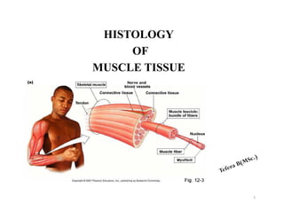
  • 10. Endomysium; Reticular fibers surrounding each muscle fibre plus the external (basal) lamina produced by the muscle fiber Perimysium; Dense connective tissue surrounding groups of fibers and dividing the muscle into fascicles Epimysium; Dense connective tissue surrounding the entire muscle, blends with the deep fascia and tendons Connective Tissue Covering Of Skeletal Muscle 10
  • 11. One of the most important roles of connective tissue is to mechanically transmit the forces generated by contracting muscle cells, because in most instances, individual muscle cells do not extend from one end of a muscle to the other 11
  • 12. 12 Fig; connective tissue covering; endomysium, perimysium and epimysium
  • 13. Myofilaments; Visible only with the electron microscope; composed primarily of actin, which forms 5-nm wide thin filaments, and myosin, which forms 15-nm wide thick filaments Myofibrils; Visible with the light microscope, 1–2 microns wide, oriented parallel to the long axis of the cell; composed of bundles of overlapping myofilaments that are arranged in register, producing an alternating light-dark, striated banding pattern Hierarchy Of Skeletal Muscle Organization 13
  • 14. Muscle fiber; Specialized term for a muscle cell, 10–100 microns wide; sarcoplasm is filled with hundreds of myofibrils, which are oriented parallel to each other and to the long axis of the muscle fiber. Muscle fascicle; Collection of muscle fibers surrounded by perimysium; collections of muscle fascicles are surrounded by the epimysium and form a muscle. 14
  • 15. Individual muscle cells(muscle fibre) are grouped together into elongated bundles – fasciculi Each fibre is separated by a thin layer of connective tissue called endomysium A group of fasciculi are surrounded by a connective tissue sheath called perimysium The epimysium is no difference from the deep fascia 15
  • 16. 16
  • 17. ACTIN FILAMENT 1. F-actin- double helix filament composed of G-actin monomeres 2. Tropomyosin – double helix peptide chain; runs in the groove of F-actin chains 3. Troponin complex (3 globular proteins - subunits): Troponin C (TnC) – binds calcium ions → contraction Troponin T (TnT) – attachment of troponin to tropomyosin Troponin I (TnI) – inhibits actin-myosin interaction 17
  • 19. A troponin complex is attached at one specific site on each tropomyosin molecule In thin filaments, each tropomyosin molecule spans seven G-actin molecules and has one troponin complex bound to its surface 19
  • 20. MYOSIN Myosin, a much larger complex, can be dissociated into two identical heavy chains and two pairs of light chains. Myosin heavy chains are thin, rodlike molecules made up of two heavy chains twisted together. Myosin head (flexible; binds to actin filament) Myosin head has: Actin binding site ATP binding site ATP-ase activity 20
  • 21. 21
  • 22. 22
  • 23. Several hundred myosin molecules are arranged within each thick filament with their rodlike portions overlapping and their globular heads directed toward either end Analysis of thin sections of striated muscle shows the presence of cross-bridges between thin and thick filaments. These bridges, which are known to be formed by the head of the myosin molecule plus a short part of its rodlike portion, are involved in the conversion of chemical energy into mechanical energy 23
  • 24. Arrangement of myofilaments within a myofibril in register result in the banding pattern seen at microscopic levels. • A band appears dark and contains actin and myosin. • I band appears light and contains actin only. 24
  • 25. • Z-line, composed of alpha-actinin, is located in the center of the I band. • H band is located in the center of the A band and represents the area where actin is not present. • M band is located in the center of the H band and represents areas of cross-connections between myosin filaments. 25
  • 26. 26
  • 27. 27
  • 28. Contractile unit of striated muscle fibers, seen in both skeletal and cardiac muscle fibers It extend from Z-line to Z-line Contains α-actinin, protein that binds actin filaments to Z-line Sarcomeres are repeated in series along the length of each myofibril. Adjacent myofibrils maintain the alignment of sarcomeres. 28 SARCOMERE
  • 29. 29
  • 30. No changes in lenght of : 1. A-band 2. actin and myosin filaments. 30 Alterations In Sarcomeres During Contraction During contraction occurs shortening of 1. Sarcomeres shorten. 2. Z-line interval narrows. 3. I-band 4. H-band (in maximal contraction could disapear
  • 31. MECHANISM OF CONTRACTION Resting sarcomeres consist of partially overlapping thick and thin filaments. During contraction, both the thick and thin filaments retain their original length. Because contraction is not caused by a shortening of individual filaments, it must be the result of an increase in the amount of overlap between the filaments. The sliding filament hypothesis of muscle contraction has received the most widespread acceptance 31
  • 32. The following is a brief description of how actin and myosin interact during a contraction cycle. At rest, ATP binds to the ATPase site on the myosin heads, but the rate of hydrolysis is very slow. Myosin requires actin as a cofactor to break down ATP rapidly and release energy. In a resting muscle, myosin cannot associate with actin, because the binding sites for myosin heads on actin molecules are covered by the troponin, tropomyosin complex on the F-actin filament 32
  • 33. When sufficiently high concentrations of calcium ions are available, however, they bind to the TnC subunit of troponin. The spatial configuration of the three troponin subunits changes and drives the tropomyosin molecule deeper into the groove of the actin helix This exposes the myosin-binding site on the globular actin components, so that actin is free to interact with the head of the myosin molecule 33
  • 34. The binding of calcium ions to the TnC unit corresponds to the stage at which myosin ATP is converted into the active complex. As a result of bridging between the myosin head and the G-actin subunit of the thin filament, the ATP is split into ADP and Pi (phosphate ion), and energy is released. This activity leads to a deformation, or bending, of the head and a part of the rodlike portion (hinge region) of the myosin Because the actin is bound to the myosin, movement of the myosin head pulls the actin past the myosin filament. The result is that the thin filament is drawn farther into the A band. 34
  • 35. Although a large number of myosin heads extends from the thick filament, at any one time during the contraction only a small number of heads aligns with available actin-binding sites. As the bound myosin heads move the actin, however, they provide for alignment of new actin myosin bridges. The old actin myosin bridges detach only after the myosin binds a new ATP molecule; This action also resets the myosin head and prepares it for another contraction cycle. If no ATP is available, the actin myosin complex becomes stable; This accounts for the extreme muscular rigidity (rigor mortis) that occurs after death. 35
  • 36. A single muscle contraction is the result of hundreds of bridge-forming and bridge-breaking cycles. The contraction activity that leads to a complete overlap between thin and thick filaments continues until Ca2+ ions are removed and the troponin “tropomyosin complex again covers the myosin-binding site 36
  • 37. During contraction, the I band decreases in size as thin filaments penetrate the A band. The H band ”the part of the A band with only thick filaments” diminishes in width as the thin filaments completely overlap the thick filaments. A net result is that each sarcomere, and consequently the whole cell (fiber), is greatly shortened 37
  • 38. Skeletal Muscle Innervation Skeletal muscle cells and the single motor neuron that innervates them constitute a motor unit. Each skeletal muscle receives at least two types of nerve fibers: motor and sensory. The motor nerve functions in eliciting contraction, whereas the sensory fibers pass to muscle spindles Additionally, autonomic fibers supply the vascular elements of skeletal muscle. The specificity of motor innervation is a function of the muscle innervated 38
  • 39. Muscle fatigue For a single muscle fiber, fatigue indicates an inability to develop tension when it is stimulated Causes: reduction in the rate of intracellular calcium release and uptake by sacroplasmic reticulum It dependent on muscle itself, exercise duration, fiber type composition, and/or pattern of motor unit activation 39
  • 40. When muscles cause a limb to move through the joint's range of motion, They usually act and are further classified into the following cooperating groups: AGONIST MUSCLES In a desired movement, the muscle mostly involved with that movement is the agonist or commonly referred to as prime movers. Agonist or prime movers may contract concentrically to lift a weight, or eccentrically to lower that weight. 40
  • 41. ANTAGONISTIC MUSCLES The muscle that lies directly opposite to the agonist or prime mover and is responsible for the opposing action of that movement is the antagonistic muscle. The triceps are the antagonist muscle to the biceps curl for instance, and the biceps would be the antagonist in a triceps extension. They are responsible for returning a limb to its initial position. 41
  • 42. SYNERGISTS These muscles perform, or assist in performing, the same set of joint motion as the agonists. Synergists are sometimes referred to as neutralizers because they help cancel out, or neutralize, extra motion from the agonists to make sure that the force generated works within the desired plane of motion. 42
  • 43. 43 Bundles form thick myocardium Cells are branch Cells join at intercalated discs 1-2 nuclei in center Inherent rhythmicity: each cell! (muscle cells beat separately without any stimulation) Myofilament organisation is similar to skeletal muscles’s hence the striations CARDIAC MUSCLES
  • 44. Smooth Muscles Muscles are spindle-shaped cells One central nucleus Grouped into sheets: often running perpendicular to each other Peristalsis No striations (no sarcomeres) Contractions are slow, sustained and resistant to fatigue Does not always require a nervous signal: can be stimulated by stretching or hormones 44
  • 45. Major Locations Inside the eye Walls of vessels Respiratory tubes Digestive tubes Urinary organs Reproductive organs 45
  • 46. Actin and myosin myofilaments are present, but they are not organized into a regular pattern, they are randomly arranged. Electron Dense bodies containing alpha actin are present in the cytoplasm of smooth muscle cells These dense bodies Serve as insertion points for myofilaments to transmit the force of filament sliding Thus, resemble Z-lines of striated muscle 46 Organization of the Contractile Proteins in Smooth Muscles
  • 47. 47