
Contenu connexe
Tendances
Tendances (20)
Similaire à Splints in DDH
Similaire à Splints in DDH (20)
Dernier
Dernier (20)
Splints in DDH
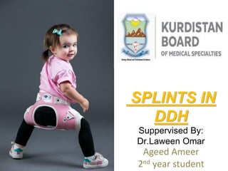
- 1. SPLINTS IN DDH Suppervised By: Dr.Laween Omar Ageed Ameer 2nd year student
- 2. What is a Splint? Harness? • A splint is defined as “a rigid or flexible device that maintains in position a displaced or movable part; also used to keep in place and protect an injured part” or as “a rigid or flexible material used to protect, immobilize, or restrict motion in a part.
- 3. What is a Splint? Harness?
- 4. History Of Splits • Evidence suggests that splint usage dates back to 1500 B.C. • Leaves, reeds, bamboo, and bark padded with linen… [and] copper." • In 1517, after the evolution of the armor trade, injuries were being treated by metal braces secured by screws. • In1592, the first written piece on splints by surgeon Hieronymus Fabricius, shows various drawings of armor-like splints for the entire body.
- 5. History Of Splits • In the mid-1700, doctors and mechanics worked with each other to create splints for certain injuries(PoP) • In the 1800s it was beginning to be recognized that rehabilitation after an injury was important. Orthopedics began to become a separate field from general surgery. A famous British Surgeon, Hugh Owen Thomas, created specialty splints that were cheap and best for injuries that were being rehabilitated.
- 6. Uses • Splints are most commonly used to immobilize broken bones or dislocated joints. • When a broken bone has been properly set. • Immobilize unset fractures . • Other injuries ; • soft tissue sprains, tendon injuries.
- 7. Indications of Splinting In DDH • 1.A hip that is dislocated and that can be reduced by the examiner (Ortolani sign) at the time the diagnosis is made. • 2. Hips that are located but that can be subluxated by the examiner (Barlow sign).
- 8. Indications of Splinting In DDH • Some of these hips will spontaneously stabilize, and some clinicians prefer to wait a few weeks and reexamine the child before initiating treatment. • When observation is chosen, steps should be taken to ensure follow-up because some of these hips will subsequently dislocate if they are left alone. • 3. Less certain are the indications for the treatment of hips that are normal on clinical examination but abnormal on ultrasonography.
- 9. Rules IN Splinting : DDH • 1. Hips must be properly reduced before splinting or in a position that reduction occures spontaneously . • 2. Extreme positions must be avoided. • 3. Hips should be able to move .(wide abduction and forced internal rotation lead to AVN).
- 10. Pavlic Harness By Dr.Arnold Pavlic(1902-1962)a Czcheh Orthopedist
- 11. Pavlic Harness/Neonates • The Pavlik harness is applied by first placing the chest strap just below the nipple line • The child’s feet are placed in the stirrups, the hips are placed in 120 degrees of flexion, and the straps are secured. • The posterior straps are fastened loosely to allow for the abduction of the hips to occur by gravity alone.
- 12. Harness/NeonatesPavlic • Weekly visit for bathing or change to a larger size(3-4weeks). • Hyperflexion leads to FNP and less than 90 deg. is inadequate for reduction. • Compliant parents needed. • On the 3rd wk U/S : if unstable hip switch to abduction orthosis(93% s.rate and no AVN). • On the 6th wk week: examination and U/S .
- 13. Harness/NeonatesPavlic • If both are toward stability start weaning.(gradual weaning is preferable by some authors). • At 3-4 months of age :radiograph. • At 1 year of age a standing radiograph. • If normal the follow up in once-twice /year till skeletal maturity (significant incidiense of asymmetric closure of the femoral head epiphysis leading to valgus and inadequate coverage of the head ).
- 14. Harness/NeonatesPavlic • If the hip remains dislocated after 3 to 4 weeks of harness wear, the use of the harness should be discontinued,and the hip should be examined while the child is under anesthesia. An arthrogram may show the cause of the instability, and the hip should be managed with either closed or open reduction.
- 15. Harness/NeonatesPavlic • If the hip is reduced at 3 weeks but dislocates during examination, the harness should be worn for 3 to 6 more weeks until the hip stabilizes. • An abduction orthosis may be used for hips that have not stabilized after 3 or more weeks of treatment in the harness.
- 16. Months6-1HarnessPavlic • To be effective, the harness must hold the hips in more than 90 degrees of flexion, with the position of the upper femoral metaphysis pointed toward the triradiate cartilage. • Higher dislocation have a higher faliure rate. • Weekly examination. • Follow up by U/S.
- 17. Months6-1HarnessPavlic • If reduction is not obtained by3-4 wks /other treatment plan. • If reduction is obtained continue for 6 wks after stability has achieved . • When harness treatment is completed, some clinicians elect to place the child in an abduction splint for several more months.
- 18. Months6-1HarnessPavlic • It is recommended for older children to have it for a longer time to encourage acetabular development. • Precise guidelines of stoppage ??? • As the harness is discontinued, another AP radiograph is obtained to assess hip reduction and acetabular development.
- 19. A notch above the acetabulum often appears after the hip is reduced, and this finding is usually followed by improved acetabular development Acetabular development may be enhanced by abduction splinting.
- 20. Months6-1HarnessPavlic • Overall, the reported rate of AVN when the Pavlik harness is used ranges from 0% to15%. • Factors that are associated with the failure of Pavlik harness treatment include • 1.Patient age of more than 7 weeks . • 2.Bilateral hip dislocation. • 3.Absent Ortolani sign.
- 21. Backs-DrawHarnessPavlic • 1. AVN (INPROPER APP VS dynamic process of dis). • 2.Failure to reduce the hip. • 3.Femoral nerve palsy. • 4.The so-called Pavlik harness disease was reported by Jones and associates, who found that prolonged positioning of the dislocated hip in flexion and abduction potentiated dysplasia and resulted in a hip that was likely to require an open reduction. • They noted a flattening of the posterolateral acetabulum in these hips and recommended discontinuing the harness if reduction had not occurred after 3 or 4 weeks. • 5. Long-term follow-up is recommended for treated hips.
- 22. A, Anteroposterior (AP) radiograph obtained at presentationwhen patient was 5 months old shows a dislocated left hip. B, AP radiograph of patient in the harness with inadequate flexion.
- 23. C, AP radiograph obtained 2 weeks later shows adequate flexion of the hip, although the hip is still dislocated. D, AP radiograph obtained 1 month later shows that the hip has been reduced.
- 24. E, AP radiograph obtained when patient was 5 years old shows good acetabular development.
- 25. Ilfeld Splint(CRAIG SPLINT) • Since October 1951 a splint • (FREDERIC W. ILFELD, M.D an American orthopedic surgeon) .with two thigh cuffs connected to an adjustable bar has been used in about 250 cases of congenital hip disease with good results.
- 26. SplintIlfeld
- 27. SplintIlfeld • With this splint the thighs are gradually and without force directed into abduction and external rotation, the "frog position." • The surgeon adjusts the splint into further abduction at weekly intervals until the desired position is obtained. • The splint is removed several times a day by the mother for rotation-abduction exercise.
- 28. SplintIlfeld • This exercise as well as the kicking and natural movement of the hips in the splint tend to improve local circulation, increase abduction, and apply gentle pressure of the femoral head against the acetabulum.
- 29. SplintIlfeld • In the frog position the thigh muscles exert a force along the femoral shaft "pulling" the head into the acetabulum. In this way the dislocation of the femoral head is reduced. • In dysplasia of the hip with delay in the development of the femoral head and acetabulum, the pressure of the femoral head in the abducted position is thought to stimulate bony growth.
- 30. SplintIlfeld • In dysplasia the splint is usually worn only at night. • In dislocation the splint is worn continuously for several months being removed daily for bathing and exercise. • The splint is then worn only at night until hip development is complete.
- 31. SplintIlfeld • In older children it may be used after closed or open reduction, even without preliminary plaster fixation. • In some cases the splint may replace the cast after 4-6 weeks thus eliminating many months of plaster immobilization.
- 32. AdvantagesSplintIlfeld • 1. Reduce a dislocation of the hip without anesthesia, hospitalization, or plaster cast • 2. Dynamic, permitting crawling, walking, and running. • 3. Adjustable for growth, cool and comfortable, light and handy. • 4. Prevents stiffness of the hips and knees, stimulates acetabular and femoral growth. • 5. Convenient. • 6. Allows mobility of the child.
- 33. Frejka pillow & Tripple diappers • Proff.Dr. Bedrich Frejka (1890-1972) a Czech Orthopedic Sx, • has a poor outcome : • 1. Forcefully abduct the hips. • 2. High rate of AVN (pressure over epiphyseal vsl) • The use of triple diapers should also be abandoned because they do not effectively position the hips, and their use may falsely suggest to parents that something positive is being accomplished.
- 34. PillowFrejka
- 35. Avascular necrosis after the use of the Frejka pillow. Anteroposterior radiograph obtained when patient was 16 years old shows a shortened femoral neck with trochanteric overgrowth. The valgus tilt of the femoral head indicates a lateral physeal injury from avascular necrosis
- 36. Von Rosen Splint • Designed by Professor Sophus Von Rosen of Sweden in 1956. • With reported 95% success rate and less than 1% risk of AVN. • Hips are held in 90 deg. flexion and 60-70 deg. abduction
- 37. Von Rosen Splint
- 38. Follow UpVon Rosen Splint • 1.The child should be seen once weekly for: • A. check for position and a possible change to a larger size(7). • B. to have a bath. • C. check for skin problems. • D. general advices for the parents.
- 39. Follow UpVon Rosen Splint • 2. U/S every 4-6 weeks • 3. Treatment continues for 6-12 weeks. Depending on the degree of displacement and the U/S finding in 6 wks.
- 40. Advices for ParentsVon Rosen Splint • 1.Never remove the splint at home. • 2.The child should lie on his back not on his tummy. • 3.Bathing: use unperfumed soap and carefully wipe the skin and dry with a towel .then use an unperfumed powder for skin. • 4.Diappers changing frequently. • 5.Contact the clinic for any concern. • 6.To lay your child on their side, support them with a rolled-up towel or blanket.
- 41. Tubingen Splint
- 42. Tubingen Splint • (By a German Professor Dr.Bernau for more than 25 yrs )in a trial to match the treated hip in a best position of being treated and safely development • “seated squat position” • The same as the child assumes in whomb before birth. • A flexion of an excess of 90 deg. of hips and spreads them slightly.
- 43. Tubingen Splint • Advantages: • 1.Freely movements of the child. • 2.Natural body posture. • 3.Easy handling. • 4.Fast conditioning, suitable for everyday use. • 5.Safe.
- 44. Tubingen Splint • Indications: • Dysplasia up to 12 months w/o instability. • Graf Type IIa,IIb and IIc .
- 45. Any Question?
- 46. Thank You