The document describes the various muscles of the back, thoracic wall, abdominal wall, and pelvic wall. It discusses the superficial and deep back muscles, including their origins, insertions, innervations, and actions. It also details the external and internal intercostal muscles of the thoracic wall, as well as the abdominal wall muscles like the external oblique, internal oblique, transversus abdominis, rectus abdominis, and pyramidalis. Finally, it briefly outlines some of the posterior abdominal wall and pelvic wall muscles.
Your abdominal muscles have many important functions, from holding organs in place to supporting your body during movement. There are five main muscles: pyramidalis, rectus abdominus, external obliques, internal obliques, and transversus abdominis. Ab strains and hernias are common, but several strategies can keep your abs safe and healthy
The document discusses the biomechanics of respiration including the structure and function of the ribs, ribcage muscles, and accessory muscles involved in breathing. The diaphragm is the primary muscle of inspiration and contracts to increase the vertical diameter of the thorax. The external intercostal muscles elevate the ribs during inspiration while the internal intercostals depress the ribs during expiration. Accessory muscles such as the scalenes, sternocleidomastoid, and pectorals assist with forced breathing by stabilizing the ribcage and sternum.
osteologyofabdomen-200315093235 best pdf of mbbs students pdfsrinathbadugu0777
This document provides information on the anatomy of the lumbar spine and abdominal wall. It describes the key features of the lumbar vertebrae, including their large kidney-shaped bodies and long slender transverse processes. It also details the muscles of the anterior and posterior abdominal wall, including the three flat muscles (external oblique, internal oblique, and transversus abdominis), two vertical muscles (rectus abdominis and pyramidalis), and muscles of the posterior wall like the quadratus lumborum, psoas major, and iliacus. Applied concepts like sacralization, lumbarization, and lumbar stenosis are also mentioned.
This document provides information on the anatomy of the abdomen, including the lumbar spine, vertebrae, muscles of the anterior and posterior abdominal walls, and joints of the lumbar region. It describes the key features of the lumbar vertebrae and distinctive characteristics of L5. It also details the layers of the anterior abdominal wall, flat and vertical muscles, and posterior muscles including the psoas major, quadratus lumborum and diaphragm. Applied concepts like sacralization and lumbarization are also mentioned.
The anterolateral abdominal wall consists of four main layers - skin, superficial fascia, muscles and associated fascia, and peritoneum. Below the umbilicus, the superficial fascia divides into two layers - Camper's fascia and Scarpa's fascia. The muscles of the abdominal wall include three flat muscles (external oblique, internal oblique, transversus abdominis) and two vertical muscles (rectus abdominis, pyramidalis). The posterior abdominal wall contains muscles like the quadratus lumborum, psoas major and minor, and is covered by fascia like the psoas and thoracolumbar fascia. Common surgical incisions of the abdominal wall
This document describes several muscles of the torso, including abdominal, postural, thoracic, and splenius muscles. It discusses the rectus abdominis, internal and external oblique, transversus abdominis muscles of the abdomen. For postural muscles it covers the erector spinae group including the spinalis, longissimus, and iliocostalis. It also mentions the serratus anterior, diaphragm, and intercostal muscles of the thorax, and the splenius capitis and cervicis muscles. The document provides details on the origin, insertion points and functions of these various muscle groups of the torso.
The document describes several muscles of the shoulder region. It discusses the deltoid muscle in detail, including its origin on the clavicle, acromion, and scapula, insertion on the humerus, nerve supply from the axillary nerve, and actions of abduction, flexion, and extension of the arm. It also summarizes the rotator cuff muscles - supraspinatus, infraspinatus, teres minor, and subscapularis - which stabilize the shoulder joint and allow rotation. Finally, it provides information on the trapezius muscle, noting its broad triangular shape and ability to elevate, retract, and depress the scapula.
This document provides an overview of muscle anatomy, including the three main types of muscle (skeletal, smooth, and cardiac), their structures and functions. It discusses the anatomy of muscles in different parts of the body like the back, thorax, upper and lower limbs, and face. Key points covered include the names and actions of major muscle groups, as well as how muscles contract and generate movement through sarcomeres and motor proteins like actin and myosin.
The intercostal muscles are arranged in three layers between the ribs. The external intercostals elevate the ribs during inspiration. The internal intercostals and innermost intercostals depress the ribs during expiration. The transversus thoracis, subcostalis, and innermost intercostals form the inner transverse thoracic muscle group. These muscles act to change the volume of the thoracic cavity during respiration.
The document summarizes several muscles of the hand, back, and lower limb. It describes the intrinsic and extrinsic muscles of the hand, noting that the extrinsic muscles are in the forearm and control crude movements, while the intrinsic muscles are within the hand and control finer movements. It then provides details on specific intrinsic hand muscles including the lumbricals and interossei. The document also summarizes the superficial, intermediate, and deep muscles of the back, giving origins, insertions, innervation and actions for some of the muscles in each group such as the trapezius, serratus posterior superior, and splenius capitis. Finally, it provides an overview of muscles in the lower limb including the gl
The document summarizes various muscles of the trunk, including:
- Intercostal and diaphragm muscles that are important for respiration and breathing.
- Three layered muscles of the abdominal wall that form a strong protective covering over the abdominal cavity.
- Back muscles that allow movement and stabilize the trunk, including erector spinae and interspinalis groups.
- Pelvic floor muscles like levator ani and coccygeus that form a hammock-like floor across the pelvis.
- Shoulder muscles that attach the upper extremity and allow movement, including pectoralis major, deltoid, serratus anterior, trapezius and levator scapulae
L9 muscles of upper limb [Autosaved].pptxssuser31c469
The document provides information on human anatomy, specifically focusing on the muscles of the upper limb. It describes the different types of muscles and their functions. It then details the specific muscles found in different regions of the upper limb, including the pectoral region, shoulder region, upper arm, and anterior and posterior compartments of the forearm. For each muscle, it provides the origin, insertion, innervation, and main actions. The document serves as a detailed reference for understanding the muscles involved in movement and stabilization of the upper limb.
This document provides an overview of the diaphragm. It discusses that the diaphragm is the main muscle of respiration that separates the chest cavity from the abdominal cavity. The diaphragm has a dome shape with a peripheral muscular part and a central tendon. It originates from the xiphoid process, lower six ribs, and vertebral columns. The diaphragm inserts into a central tendon that is fused to the pericardium. It is the most important muscle for breathing movements.
Muscles of head, Muscles of face, Muscles of neck, Muscles of shoulder girdle, Muscles of upper limbs, Muscles of thorax, Diaphragm, Muscles of abdomen, Muscles of back, Muscles of perineum, Muscles of pelvis, Muscles of lower limb, Muscles of leg, Muscles of foot
1 GNM anatomy Unit _ 14 Muscular System.pptxthiru murugan
By:M. Thiru murugan
Unit XIV
Type, structure and functions of muscle
Origin, Insertion, and action of muscles
Muscle:
Muscle is a soft tissue and it is one of the 4 basic tissues, along with nervous tissue, epithelium, and connective tissue.
Muscles helps in movement, support and protection of internal organs.
Muscle cells or myocytes contain protein filaments called myofilaments actin & myosin that producing a contraction that changes both the length and the shape of the cell.
Types of Muscles:
There are 3 types of muscles: skeletal, cardiac & smooth muscle
Skeletal muscle or “voluntary muscle” or striated Muscles is attached to bone, helps in movement & in maintaining posture.
Smooth muscle or “involuntary muscle” or non striated muscles is found within the walls of organs and structures
Cardiac muscle is also an "involuntary muscle" found only in the heart.
Structure of Skeletal muscle:
Skeletal muscle consist plasma membrane is called sarcolemma, the cytoplasm is called sarcoplasm & specialized sarcoplasmic reticulum that play important role in regulation of Calcium.
Cells contain many nucleus peripherally
It consist of number of elongated cells called muscle fibers or muscle cell , that consist of myofibrils (elongated protein molecules).
These Muscle fibers are arranged in bundle called fasciculi
Each muscle fibers covered by endomysium, each fasciculi covered by perimysium & the whole muscle is covered by epimysium.
Neuromuscular junction (NMJ) or myoneural junction is a chemical synapse between a motor neuron and a muscle fiber.
It allows the motor neuron to transmit a signal to the muscle fiber, causing muscle contraction.
Properties of skeletal muscle:
Excitability: this refers to muscle tissue being able to react to nervous stimulation.
Extensibility: this refers to the ability of muscle tissue to lengthen when contracting
Elasticity: this refers to the ability of muscle tissue to return to its normal resting length once it has been stretched.
Contractility: this refers to the capacity of a muscle to contract or shorten forcibly when stimulated by nerves and hormones
Muscle contraction:
Muscle use the movement of actin against myosin to create contraction.
In skeletal muscle, contraction is stimulated by electrical impulses transmitted by the nerves
Cardiac and smooth muscle contractions are stimulated by internal pacemaker cells which regularly contract, and propagate contractions to other muscle cells they are in contact with.
All skeletal muscle and many smooth muscle contractions are facilitated by the neurotransmitter acetylcholine.
Types of muscle contraction:
Isotonic contractions maintain constant force or tone in the muscle as the muscle length changes
Isometric contractions changes in force without changing the length of the muscle
Muscle tone is defined as the tension in a muscle at rest.
Function of skeletal muscles
Body movement (Locomotion)
Maintenance of posture
Respiration
Constriction of organs and vess
Surgery & anatomy of vertebral column and spinal nervesKapil Sharma
The document provides an overview of the topographic anatomy of the vertebral column, spinal cord, and associated membranes. It describes the basic structure and features of different types of vertebrae, including cervical, thoracic, lumbar, sacral and coccygeal vertebrae. It also outlines the joints and ligaments of the vertebral column. Furthermore, it categorizes the muscles of the back into superficial, intermediate and deep groups, and provides details on origin, insertion, innervation and actions of representative muscles within each group, such as the trapezius, erector spinae, and multifidus muscles.
The gluteal region contains important muscles that control movement of the hip and thigh. The piriformis muscle divides the greater sciatic foramen, with structures passing above or below it. Deep muscles like piriformis, obturator internus and quadratus femoris laterally rotate the femur. Superficial muscles like gluteus maximus, medius and minimus abduct and extend the hip. Nerves like the superior and inferior gluteal innervate these muscles. The sciatic nerve passes through the gluteal region en route to the lower limb. Landmarks are used to safely administer intramuscular injections into the gluteus maximus muscle.
The document describes the muscles of the thigh and gluteal region. It discusses:
1. The thigh region is divided into four compartments - anterior, medial, posterior, and lateral. The anterior compartment contains muscles that flex the hip and extend the knee. The medial compartment contains adductor muscles. The posterior compartment contains hamstring muscles that extend the hip and flex the knee.
2. The gluteal region contains the gluteal muscles - gluteus maximus, medius, and minimus. It also contains smaller lateral rotator muscles.
3. Several important structures pass through openings in the pelvis. The sciatic nerve passes through the greater and lesser sciatic foramina.
The document describes the anatomy of the thoracic cavity and its contents. It is the region between the neck and abdomen, bounded by ribs, vertebrae, and sternum. It contains the lungs, heart, blood vessels, esophagus, and lymph nodes. There are openings at the top (thoracic inlet) and bottom. The thoracic wall is formed by the vertebrae, sternum, ribs, and diaphragm. Numerous muscles are also described, including intercostal muscles, pectoralis muscles, serratus muscles, and others that help with breathing and chest/shoulder movement.
The document provides an overview of the gross anatomy of skeletal muscles presented by Nikhil Vaishnav. It begins with an outline listing the major muscle groups that will be discussed, including muscles of the head, neck, thorax, abdomen, pelvis, shoulder girdle and upper limb, and lower limb. The presentation then covers the muscles within each of these groups in further detail over multiple slides, describing the origin, insertion, innervation, and action of each muscle. The focus is on identifying the individual muscles and briefly explaining their structure and function.
The document describes the anatomy of the anterior abdominal wall. It is divided into nine quadrants and contains skin, superficial fascia with fatty and membranous layers, deep fascia, three muscle layers (external oblique, internal oblique, transversus abdominis), rectus abdominis muscles, pyramidalis muscle, extraperitoneal fascia, and parietal peritoneum from external to internal. Key nerves are branches of thoracic and lumbar nerves, and arteries include the superior and inferior epigastric arteries. Lymphatic drainage is to axillary nodes above the umbilicus and inguinal nodes below.
The neck contains many vital structures and connects the head to the torso. The skeleton of the neck includes 7 cervical vertebrae, the hyoid bone, manubrium, and clavicles. The neck separates into anterior and posterior triangles divided by the sternocleidomastoid muscle. The triangles contain muscles, vessels, and nerves. Conditions like torticollis can cause neck twisting that is treated with physiotherapy including massage, stretches, and exercises to improve range of motion.
This document provides an overview of the muscles of mastication. It begins with definitions of muscle and mastication. The muscles are classified as primary muscles of mastication (masseter, temporalis, lateral pterygoid, medial pterygoid) or accessory muscles. The anatomy and development of skeletal muscle and the specific muscles of mastication are described. The document outlines the origin, insertion, blood supply, nerve supply, actions, and palpation of each primary muscle of mastication. It provides clinical characteristics and functions of the muscles in mandibular movement and importance in dentofacial growth.
Anterior abdominal wall , Rectus sheath and Inguinal.pptxJudeChinecherem
In this detailed lecture note, we embark on a comprehensive journey through the complex and crucial anatomy of the abdominal wall. The abdominal wall is not just a physical barrier; it is a dynamic structure with multiple layers, muscles, and intricate structures that play a fundamental role in protecting our internal organs, providing support, and enabling various bodily functions.
We will delve deep into the layers of the abdominal wall, understanding the significance of each component - from the outermost skin to the innermost peritoneum. Through detailed illustrations, diagrams, and explanations, you will gain a profound insight into the anatomical intricacies of this region.
Moreover, this lecture note provides valuable insights into the clinical relevance of the abdominal wall. Learn about common medical conditions and surgical procedures related to the abdominal wall, including hernias, trauma, and abdominal wall reconstruction. Whether you are a medical student, healthcare professional, or simply intrigued by the wonders of the human body, this resource will enrich your knowledge and understanding of this vital anatomical structure.
Join us on this educational journey as we unravel the mysteries of the abdominal wall, exploring its anatomy, functions, and clinical significance. Whether you're studying medicine, pursuing a career in healthcare, or just eager to expand your knowledge, this lecture note is a valuable resource for anyone interested in the fascinating world of human anatomy."
The abdominal wall has several layers including skin, superficial fascia, muscles and fascia. The superficial fascia below the umbilicus divides into two layers: Camper's fascia and Scarpa's fascia. The muscles of the abdominal wall include three flat muscles on each side (external oblique, internal oblique, transversus abdominis) and two vertical muscles near the midline (rectus abdominis). The aponeuroses of the flat muscles come together to form the rectus sheath surrounding the rectus abdominis muscle. Below the arcuate line, the posterior wall of the rectus sheath is deficient.
Similaire à Vertebral column4physiotherapy.stts2.pptx (20)
The Importance of Black Women Understanding the Chemicals in Their Personal C...bkling
Certain chemicals, such as phthalates and parabens, can disrupt the body's hormones and have significant effects on health. According to data, hormone-related health issues such as uterine fibroids, infertility, early puberty and more aggressive forms of breast and endometrial cancers disproportionately affect Black women. Our guest speaker, Jasmine A. McDonald, PhD, an Assistant Professor in the Department of Epidemiology at Columbia University in New York City, discusses the scientific reasons why Black women should pay attention to specific chemicals in their personal care products, like hair care, and ways to minimize their exposure.
Joker Wigs has been a one-stop-shop for hair products for over 26 years. We provide high-quality hair wigs, hair extensions, hair toppers, hair patch, and more for both men and women.
Exploring the Benefits of Binaural Hearing: Why Two Hearing Aids Are Better T...Ear Solutions (ESPL)
Binaural hearing using two hearing aids instead of one offers numerous advantages, including improved sound localization, enhanced sound quality, better speech understanding in noise, reduced listening effort, and greater overall satisfaction. By leveraging the brain’s natural ability to process sound from both ears, binaural hearing aids provide a more balanced, clear, and comfortable hearing experience. If you or a loved one is considering hearing aids, consult with a hearing care professional at Ear Solutions hearing aid clinic in Mumbai to explore the benefits of binaural hearing and determine the best solution for your hearing needs. Embracing binaural hearing can lead to a richer, more engaging auditory experience and significantly improve your quality of life.
TEST BANK FOR Health Assessment in Nursing 7th Edition by Weber Chapters 1 - ...rightmanforbloodline
TEST BANK FOR Health Assessment in Nursing 7th Edition by Weber Chapters 1 - 34.
We are one of the top Massage Spa Ajman Our highly skilled, experienced, and certified massage therapists from different corners of the world are committed to serving you with a soothing and relaxing experience. Luxuriate yourself at our spas in Sharjah and Ajman, which are indeed enriched with an ambiance of relaxation and tranquility. We could confidently claim that we are one of the most affordable Spa Ajman and Sharjah as well, where you can book the massage session of your choice for just 99 AED at any time as we are open 24 hours a day, 7 days a week.
Visit : https://massagespaajman.com/
Call : 052 987 1315
Sectional dentures for microstomia patients.pptxSatvikaPrasad
Microstomia, characterized by an abnormally small oral aperture, presents significant challenges in prosthodontic treatment, including limited access for examination, difficulties in impression making, and challenges with prosthesis insertion and removal. To manage these issues, customized impression techniques using sectional trays and elastomeric materials are employed. Prostheses may be designed in segments or with flexible materials to facilitate handling. Minimally invasive procedures and the use of digital technologies can enhance patient comfort. Education and training for patients on prosthesis care and maintenance are crucial for compliance. Regular follow-up and a multidisciplinary approach, involving collaboration with other specialists, ensure comprehensive care and improved quality of life for microstomia patients.
R3 Stem Cell Therapy: A New Hope for Women with Ovarian FailureR3 Stem Cell
Discover the groundbreaking advancements in stem cell therapy by R3 Stem Cell, offering new hope for women with ovarian failure. This innovative treatment aims to restore ovarian function, improve fertility, and enhance overall well-being, revolutionizing reproductive health for women worldwide.
At Malayali Kerala Spa Ajman, Full Service includes individualized care for every client. We specifically design each massage session for the individual needs of the client. Our therapists are always willing to adjust the treatments based on the client's instruction and feedback. This guarantees that every client receives the treatment they expect.
By offering a variety of massage services, our Ajman Spa Massage Center can tackle physical, mental, and emotional illnesses. In addition, efficient identification of specific health conditions and designing treatment plans accordingly can significantly enhance the quality of massaging.
At Malayali Kerala Spa Ajman, we firmly believe that everyone should have the option to experience top-quality massage services regularly. To achieve that goal we offer cheap massage services in Ajman.
If you are interested in experiencing transformative massage treatment at Malayali Kerala Spa Ajman, you can use our Ajman Massage Center WhatsApp Number to schedule your next massage session.
Contact @ +971 529818279
Visit @ https://malayalikeralaspaajman.com/
NURSING MANAGEMENT OF PATIENT WITH EMPHYSEMA .PPTblessyjannu21
Prepared by Prof. BLESSY THOMAS, VICE PRINCIPAL, FNCON, SPN.
Emphysema is a disease condition of respiratory system.
Emphysema is an abnormal permanent enlargement of the air spaces distal to terminal bronchioles, accompanied by destruction of their walls and without obvious fibrosis.
Emphysema of lung is defined as hyper inflation of the lung ais spaces due to obstruction of non respiratory bronchioles as due to loss of elasticity of alveoli.
It is a type of chronic obstructive
pulmonary disease.
It is a progressive disease of lungs.
VEDANTA AIR AMBULANCE SERVICES IN REWA AT A COST-EFFECTIVE PRICE.pdfVedanta A
Air Ambulance Services In Rewa works in close coordination with ground-based emergency services, including local Emergency Medical Services, fire departments, and law enforcement agencies.
More@: https://tinyurl.com/2shrryhx
More@: https://tinyurl.com/5n8h3wp8
Unlocking the Secrets to Safe Patient Handling.pdfLift Ability
Furthermore, the time constraints and workload in healthcare settings can make it challenging for caregivers to prioritise safe patient handling Australia practices, leading to shortcuts and increased risks.
Unlocking the Secrets to Safe Patient Handling.pdf
Vertebral column4physiotherapy.stts2.pptx
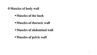
1. Muscles of body wall
Muscles of the back
Muscles of thoracic wall
Muscles of abdominal wall
Muscles of pelvic wall
1
2. Muscles of the back
• There are two major groups of muscles in the back:
The extrinsic back muscles include:
Superficial muscles that produce and control limb movement
Intermediate back muscles: produce & control respiratory
movements
The intrinsic (deep) back muscles include muscles that specifically act
on the vertebral column, producing its movements and maintaining
posture.
2
3. Superficial group of back muscles
• The muscles in the superficial group are
immediately deep to the skin and superficial
fascia.
• They attach the superior part of the appendicular
skeleton (clavicle, scapula, and humerus) to the
axial skeleton (skull, ribs, and vertebral column).
• Because these muscles are primarily involved
with movements of this part of the appendicular
skeleton, they are sometimes referred to as the
appendicular group.
3
4. Trapezius: is flat and triangularly shaped muscle
Origin- skull and upper portion of the vertebral
column
Insertion-attach to the lateral third of the clavicle
and to the acromion of the scapula
Innervation- the spinal accessory nerve (CN
XI).
Action-assists in rotating the scapula during
abduction of humerus; upper fibers elevate,
middle fibers adduct, and lower fibers depress
scapula 4
5. As a result, movements associated with this
muscle include extension, adduction, and
medial rotation of the upper limb.
• Latissimus dorsi can also depress the shoulder,
preventing its upward movement
• The thoracodorsal nerve innervates the
latissimus dorsi muscle
5
Latissimus dorsi: is a large, flat triangular muscle that
attaches the back and to the humerus
6. • It elevates the scapula and may assist
other muscles in rotating the scapula
inferiorly
• Innervation; from the anterior rami
of spinal nerves C3, C4 and the dorsal
scapular nerve
6
Levator scapulae; descends from the transverse processes
of the upper cervical vertebrae to the upper portion of the
scapula.
7. Rhomboid minor and major are inferior to
levator scapulae .
arises from the ligamentum nuchae of the neck
and the spinous processes of vertebrae CVII & TI
and attaches to the medial scapular border
The two rhomboid muscles work together to
retract or pull the scapula toward.
• Innervation- dorsal scapular nerve,
7
8. Intrinsic Back Muscles
• The intrinsic back muscles (muscles of back proper, deep back
muscles) are innervated by the posterior rami of spinal nerves and
act to maintain posture and control movements of the vertebral
column.
• The intrinsic back muscles are grouped into superficial,
intermediate, and deep layers according to their relationship to the
surface.
8
9. Superficial layer of intrinsic back muscles
• The splenius muscles are thick and
flat and lie on the lateral and
posterior aspects of the neck
• The splenii arise from the midline
and extend superolaterally to the
cervical vertebrae (splenius
cervicis) and cranium (splenius
capitis)
9
10. Intermediate layer of intrinsic back muscles
10
The erector spinae muscles lie
in a groove on each side of the
vertebral column centrally and the
angles of the ribs laterally.
The iliocostalis forms the
lateral column, the longissimus
forms the intermediate column,
and the spinalis forms the
medial column.
11. Deep layer of intrinsic back muscles
• Deep to the erector spinae is an obliquely disposed group of
much shorter muscles called the transversospinal muscle
group, consisting of the semispinalis, multifidus, and rotators
• These muscles originate from transverse processes of
vertebrae & pass to spinous processes of more superior
vertebrae
11
12. Deep layer of intrinsic back muscles,,,
Semispinalis
• the superficial member of the group
• as its name indicates, it arises from approximately half of the vertebral
column (spine)
• it is divided into three parts according to the superior attachments:
semispinalis capitis, semispinalis thoracis, and semispinalis cervicis
Multifidus
• the middle layer of the group
• consists of short, triangular muscular bundles that are thickest in the lumbar
region
Rotatores, or rotator muscles
• the deepest of the three layers of transversospinal muscles
• best developed in the thoracic region 12
17. Muscles of thoracic wall
Pectoralis major and pectoralis minor.
Subclavius.
Serratus anterior muscles anteriorly.
Latissimus dorsi muscles posteriorly.
Anterolateral abdominal muscles and.
Some back and neck muscles. 17
Several upper limb (thoracoappendicular) muscles attach to the thoracic
cage – including:
18. Muscles of thoracic wall,,,
Axio-appendicular, neck &
anterolateral abdominal muscles
overlying thoracic wall.
Muscles act primarily on the upper
limbs
The pectoralis major has been
removed on the left side to expose
the pectoralis minor, subclavius, and
external intercostal muscles.
18
19. Intercostal muscles
The Extercostal muscles occupy the
intercostal spaces.
Originate from the inferior border of one
rib, course infero-medially and insert to
the superior border of the immediate rib
below.
Anteriorly, the muscle fibers are replaced
by the external intercostal membranes at
the costochondral junctions.
19
21. Intercostal muscles,,,
The internal intercostal muscles
(11 pairs) run deep to and at right
angles to the external
intercostals.
Their fibers run inferoposteriorly
from the floors of the costal
grooves to the superior borders of
the ribs inferior to them.
21
22. Intercostal muscles,,,
Between the ribs posteriorly,
medial to the angles, the internal
intercostals are replaced by the
internal intercostal
membranes.
The external intercostals are
most active during inspiration.
The internal intercostals are
most active during expiration
22
23. Intercostal muscles,,,
The innermost intercostal muscles are
similar to the internal intercostals.
The innermost intercostal muscle forms the
deepest layer
The innermost intercostals are separated
from the internal intercostals by intercostal
nerves and vessels.
Actions innermost intercostals are the
same as those of the internal intercostal
muscles.
23
24. Intercostal muscles,,,
The subcostal muscles are variable in size
and shape, usually being well developed
only in the lower thoracic wall..
They extend from the internal surfaces of
one rib to the internal surface of the second
(next) or third rib below.
Crossing one or two intercostal spaces, the
subcostals run in the same direction as the
internal intercostals and blend with them. 24
25. Muscles of anterolateral abdominal wall
• There are five (bilaterally paired)muscles in the anterolateral group
of abdominal wall muscles:
The 3 flat muscles are; external oblique, internal oblique, and
transversus abdominis muscles
Two vertical muscles, near the midline, which are enclosed within a
tendinous sheath formed by the aponeuroses of the flat muscles-
Rectus abdominus and pyramidalis
25
26. • All three flat muscles are continued anteriorly and
medially as strong, sheet-like aponeuroses.
• B/n the midclavicular line & the midline, the
aponeuroses form the tough, aponeurotic, tendinous
rectus sheath enclosing the rectus abdominis
muscle
• The aponeuroses then interweave with their
fellows of the opposite side, forming a midline
raphe the linea alba which extends from the
xiphoid process to the pubic symphysis.
26
27. External oblique muscle
• The external oblique muscle is the largest &
most superficial of the 3 flat anterolateral
abdominal muscles
• Its muscle fibers pass in an inferomedial
direction,
• The aponeuroses then interweave with their
fellows of the opposite side, forming a midline
raphe, the linea alba
27
28. External oblique muscle,,,
• Origin: Muscular slips from the outer surfaces of the lower
8 ribs(5th–12th ribs)
• Insertion: Lateral lip of iliac crest; aponeurosis ending in
midline (linea alba),pubic tubercle
• Nerve supply: Thoraco-abdominal nerves (T7–T11 spinal
nerves) and subcostal nerve
• Main Action: Compress abdominal contents; both muscles
flex trunk; each muscle bends trunk to same side, turning
anterior part of abdomen to opposite side
28
29. Internal oblique muscle
• The intermediate of the three flat abdominal muscles.
• Its fleshy fibers run perpendicular to those of the
external oblique, running superomedially
• Origin: Thoracolumbar fascia; iliac crest & lateral
2/3rd of inguinal ligament
• Insertion: Inferior border of the lower three or four
ribs; linea alba; pubic crest
• Nerve supply: anterior rami of T6–T12 and first
lumbar nerves
• Main Action: Compress abdominal contents; both
muscles flex trunk; each muscle bends trunk 29
30. Transversus abdominis muscle
• The innermost of the 3 flat abdominal
muscles
• Its fibers run transversally, except for the
inferior ones.
• This muscle is ideal for compressing the
abdominal contents, increasing intra-
abdominal pressure.
• B/n the internal oblique & the transversus
abdominis muscles is a neurovascular plane,
30
31. Origin: Thoracolumbar fascia; medial lip of iliac
crest; lateral one-third of inguinal ligament;
internal surfaces of 7th–12th costal cartilages,
Insertion: Aponeurosis ending in linea alba; pubic
crest and pectineal line via conjoint tendon
Nerve supply: Thoraco-abdominal nerves
(anterior rami of T6–T12 spinal nerves) and first
lumbar nerves
• Main action: Compresses and supports
abdominal viscera 31
33. Rectus abdominis muscle
• A long, broad, strap-like muscle, the rectus
abdominis is the principal vertical muscle
• Is a paired muscle, separated by the linea alba
• The rectus abdominis is 3 times as wide
superiorly as inferiorly; it is broad and thin
superiorly and narrow and thick inferiorly..
• Most of the rectus abdominis is enclosed in the
rectus sheath.
• Along its course, it is intersected by 3 or 4
transverse fibrous bands or tendinous
intersections
33
34. Origin: Pubic crest, pubic tubercle, and
pubic symphysis
Insertion: Xiphoid process and 5th– 7th
costal cartilages
• Nerve supply:
• Anterior rami of lower seven thoracic
spinal nerves (T7 to T12)
Main Action: Compress abdominal
contents; flex vertebral column; tense
abdominal wall
34
35. Pyramidalis muscle
• Is small, triangular-shaped muscle
• Which may be absent in some individuals
• Is anterior to the rectus abdominis
• Has its base on the pubis, and its apex is
attached superiorly and medially to the linea
alba
• Origin: Front of pubis and pubic symphysis
• Insertion: Into linea alba
• Nerve supply: Anterior ramus of T12
• Action: Tenses the linea alba
35
37. Functions and actions of anterolateral abdominal muscles
Form a strong expandable support for the anterolateral abdominal wall.
Support the abdominal viscera & protect them from injuries.
Compress the abdominal contents to maintain/increase the intra-
abdominal pressure & in so doing, oppose the diaphragm
Move the trunk and help to maintain posture.
37
39. Psoas major and minor
• Medially, the psoas major muscles cover the anterolateral surface of the
bodies of the lumbar vertebrae, filling in the space between the vertebral
bodies and the transverse processes
• Each of these muscles arises from the bodies of vertebra TXII and all five
lumbar vertebrae, from the intervertebral discs between each vertebra, and
from the transverse processes of the lumbar vertebrae
• Passing inferiorly along the pelvic brim, each muscle continues into the
anterior thigh, under the inguinal ligament, to attach to the lesser trochanter
of the femur 39
40. Quadratus lumborum
Laterally, the quadratus lumborum muscles fill the space between
ribs XII and the iliac crest on both sides of the vertebral column
They are overlapped medially by the psoas major muscles; along
their lateral borders are the transversus abdominis muscles
The quadratus lumborum muscles arise from the transverse process
of vertebra LV, the iliolumbar ligament, and the adjoining part of
the iliac crest
40
41. Quadratus lumborum,,,
• The quadratus lumborum muscles depress
and stabilize rib XII and contribute to lateral
bending of the trunk. Acting together, the
muscles may extend the lumbar part of the
vertebral column
• They are innervated by anterior rami of T12
and L1 to L4 spinal nerves
41
42. Diaphragm
• The diaphragm is a double-domed musculotendinous sheet, located at the
inferior-most aspect of the rib cage.
It serves two main functions:
1. Separates the thoracic cavity from the abdominal cavity
Note: the word diaphragm is derived from the Greek ‘diáphragma’, meaning partition.
2. Undergoes contraction and relaxation, altering the volume of the thoracic
cavity and the lungs, producing inspiration and expiration.
It is the principal muscle of respiration.
The pericardium, containing the heart, lies on the central part of the
diaphragm.
42
44. Muscles of the pelvic wall,,,
Medial view of hemisected pelvis
Posterior view
44
45. Muscles of the pelvic wall,,,
Obturator internus muscle
Origin - Pelvic surfaces of ilium and
ischium; obturator membrane
Insertion - greater trochanter of the
femur
Action - Rotates thigh laterally;
assists in holding head of femur in
acetabulum
Nerve supply - nerve to obturator
internus (L5, S1, S2) 45
46. Muscles of the pelvic wall,,,
Piriformis muscle: is triangular muscle.
• It originates in the bridges of bone and passes
through the greater sciatic foramen.
Origin-Pelvic surface of S2 - S4 segments
-superior margin of greater sciatic notch and
sacrotuberous ligament
Insertion - greater trochanter of femur
Nerve supply – nerve to piriformis from sacral
plexus (S1 and S2)
Action – Rotates thigh laterally; abducts
thigh; assists in holding head of femur in
acetabulum 46