SlideShare une entreprise Scribd logo
1  sur  31
Télécharger pour lire hors ligne
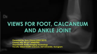
VIEWS FOR FOOT, CALCANEUM
AND ANKLE JOINT
Presented By: Ram Chand (220513019)
Course: MSc RIT (2nd SEMESTER)
Department: Radio-Imaging Technology
Faculty: Allied Health Sciences, SGT University, Gurugram
INTRODUCTION
A foot, ankle, and calcaneum X-ray is a diagnostic tool used
to assess the bones and joints in the lower leg and foot. It is a
safe and effective way to identify conditions or injuries that
may require treatment.
ANATOMY
Image Sources: Research gate, Pinterest
INDICATIONS
• foot trauma
• bony tenderness at the base of the metatarsal
• bony tenderness at the navicular bone
• inability to weight-bear more than four steps
• non-traumatic foot pain
PREPARATION
• Patient history is collected
• Patient is asked to remove all radiopaque materials
• Wear hospital gown
• Chaperone is necessary for female patient
• Shielding is provided to patient
FOOT SERIES
• Dorsoplantar view
• Medial Oblique view
• Lateral view
FOOT (DORSOPLANTER VIEW) POSITIONING
• the patient may be supine or
upright depending on comfort
• the affected leg must be flexed
enough that the plantar aspect of
the foot is resting on the image
receptor
Image Sources: Department of Radiology, Apollo Hospital, Bhubaneswar
TECHNICAL FACTORS
• centring point
• x-ray beam centred to the base of the 3rd metatarsal
• Orientation: Portrait
• detector size: 18 cm x 24 cm
• exposure
• 50-55 kVp
• 3-4 mAs
• SID: 100 cm
• Grid: no
Image Sources: Bontrager’s
FOOT MEDIAL OBLIQUE VIEW POSITIONING
• the patient may be supine or upright
depending on comfort
• the affected leg must be flexed enough
that the plantar aspect of the foot is resting
on the image receptor
• the foot is medially rotated until the
plantar surface sits at a 45° angle to the
image receptor
Image Sources: Department of Radiology, Apollo Hospital, Bhubaneswar
TECHNICAL FACTORS
• centring point
• x-ray beam centred to the base of the 3rd metatarsal
• the beam will be perpendicular to the image receptor
• Orientation: Portrait
• detector size: 18 cm x 24 cm
• exposure
• 50-55 kVp
• 3-4 mAs
• SID: 100 cm
• Grid: no
Image Sources: Bontrager’s
FOOT LATERAL VIEW POSITIONING
• the patient may be supine or upright depending on
comfort
• the affected leg is externally rotated until the distal limb is
parallel to the table, in many cases, the patient will have
to half roll onto the affected side
• the lateral aspect of the foot will be in contact with the
image receptor
• the non-affected side is kept posterior to prevent over
rotation
• the foot is in slight dorsiflexion
• the planter surface should be perpendicular to the image
receptor
Image Sources: Department of Radiology, Apollo Hospital, Bhubaneswar
TECHNICAL FACTORS
• centring point
• base of metatarsals or midfoot
• Orientation: Landscape
• detector size: 18 cm x 24 cm
• exposure
• 50-60 kVp
• 4-6 mAs
• SID: 100 cm
• Grid: no
Image Sources: Bontrager’s
CALCANEUM
SERIES
• Axial view
• Lateral view
AXIAL VIEW OF CALCANIUM POSITIONING
• The patient is seated on x ray table
with legs extended.
• Affected side ankle is dorsiflexed
placing the heel on the cassette.
• The patient is asked to hold the
ankle by the help of a bandage
cloth sling over the foot.
• Patient is immobilized side is
marked and beam is collimated.
TECHNICAL FACTORS
• centring point
• Middle sole of the foot
• Orientation: Portrait
• detector size: 18 cm x 24 cm
• exposure
• 50-60 kVp
• 3-5 mAs
• SID: 100 cm
• Grid: no
Image Sources: Bontrager’s
LATERAL VIEW OF CALCANIUM POSITIONING
• The patient is seated on x ray table
with legs extended.
• Recumbent, on affected side, knee
flexed with unaffected limb behind,
to prevent over-rotation
• Place support under knee and leg
as needed for a true lateral
• Dorsiflex foot so the plantar surface
is near 90° to leg if possible.
TECHNICAL FACTORS
• centring point
• Mid calcaneus, inferior to medial malleolus
• Orientation: Portrait
• detector size: 18 cm x 24 cm
• exposure
• 50-60 kVp
• 3-5 mAs
• SID: 100 cm
• Grid: no
Image Sources: Bontrager’s
ANKLE SERIES
• AP view
• Lateral view
•Special projections
• Mortise view
• Stress view
ANKLE AP VIEW POSITIONG
• the patient may be supine or sitting
upright with their leg straighten on the
table
• the foot is in dorsiflexion
• the toes will be pointing directly
toward the ceiling
Image Sources: Department of Radiology, Apollo Hospital, Bhubaneswar
TECHNICAL FACTORS
• centring point
• the midpoint of the lateral and medial malleoli
• Orientation: Portrait
• detector size: 24 cm x 30 cm
• exposure
• 50-60 kVp
• 3-5 mAs
• SID: 100 cm
• Grid: no
Image Sources: Bontrager’s
ANKLE LATERAL VIEW POSITIONG
• patient is in a lateral recumbent position on the
table
• the lateral aspect of the knee and ankle joint
should be in contact with the table resulting in the
tibia lying parallel to the table
• the leg can be bent or straight
• foot in dorsiflexion
• place the opposite leg behind the injured limb to
avoid over-rotation
Image Sources: Department of Radiology, Apollo Hospital, Bhubaneswar
TECHNICAL FACTORS
• centring point
• the bony prominence of the medial malleolus of the distal tibia
• Orientation: Portrait
• detector size: 18 cm x 24 cm
• exposure
• 50-60 kVp
• 3-5 mAs
• SID: 100 cm
• Grid: no
Image Sources: Bontrager’s
SPECIAL PROJECTIONS
MORTISE VIEW STRESS VIEW
This projection is use to assess the
articulation of the Tibial Plafond and tow
Malleoli with the Talar Dome
Stress view is important in the evaluation of
the Ligamentous Tears, joint stability and
fracture unions
Image Sources: Bontrager’s
ANKLE MORTISE VIEW POSITIONG
• the patient may be supine or sitting upright with
the leg straightened on the table
• the leg must be rotated internally 15° to 20°, thus
aligning the intermalleolar line parallel to the
detector. This usually results in the 5th toe being
directly in line with the centre of the calcaneum
• internal rotation must be from the hip; isolated
rotation of the ankle will result in a non-diagnostic
image
• foot should be in slight dorsiflexion
Image Sources: Bontrager’s
TECHNICAL FACTORS
• centring point
• the midpoint of the lateral and medial malleoli
• Orientation: Portrait
• detector size: 24 cm x 30 cm
• exposure
• 50-60 kVp
• 3-5 mAs
• SID: 100 cm
• Grid: no
Image Sources: Bontrager’s
ANKLE STRESS VIEW POSITIONG
• the patient may be supine or sitting
upright with the leg straightened on the
table
• the leg must be rotated internally 15° to
20°
• the second person (often requesting
physician) will then place the ankle into
supination and external rotation
Image Sources: Bontrager’s
TECHNICAL FACTORS
• centring point
• the midpoint of the lateral and medial malleoli
• Orientation: Landscape
• detector size: 24 cm x 30 cm
• exposure
• 50-60 kVp
• 3-5 mAs
• SID: 100 cm
• Grid: no
Image Sources: Bontrager’s
BIBLIOGRAPHY
https://courseware.cutm.ac.in/teachers/sambangi-
satyananda-shiva-sagar/
https://radiopaedia.org/articles/
Bontrager’s HANDBOOK OF RADIOGRAPHIC POSITIONING AND
TECHNIQUES
FOOTNOTES
Disclosure. This topic is more often based on the practical
aspects under authorized personnel.
QUESTIONS
Can you identify the following figure positionings?
A B
Image Sources: Department of Radiology, Apollo Hospital, Bhubaneswar
QUESTIONS
What is Tibial Plafond?
Image Sources: Bontrager’s
A tibial plafond fracture (also known as a pilon fracture) is a fracture of the
distal end of the tibia
THANK
YOU

Contenu connexe

Similaire à X-RAY views for foot, calcaneum, and ankle joint.

Portals in a'scopy
Portals in a'scopyPortals in a'scopy
Portals in a'scopyRaj Kishore
 
General Principles of Arthroscopy Pramesh.pptx
General Principles of Arthroscopy Pramesh.pptxGeneral Principles of Arthroscopy Pramesh.pptx
General Principles of Arthroscopy Pramesh.pptxBirajkc5
 
Radiography Positioning Spine
Radiography Positioning SpineRadiography Positioning Spine
Radiography Positioning SpineDeepak Prasath
 
Hip and sacroiliac joints_compressed.pdf
Hip and sacroiliac joints_compressed.pdfHip and sacroiliac joints_compressed.pdf
Hip and sacroiliac joints_compressed.pdfchristapherjohn1433
 
Radiographic views of proximal femur and pelvis
Radiographic views of proximal femur and pelvisRadiographic views of proximal femur and pelvis
Radiographic views of proximal femur and pelvisChandan Prasad
 
X-RAY PELVIS PROJECTIONS
X-RAY PELVIS PROJECTIONSX-RAY PELVIS PROJECTIONS
X-RAY PELVIS PROJECTIONSJai Kumar
 
RADIOGRAPHIC VIEWS OF ANKLE JOINT
RADIOGRAPHIC VIEWS OF ANKLE JOINTRADIOGRAPHIC VIEWS OF ANKLE JOINT
RADIOGRAPHIC VIEWS OF ANKLE JOINTGanesan Yogananthem
 
Radiographic evaluation of shoulder
Radiographic evaluation of shoulderRadiographic evaluation of shoulder
Radiographic evaluation of shouldermrinal joshi
 
Technique 1 Lower limbs 2
Technique 1 Lower limbs 2Technique 1 Lower limbs 2
Technique 1 Lower limbs 2Behzad Ommani
 
Knee joint (Radiographic projection's)
Knee joint  (Radiographic  projection's)Knee joint  (Radiographic  projection's)
Knee joint (Radiographic projection's)harshit chauhan
 
Final Image Evaluation: Left Lateral Lumbar Spine
Final Image Evaluation: Left Lateral Lumbar SpineFinal Image Evaluation: Left Lateral Lumbar Spine
Final Image Evaluation: Left Lateral Lumbar SpineNBlankz
 
Technique 1 Lower limbs 3
Technique 1 Lower limbs 3Technique 1 Lower limbs 3
Technique 1 Lower limbs 3Behzad Ommani
 
Extraoral radiograph lecture
Extraoral radiograph lectureExtraoral radiograph lecture
Extraoral radiograph lectureLama K Banna
 
dental operating microscope.ppt
dental operating microscope.pptdental operating microscope.ppt
dental operating microscope.pptRoopaBabannavar1
 
Extraoral Imaging Techniques
Extraoral Imaging TechniquesExtraoral Imaging Techniques
Extraoral Imaging TechniquesSarbajit Halder
 

Similaire à X-RAY views for foot, calcaneum, and ankle joint. (20)

Portals in a'scopy
Portals in a'scopyPortals in a'scopy
Portals in a'scopy
 
General Principles of Arthroscopy Pramesh.pptx
General Principles of Arthroscopy Pramesh.pptxGeneral Principles of Arthroscopy Pramesh.pptx
General Principles of Arthroscopy Pramesh.pptx
 
Radiography Positioning Spine
Radiography Positioning SpineRadiography Positioning Spine
Radiography Positioning Spine
 
Hip and sacroiliac joints_compressed.pdf
Hip and sacroiliac joints_compressed.pdfHip and sacroiliac joints_compressed.pdf
Hip and sacroiliac joints_compressed.pdf
 
Radiographic views of proximal femur and pelvis
Radiographic views of proximal femur and pelvisRadiographic views of proximal femur and pelvis
Radiographic views of proximal femur and pelvis
 
X-RAY PELVIS PROJECTIONS
X-RAY PELVIS PROJECTIONSX-RAY PELVIS PROJECTIONS
X-RAY PELVIS PROJECTIONS
 
RADIOGRAPHIC VIEWS OF ANKLE JOINT
RADIOGRAPHIC VIEWS OF ANKLE JOINTRADIOGRAPHIC VIEWS OF ANKLE JOINT
RADIOGRAPHIC VIEWS OF ANKLE JOINT
 
Seminar new
Seminar newSeminar new
Seminar new
 
Various X-ray views of Knee Joint
Various X-ray views of Knee JointVarious X-ray views of Knee Joint
Various X-ray views of Knee Joint
 
Radiographic evaluation of shoulder
Radiographic evaluation of shoulderRadiographic evaluation of shoulder
Radiographic evaluation of shoulder
 
Technique 1 Lower limbs 2
Technique 1 Lower limbs 2Technique 1 Lower limbs 2
Technique 1 Lower limbs 2
 
Knee joint (Radiographic projection's)
Knee joint  (Radiographic  projection's)Knee joint  (Radiographic  projection's)
Knee joint (Radiographic projection's)
 
Final Image Evaluation: Left Lateral Lumbar Spine
Final Image Evaluation: Left Lateral Lumbar SpineFinal Image Evaluation: Left Lateral Lumbar Spine
Final Image Evaluation: Left Lateral Lumbar Spine
 
Technique 1 Lower limbs 3
Technique 1 Lower limbs 3Technique 1 Lower limbs 3
Technique 1 Lower limbs 3
 
Ortho Journal Club 2 by Dr Saumya Agarwal
Ortho Journal Club 2 by Dr Saumya AgarwalOrtho Journal Club 2 by Dr Saumya Agarwal
Ortho Journal Club 2 by Dr Saumya Agarwal
 
Extraoral radiograph lecture
Extraoral radiograph lectureExtraoral radiograph lecture
Extraoral radiograph lecture
 
Extraoral radiography
Extraoral radiographyExtraoral radiography
Extraoral radiography
 
dental operating microscope.ppt
dental operating microscope.pptdental operating microscope.ppt
dental operating microscope.ppt
 
Fracture Neck Of Femur
Fracture Neck Of FemurFracture Neck Of Femur
Fracture Neck Of Femur
 
Extraoral Imaging Techniques
Extraoral Imaging TechniquesExtraoral Imaging Techniques
Extraoral Imaging Techniques
 

Dernier

Contoh Aksi Nyata Refleksi Diri ( NUR ).pdf
Contoh Aksi Nyata Refleksi Diri ( NUR ).pdfContoh Aksi Nyata Refleksi Diri ( NUR ).pdf
Contoh Aksi Nyata Refleksi Diri ( NUR ).pdfcupulin
 
HMCS Vancouver Pre-Deployment Brief - May 2024 (Web Version).pptx
HMCS Vancouver Pre-Deployment Brief - May 2024 (Web Version).pptxHMCS Vancouver Pre-Deployment Brief - May 2024 (Web Version).pptx
HMCS Vancouver Pre-Deployment Brief - May 2024 (Web Version).pptxmarlenawright1
 
Stl Algorithms in C++ jjjjjjjjjjjjjjjjjj
Stl Algorithms in C++ jjjjjjjjjjjjjjjjjjStl Algorithms in C++ jjjjjjjjjjjjjjjjjj
Stl Algorithms in C++ jjjjjjjjjjjjjjjjjjMohammed Sikander
 
Spring gala 2024 photo slideshow - Celebrating School-Community Partnerships
Spring gala 2024 photo slideshow - Celebrating School-Community PartnershipsSpring gala 2024 photo slideshow - Celebrating School-Community Partnerships
Spring gala 2024 photo slideshow - Celebrating School-Community Partnershipsexpandedwebsite
 
UGC NET Paper 1 Unit 7 DATA INTERPRETATION.pdf
UGC NET Paper 1 Unit 7 DATA INTERPRETATION.pdfUGC NET Paper 1 Unit 7 DATA INTERPRETATION.pdf
UGC NET Paper 1 Unit 7 DATA INTERPRETATION.pdfNirmal Dwivedi
 
24 ĐỀ THAM KHẢO KÌ THI TUYỂN SINH VÀO LỚP 10 MÔN TIẾNG ANH SỞ GIÁO DỤC HẢI DƯ...
24 ĐỀ THAM KHẢO KÌ THI TUYỂN SINH VÀO LỚP 10 MÔN TIẾNG ANH SỞ GIÁO DỤC HẢI DƯ...24 ĐỀ THAM KHẢO KÌ THI TUYỂN SINH VÀO LỚP 10 MÔN TIẾNG ANH SỞ GIÁO DỤC HẢI DƯ...
24 ĐỀ THAM KHẢO KÌ THI TUYỂN SINH VÀO LỚP 10 MÔN TIẾNG ANH SỞ GIÁO DỤC HẢI DƯ...Nguyen Thanh Tu Collection
 
What is 3 Way Matching Process in Odoo 17.pptx
What is 3 Way Matching Process in Odoo 17.pptxWhat is 3 Way Matching Process in Odoo 17.pptx
What is 3 Way Matching Process in Odoo 17.pptxCeline George
 
Analyzing and resolving a communication crisis in Dhaka textiles LTD.pptx
Analyzing and resolving a communication crisis in Dhaka textiles LTD.pptxAnalyzing and resolving a communication crisis in Dhaka textiles LTD.pptx
Analyzing and resolving a communication crisis in Dhaka textiles LTD.pptxLimon Prince
 
Graduate Outcomes Presentation Slides - English (v3).pptx
Graduate Outcomes Presentation Slides - English (v3).pptxGraduate Outcomes Presentation Slides - English (v3).pptx
Graduate Outcomes Presentation Slides - English (v3).pptxneillewis46
 
Pharmaceutical Biotechnology VI semester.pdf
Pharmaceutical Biotechnology VI semester.pdfPharmaceutical Biotechnology VI semester.pdf
Pharmaceutical Biotechnology VI semester.pdfBALASUNDARESAN M
 
Model Attribute _rec_name in the Odoo 17
Model Attribute _rec_name in the Odoo 17Model Attribute _rec_name in the Odoo 17
Model Attribute _rec_name in the Odoo 17Celine George
 
When Quality Assurance Meets Innovation in Higher Education - Report launch w...
When Quality Assurance Meets Innovation in Higher Education - Report launch w...When Quality Assurance Meets Innovation in Higher Education - Report launch w...
When Quality Assurance Meets Innovation in Higher Education - Report launch w...Gary Wood
 
UChicago CMSC 23320 - The Best Commit Messages of 2024
UChicago CMSC 23320 - The Best Commit Messages of 2024UChicago CMSC 23320 - The Best Commit Messages of 2024
UChicago CMSC 23320 - The Best Commit Messages of 2024Borja Sotomayor
 
8 Tips for Effective Working Capital Management
8 Tips for Effective Working Capital Management8 Tips for Effective Working Capital Management
8 Tips for Effective Working Capital ManagementMBA Assignment Experts
 
An overview of the various scriptures in Hinduism
An overview of the various scriptures in HinduismAn overview of the various scriptures in Hinduism
An overview of the various scriptures in HinduismDabee Kamal
 
How to Send Pro Forma Invoice to Your Customers in Odoo 17
How to Send Pro Forma Invoice to Your Customers in Odoo 17How to Send Pro Forma Invoice to Your Customers in Odoo 17
How to Send Pro Forma Invoice to Your Customers in Odoo 17Celine George
 

Dernier (20)

Contoh Aksi Nyata Refleksi Diri ( NUR ).pdf
Contoh Aksi Nyata Refleksi Diri ( NUR ).pdfContoh Aksi Nyata Refleksi Diri ( NUR ).pdf
Contoh Aksi Nyata Refleksi Diri ( NUR ).pdf
 
HMCS Vancouver Pre-Deployment Brief - May 2024 (Web Version).pptx
HMCS Vancouver Pre-Deployment Brief - May 2024 (Web Version).pptxHMCS Vancouver Pre-Deployment Brief - May 2024 (Web Version).pptx
HMCS Vancouver Pre-Deployment Brief - May 2024 (Web Version).pptx
 
Stl Algorithms in C++ jjjjjjjjjjjjjjjjjj
Stl Algorithms in C++ jjjjjjjjjjjjjjjjjjStl Algorithms in C++ jjjjjjjjjjjjjjjjjj
Stl Algorithms in C++ jjjjjjjjjjjjjjjjjj
 
Spring gala 2024 photo slideshow - Celebrating School-Community Partnerships
Spring gala 2024 photo slideshow - Celebrating School-Community PartnershipsSpring gala 2024 photo slideshow - Celebrating School-Community Partnerships
Spring gala 2024 photo slideshow - Celebrating School-Community Partnerships
 
UGC NET Paper 1 Unit 7 DATA INTERPRETATION.pdf
UGC NET Paper 1 Unit 7 DATA INTERPRETATION.pdfUGC NET Paper 1 Unit 7 DATA INTERPRETATION.pdf
UGC NET Paper 1 Unit 7 DATA INTERPRETATION.pdf
 
Mattingly "AI and Prompt Design: LLMs with NER"
Mattingly "AI and Prompt Design: LLMs with NER"Mattingly "AI and Prompt Design: LLMs with NER"
Mattingly "AI and Prompt Design: LLMs with NER"
 
24 ĐỀ THAM KHẢO KÌ THI TUYỂN SINH VÀO LỚP 10 MÔN TIẾNG ANH SỞ GIÁO DỤC HẢI DƯ...
24 ĐỀ THAM KHẢO KÌ THI TUYỂN SINH VÀO LỚP 10 MÔN TIẾNG ANH SỞ GIÁO DỤC HẢI DƯ...24 ĐỀ THAM KHẢO KÌ THI TUYỂN SINH VÀO LỚP 10 MÔN TIẾNG ANH SỞ GIÁO DỤC HẢI DƯ...
24 ĐỀ THAM KHẢO KÌ THI TUYỂN SINH VÀO LỚP 10 MÔN TIẾNG ANH SỞ GIÁO DỤC HẢI DƯ...
 
What is 3 Way Matching Process in Odoo 17.pptx
What is 3 Way Matching Process in Odoo 17.pptxWhat is 3 Way Matching Process in Odoo 17.pptx
What is 3 Way Matching Process in Odoo 17.pptx
 
ESSENTIAL of (CS/IT/IS) class 07 (Networks)
ESSENTIAL of (CS/IT/IS) class 07 (Networks)ESSENTIAL of (CS/IT/IS) class 07 (Networks)
ESSENTIAL of (CS/IT/IS) class 07 (Networks)
 
Analyzing and resolving a communication crisis in Dhaka textiles LTD.pptx
Analyzing and resolving a communication crisis in Dhaka textiles LTD.pptxAnalyzing and resolving a communication crisis in Dhaka textiles LTD.pptx
Analyzing and resolving a communication crisis in Dhaka textiles LTD.pptx
 
Graduate Outcomes Presentation Slides - English (v3).pptx
Graduate Outcomes Presentation Slides - English (v3).pptxGraduate Outcomes Presentation Slides - English (v3).pptx
Graduate Outcomes Presentation Slides - English (v3).pptx
 
Pharmaceutical Biotechnology VI semester.pdf
Pharmaceutical Biotechnology VI semester.pdfPharmaceutical Biotechnology VI semester.pdf
Pharmaceutical Biotechnology VI semester.pdf
 
Including Mental Health Support in Project Delivery, 14 May.pdf
Including Mental Health Support in Project Delivery, 14 May.pdfIncluding Mental Health Support in Project Delivery, 14 May.pdf
Including Mental Health Support in Project Delivery, 14 May.pdf
 
Model Attribute _rec_name in the Odoo 17
Model Attribute _rec_name in the Odoo 17Model Attribute _rec_name in the Odoo 17
Model Attribute _rec_name in the Odoo 17
 
When Quality Assurance Meets Innovation in Higher Education - Report launch w...
When Quality Assurance Meets Innovation in Higher Education - Report launch w...When Quality Assurance Meets Innovation in Higher Education - Report launch w...
When Quality Assurance Meets Innovation in Higher Education - Report launch w...
 
UChicago CMSC 23320 - The Best Commit Messages of 2024
UChicago CMSC 23320 - The Best Commit Messages of 2024UChicago CMSC 23320 - The Best Commit Messages of 2024
UChicago CMSC 23320 - The Best Commit Messages of 2024
 
8 Tips for Effective Working Capital Management
8 Tips for Effective Working Capital Management8 Tips for Effective Working Capital Management
8 Tips for Effective Working Capital Management
 
Mattingly "AI & Prompt Design: Named Entity Recognition"
Mattingly "AI & Prompt Design: Named Entity Recognition"Mattingly "AI & Prompt Design: Named Entity Recognition"
Mattingly "AI & Prompt Design: Named Entity Recognition"
 
An overview of the various scriptures in Hinduism
An overview of the various scriptures in HinduismAn overview of the various scriptures in Hinduism
An overview of the various scriptures in Hinduism
 
How to Send Pro Forma Invoice to Your Customers in Odoo 17
How to Send Pro Forma Invoice to Your Customers in Odoo 17How to Send Pro Forma Invoice to Your Customers in Odoo 17
How to Send Pro Forma Invoice to Your Customers in Odoo 17
 

X-RAY views for foot, calcaneum, and ankle joint.

  • 1. VIEWS FOR FOOT, CALCANEUM AND ANKLE JOINT Presented By: Ram Chand (220513019) Course: MSc RIT (2nd SEMESTER) Department: Radio-Imaging Technology Faculty: Allied Health Sciences, SGT University, Gurugram
  • 2. INTRODUCTION A foot, ankle, and calcaneum X-ray is a diagnostic tool used to assess the bones and joints in the lower leg and foot. It is a safe and effective way to identify conditions or injuries that may require treatment.
  • 4. INDICATIONS • foot trauma • bony tenderness at the base of the metatarsal • bony tenderness at the navicular bone • inability to weight-bear more than four steps • non-traumatic foot pain
  • 5. PREPARATION • Patient history is collected • Patient is asked to remove all radiopaque materials • Wear hospital gown • Chaperone is necessary for female patient • Shielding is provided to patient
  • 6. FOOT SERIES • Dorsoplantar view • Medial Oblique view • Lateral view
  • 7. FOOT (DORSOPLANTER VIEW) POSITIONING • the patient may be supine or upright depending on comfort • the affected leg must be flexed enough that the plantar aspect of the foot is resting on the image receptor Image Sources: Department of Radiology, Apollo Hospital, Bhubaneswar
  • 8. TECHNICAL FACTORS • centring point • x-ray beam centred to the base of the 3rd metatarsal • Orientation: Portrait • detector size: 18 cm x 24 cm • exposure • 50-55 kVp • 3-4 mAs • SID: 100 cm • Grid: no Image Sources: Bontrager’s
  • 9. FOOT MEDIAL OBLIQUE VIEW POSITIONING • the patient may be supine or upright depending on comfort • the affected leg must be flexed enough that the plantar aspect of the foot is resting on the image receptor • the foot is medially rotated until the plantar surface sits at a 45° angle to the image receptor Image Sources: Department of Radiology, Apollo Hospital, Bhubaneswar
  • 10. TECHNICAL FACTORS • centring point • x-ray beam centred to the base of the 3rd metatarsal • the beam will be perpendicular to the image receptor • Orientation: Portrait • detector size: 18 cm x 24 cm • exposure • 50-55 kVp • 3-4 mAs • SID: 100 cm • Grid: no Image Sources: Bontrager’s
  • 11. FOOT LATERAL VIEW POSITIONING • the patient may be supine or upright depending on comfort • the affected leg is externally rotated until the distal limb is parallel to the table, in many cases, the patient will have to half roll onto the affected side • the lateral aspect of the foot will be in contact with the image receptor • the non-affected side is kept posterior to prevent over rotation • the foot is in slight dorsiflexion • the planter surface should be perpendicular to the image receptor Image Sources: Department of Radiology, Apollo Hospital, Bhubaneswar
  • 12. TECHNICAL FACTORS • centring point • base of metatarsals or midfoot • Orientation: Landscape • detector size: 18 cm x 24 cm • exposure • 50-60 kVp • 4-6 mAs • SID: 100 cm • Grid: no Image Sources: Bontrager’s
  • 14. AXIAL VIEW OF CALCANIUM POSITIONING • The patient is seated on x ray table with legs extended. • Affected side ankle is dorsiflexed placing the heel on the cassette. • The patient is asked to hold the ankle by the help of a bandage cloth sling over the foot. • Patient is immobilized side is marked and beam is collimated.
  • 15. TECHNICAL FACTORS • centring point • Middle sole of the foot • Orientation: Portrait • detector size: 18 cm x 24 cm • exposure • 50-60 kVp • 3-5 mAs • SID: 100 cm • Grid: no Image Sources: Bontrager’s
  • 16. LATERAL VIEW OF CALCANIUM POSITIONING • The patient is seated on x ray table with legs extended. • Recumbent, on affected side, knee flexed with unaffected limb behind, to prevent over-rotation • Place support under knee and leg as needed for a true lateral • Dorsiflex foot so the plantar surface is near 90° to leg if possible.
  • 17. TECHNICAL FACTORS • centring point • Mid calcaneus, inferior to medial malleolus • Orientation: Portrait • detector size: 18 cm x 24 cm • exposure • 50-60 kVp • 3-5 mAs • SID: 100 cm • Grid: no Image Sources: Bontrager’s
  • 18. ANKLE SERIES • AP view • Lateral view •Special projections • Mortise view • Stress view
  • 19. ANKLE AP VIEW POSITIONG • the patient may be supine or sitting upright with their leg straighten on the table • the foot is in dorsiflexion • the toes will be pointing directly toward the ceiling Image Sources: Department of Radiology, Apollo Hospital, Bhubaneswar
  • 20. TECHNICAL FACTORS • centring point • the midpoint of the lateral and medial malleoli • Orientation: Portrait • detector size: 24 cm x 30 cm • exposure • 50-60 kVp • 3-5 mAs • SID: 100 cm • Grid: no Image Sources: Bontrager’s
  • 21. ANKLE LATERAL VIEW POSITIONG • patient is in a lateral recumbent position on the table • the lateral aspect of the knee and ankle joint should be in contact with the table resulting in the tibia lying parallel to the table • the leg can be bent or straight • foot in dorsiflexion • place the opposite leg behind the injured limb to avoid over-rotation Image Sources: Department of Radiology, Apollo Hospital, Bhubaneswar
  • 22. TECHNICAL FACTORS • centring point • the bony prominence of the medial malleolus of the distal tibia • Orientation: Portrait • detector size: 18 cm x 24 cm • exposure • 50-60 kVp • 3-5 mAs • SID: 100 cm • Grid: no Image Sources: Bontrager’s
  • 23. SPECIAL PROJECTIONS MORTISE VIEW STRESS VIEW This projection is use to assess the articulation of the Tibial Plafond and tow Malleoli with the Talar Dome Stress view is important in the evaluation of the Ligamentous Tears, joint stability and fracture unions Image Sources: Bontrager’s
  • 24. ANKLE MORTISE VIEW POSITIONG • the patient may be supine or sitting upright with the leg straightened on the table • the leg must be rotated internally 15° to 20°, thus aligning the intermalleolar line parallel to the detector. This usually results in the 5th toe being directly in line with the centre of the calcaneum • internal rotation must be from the hip; isolated rotation of the ankle will result in a non-diagnostic image • foot should be in slight dorsiflexion Image Sources: Bontrager’s
  • 25. TECHNICAL FACTORS • centring point • the midpoint of the lateral and medial malleoli • Orientation: Portrait • detector size: 24 cm x 30 cm • exposure • 50-60 kVp • 3-5 mAs • SID: 100 cm • Grid: no Image Sources: Bontrager’s
  • 26. ANKLE STRESS VIEW POSITIONG • the patient may be supine or sitting upright with the leg straightened on the table • the leg must be rotated internally 15° to 20° • the second person (often requesting physician) will then place the ankle into supination and external rotation Image Sources: Bontrager’s
  • 27. TECHNICAL FACTORS • centring point • the midpoint of the lateral and medial malleoli • Orientation: Landscape • detector size: 24 cm x 30 cm • exposure • 50-60 kVp • 3-5 mAs • SID: 100 cm • Grid: no Image Sources: Bontrager’s
  • 28. BIBLIOGRAPHY https://courseware.cutm.ac.in/teachers/sambangi- satyananda-shiva-sagar/ https://radiopaedia.org/articles/ Bontrager’s HANDBOOK OF RADIOGRAPHIC POSITIONING AND TECHNIQUES FOOTNOTES Disclosure. This topic is more often based on the practical aspects under authorized personnel.
  • 29. QUESTIONS Can you identify the following figure positionings? A B Image Sources: Department of Radiology, Apollo Hospital, Bhubaneswar
  • 30. QUESTIONS What is Tibial Plafond? Image Sources: Bontrager’s A tibial plafond fracture (also known as a pilon fracture) is a fracture of the distal end of the tibia