
Recommandé
Contenu connexe
Tendances
Tendances (20)
Similaire à Operative Vaginal delivery
Similaire à Operative Vaginal delivery (20)
Plus de SREEVIDYA UMMADISETTI
Plus de SREEVIDYA UMMADISETTI (20)
Dernier
Dernier (20)
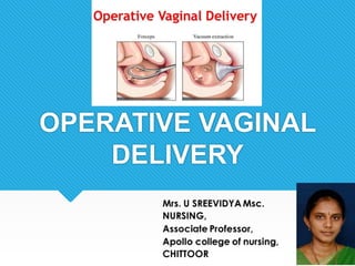
Operative Vaginal delivery
- 2. Definition: A surgically planned incision on the perineum and the posterior vaginal wall during the second stage of labour is called episiotomy. It is also called as periniotomy. It is the most common obstetric operation performed during vaginal delivery. EPISIOTOMY
- 4. Objectives: To enlarge the vaginal introitus, so as to facilitate easy and safe delivery. To prevent perineal tear.
- 5. Indications: Rigid or inelastic perineum – usually in primi and elderly primi gravidae. Suspected perineal tear – in case of big baby, breech delivery, shoulder dystocia, etc., Operative delivery such as forceps delivery, ventouse delivery. Previous perineal surgery – Pelvic floor repair, Perineal reconstructive surgery.
- 7. Timing of episiotomy If episiotomy done early, thee blood loss will be more. If done late, it fails to prevent the perineal lacerations. Bulging thinned perineum during contractions, just prior to crowning is the ideal time.
- 8. Advantages: Clear & controlled incision is easy to repair and heals better than a lacerated wound. Reduces the duration of second stage of labour. Prevents perineal tear. Minimises intracranial injuries specially in premature babies. MATERNAL FETAL
- 9. Types of episiotomy: There are four varieties in the episiotomy. MEDIO-LATERAL MEDIAN LATERAL J- SHAPED
- 10. 1. MEDIO-LATERAL: The incision is made downwards and outwards from the midpoint of the fourchette either to the right or to the left. Right- Right medio-lateral episiotomy (RMLE) Left- Left medio-lateral episiotomy (LMLE) 2. MEDIAN: The incision commences from the centre of the fourchette and extends posteriorly along the midline for about 2.5 cm.
- 11. EPISIOTOMY TYPES
- 12. • LATERAL: The incision starts from about 1 cm away from the center of the fourchette and extends laterally. It has got many drawbacks including chance of injury to the Bartholin’s duct. It is totally condemned. • ‘J’ SHAPED: The incision begins in the center of the fourchette and is directed posteriorly along the midline for about 1.5 cm and then directed downwards and outwards along 5 or 7 O’clock position to avoid the anal sphincter. This is also not done widely.
- 16. STEP I Preliminaries • The perineum is thoroughly swabbed with antiseptic (povidone-iodine) lotion and draped properly. Local anesthesia • The perineum, in the line of proposed incision is infiltrated with 10 mL of 1% solution of lignocaine
- 17. STEP II Incision Two fingers are placed in the vagina between the presenting part and the posterior vaginal wall. The incision is made by a curved or straight blunt pointed sharp scissors (scalpel may also be used) One blade of which is placed inside, in between the fingers and the posterior vaginal wall and the other on the skin. The incision should be made at the height of an uterine contraction when an accurate idea of the extent of incision can be better judged from the stretched perineum. Deliberate cut should be made starting from the center of the fourchette extending laterally either to the right or to the left. It is directed diagonally in a straight line which runs about 2.5 cm away from the anus. The incision ought to be adequate to serve the purpose for which it is needed, The bleeding is usually not sufficient to use artery forceps unless the operation is done too early or the perineum is thick.
- 19. STRUCTURES CUT ARE (1) Posterior vaginal wall (2) Superficial and deep transverse perineal muscles, bulbospongiosus and part of levator ani (3) Fascia covering those muscles (4) Transverse perineal branches of pudendal vessels and nerves (5) Subcutaneous tissue and skin.
- 22. STEP III Timing of repair 1. The repair is done soon after expulsion of placenta. 2. If repair is done prior to that, disruption of the wound is inevitable, if subsequent manual removal or exploration of the genital tract is needed. 3. Oozing during this period should be controlled by pressure with a sterile gauze swab and bleeding by the artery forceps. 4. Early repair prevents sepsis and eliminates the patient’s prolonged apprehension of “stitches”.
- 23. REPAIR STEPS Preliminaries • Lithotomy position. • A good light source • Cleansed with antiseptic solution. • Blood clots are removed from the vagina and the wound area. • The patient is draped properly and repair should be done under strict aseptic precautions. • If the repair field is obscured by oozing of blood from above, a vaginal pack may be inserted and is placed high up.
- 24. Repair • Done in three layers. • The principles to be followed are: (1) perfect hemostasis (2) to obliterate the dead space and (3) suture without tension.
- 25. LAYERS • The repair is to be done in the following order: (1) Vaginal mucosa and submucosal tissues (2) Perineal muscles (3) Skin and subcutaneous
- 27. REPAIR STEPS • The vaginal mucosa is sutured first. • The first suture is placed at or just above the apex of the tear. • Thereafter, the vaginal walls are apposed by interrupted sutures with polyglycolic acid suture (Dexon) or No. “0” chromic catgut, from above downwards till the fourchette is reached. • The suture should include the deep tissues to obliterate the dead space. • A continuous suture may cause puckering and shortening of the posterior vaginal wall. • Care should be taken not to injure the rectum.
- 28. POSTOPERATIVE CARE • Dressing • Comfort – MgSo4 compression – Infrared heat – Ice pack – Analgesic (ibuprofen) • Ambulance • Removal of stitches – Non-absorbable (6th day)
- 29. IMMEDIATE COMPLICATIONS (1) Extension of the incision to involve the rectum. (2) Vulval hematoma (3) Infection: (A) throbbing pain on the perineum (B) rise in temperature (C) the wound area looks moist, red and swollen and (D) offensive discharge TREATMENT: (a) Tofacilitate drainage of pus (b) Local dressing with antiseptic powder or ointment (c) MgSO4 compression or application of infrared heat to the area to reduce edema and pain (d) Systemic antibiotic (IV).
- 30. CONT… (4) Wound dehiscence (5)Injury to anal sphincter causing incontinence of flatus or feces. (6) Rectovaginal fistula and rarely. (7)Necrotizing fasciitis (rare) in a woman who is diabetic or immunocompromised
- 31. REMOTE COMPLICATION (1) Dyspareunia (2)Chance of perineal lacerations in subsequent labor (3) Scar endometriosis (rare).
- 32. FORCEPS DELIVERY
- 33. Operative vaginal delivery refers to any delivery process which is assisted by vaginal operations. Delivery by forceps, ventouse and destructive operations are generally included. FORCEPS DELIVERY: means extracting the fetus with the aid of obstetric forceps when it is inadvisable or impossible for the mother to complete the delivery by her own efforts. Forceps are also used to assist the delivery after coming head in breech presentation and on occasion to withdraw the head up and out of the pelvis at cesarean section.
- 35. Obstetric forceps is a pair of instruments specially designed to assist extraction of the fetal head and thereby accomplishing delivery of the fetus. VARIETIES OF OBSTETRIC FORCEPS: • Ever since either Peter I or Peter II of the Chamberlin family invented the forceps around AD 1600, more than 700 varieties were invented or modified. • Most of them are of historical interest only. But only three varieties are commonly used in present day obstetric practice.
- 36. These are:- 1.Long-curved forceps with or without axis traction device 2. Short-curved forceps 3. Kielland’s forceps The basic construction of these forceps is the same in that each consists of two halves (blades) articulated by a lock.
- 39. Long-curved obstetric forceps is relatively heavy and is about 37cm (15”) long. In India, Das’s variety (named after Sir Kedar Nath Das) is commonly used with advantages. It is comparatively lighter and slightly shorter than its Western counterpart but is quite suited for the comparatively small pelvis and small baby of Indian women. Measurements:-Length is 37cm (15”);distance in between the tips is 2.5 cm and widest diameter between the blades is 9 cm. i. BLADES: There are two blades and are named right or left in relation to maternal pelvis in which they lie when applied. ii. Shank iii. Lock iv. Handle with or without screw.
- 41. i. Blade: - The blade is fenestrated to facilitate a good grip of the fetal head. There is usually a slot in the lower part of the fenestrum of the blades to allow the upper end of the axis traction rod to be fitted. The toe of the blade refers to the tip and the heel to the end of the blade that is attached to the shank. The blade has got two curves: - Pelvic curve:-The curve on the edge is to fit more or less the curve on the axis of the birth canal (curve of Carus). The front of the forceps is the concave side of the pelvic curve. Pelvic curve permits ease of application along the maternal pelvic axis. Cephalic curve:-It is the curve on the flat surface which when articulated grasps the fetal head without compression.
- 42. ii.Shank:-It is the part between the blade and the lock and usually measures 6.25 cm(2.5”).It increases the length of the instrument and thereby, facilitates locking of the blades outside the vulva. iii.Lock: -The common method of articulation consists of a socket system located on the shank at its junction with the handle (English lock). Such type of lock requires introduction of the left blade first. iv.Handle: -The handles are apposed when the blades are articulated. It measures 12.5 cm(5”). There is a finger guard on which a finger can be placed during traction. A screw may be attached usually at the end (or at the base) of one blade (commonly left). It helps to keep the blades in position.
- 46. 3.Kielland’s Forceps It is a long almost straight (very slight pelvic curve) obstetric forceps without any axis traction device. It has got a sliding lock which facilitates correction of the head. One small knob on each blade is directed towards the occiput.
- 47. IDENTIFICATION OF BLADE OF FORCEPS Take the blade of forceps Place it in front of maternal pelvis, tip of the forceps directed towards fetal head, concavity of pelvic curve directed toward the midline of pelvis The tip point of blade should be upwards. The cephalic curve is to be directed inwards and the pelvic curve forwards. The blade which correspond to left side of mother is left blade and right side right blade.
- 48. Type of procedure (Application) Criteria Outlet Forceps Operation (1) Scalp is visible at the introitus without separating the labia (2) Fetal skull has reached the level of the pelvic floor (3) Sagittal suture is in direct anteroposterior diameter. ( Wrigley's forceps) Low Forceps Operation Leading point of the fetal skull (station) is at +2 cm or more but has not yet reached the pelvic floor. (Long curved obstetric forceps) Mid Forceps Operation Fetal head is engaged. Leading point of the fetal skull (station) is at +2 cm or less above the spine.(Kielland's forceps) High(Excluded) High Head is not engaged. This type is not included in classification Types of application of forceps: Forceps application is classified according to the station and rotation of the fetal head.
- 51. Choices of forceps Mid forceps – 10% Low forceps & Outlet forceps – 90%
- 52. INDICATIONS OF FORCEPS DELIVERY Post caesarean pregnancy Prolonged 2nd stage It is the prolongation for more than 1 hour in primigravidae or 30 mins in multiparae. This may be due to: Poor voluntary bearing down Large fetus Rigid perineum Malposition: persistent occipito posterior and deep transverse arrest.
- 53. MATERNALINDICATIONS Maternal distress are manifested by Exhaustion Pulse greater than 100 beats per min Temperature greater than 38 C Sign of dehydration Maternal diseases as: Heart disease Pulmonary TB Pre eclampsia and eclampsia
- 54. FOETALINDICATIONS Fetal distress Prolapsed pulsating cord Low birth weight baby Post maturity After coming head in breech delivery
- 55. PREREQUISITES FOR FORCEPS APPLICATION There are certain conditions which must exist before delivery can be performed. • The cervix must be completely dilated. • The membranes must be ruptured. • The head must be engaged. • No appreciable Cephalopelvic disproportion. • The bladder must be emptied. • Presence of good uterine contractions as a safeguard to postpartum hemorrhage. • The fetus must be vertex, or present a face with chin anterior. • The position of the fetal head must be known. Verbal or written consent need to be obtained in some conditions.
- 56. TYPES OF FORCEPS APPLICATION CEPHALICAPPLICATION: The forceps is applied on the sides of the foetal head in the mento- vertical diameter so, injury of the fetal face, eyes and facial nerve is avoided. PELVICAPPLICATION: The forceps is applied along the maternal pelvic wall irrespective to the position of the head.it is easier for application but carries a great risk of foetal injuries. CEPHALO-PELVIC APPLICATION: It is the ideal and possible application when the occiput is directly anterior or in mento-anterior diameter position.
- 58. PROCEDURE (LOW FORCEPS OPERATION) Preparation of mother- Clear explanation should be given and informed consent obtained. Appropriate analgesia is in place for mid-cavity rotational deliveries. This will usually be a regional block. A pudendal block may be appropriate, particularly in the context of urgent delivery. Maternal bladder has been emptied recently. Aseptic technique.
- 59. Preparation of staff- Operator must have the knowledge, experience and skill necessary. Adequate facilities are available (appropriate equipment, bed, lighting). Back-up plan in place in case of failure to deliver. When conducting mid-cavity deliveries, theatre staff should be immediately available to allow a caesarean section to be performed without delay (less than 30 minutes).
- 60. A senior obstetrician competent in performing mid- cavity deliveries should be present if a junior trainee is performing the delivery. Anticipation of complications that may arise (e.g. shoulder dystocia, postpartum haemorrhage) Personnel present that are trained in neonatal resuscitation.
- 61. The women should be prepared in advances for the possibility of a forceps delivery. The women should be placed in lithotomy position. Both legs must be placed simultaneously to avoid strain on the woman’s back and hips. Anaesthesia- can be given by perineal infiltration with 1% lignocaine as local anaesthesia. If bladder was not emptied, catheterisation to be done. Internal examination to be done to assess- • state of the cervix • Membranes status • Presentation and position of the head (station) Episiotomy- It can be done prior to application of forceps to prevent tear. PRELIMINARIES
- 62. Steps of forceps delivery The operation consists of the following steps: Identification of the blades and their application Locking of the blades Traction Removal of the blades
- 63. 1. Identification of the blades and their application The women’s vulval area is thoroughly cleaned and draped with sterile towels using aseptic technique. The forceps are identified as left or right by assembling them briefly before proceeding. The left blade is passed gently between the perineum and fetal head with the first two figures of the operator’s hand lying alongside the fetal head protecting the maternal tissue. The tip of the forceps blade slides lightly over the head, in to the hollow of the sacrum and is then ‘wandered’ to the left side of the pelvis where it should sit alongside the head. The procedure is repeated with the right blade until it sits on the right of the pelvis.
- 65. Application of right blade
- 66. 2. Locking of the blades It should then be easy to lock the two blades and there should be little or no gap between the handles. Minor difficulty in locking can be corrected by depressing the handles on the perineum. A significant gap suggests that the forceps are wrongly positioned and they should be reapplied after carefully checking the position of head. The handles should never be forced to lock them.
- 67. 2. Traction & Removal of the blades As soon as the operator is ready and the uterus contracts, the woman is encouraged to push. To supplement her efforts the obstetrician exerts steady, downwards traction on the forceps. Traction is released between contractions. Intermittent traction is continued in a downward and backward direction until the head comes to the perineum. The pull is then directed horizontally straight towards the operator until the head is almost crowned. The direction of pull is gradually changed towards the mother’s abdomen to deliver the head by extension. The blades are removed one after the other, the right one first. Following the birth of the head, usual procedures are to be followed as in normal delivery. Intravenous methergine 0.2mg is to be administered with the delivery of the anterior shoulder. Episiotomy is repaired as quickly as possible and the woman made comfortable.
- 69. Forceps Delivery
- 70. OUTLET FORCEPS OPERATION Wrigley's forceps are used exclusively in outlet forceps operation. Procedure is same as low forceps operation except the traction – the direction of the pull is straight horizontal and then upwards & forwards.
- 71. CONTRAINDICATIONS FOR FORCEPS -Absence of full dilatation of cervix. -In case of cephalopelvic disproportion. -High station of fetal head. -If uterine contraction cease. -Lack of experience of operator. -Mentum posterior face presentation. -Hydrocephalic infant. -Brow presentation. -Fetal prematurity
- 72. Difficulties in forceps operation The difficulties are encountered mainly due to faulty assessment of the case. Difficulties occurs in four areas. During application of blades – due to incompletely dilated cervix and non-engaged head. Difficulty in locking – due to improper insertion of blades and failure to depress the handle. Difficulty in traction – due to faulty cephalic application, wrong direction of traction and constriction ring. Slipping of blades – due to faulty application.
- 73. The hazards of the forceps operation are mostly related to the faulty technique and to the indication for which the forceps are applied. MATERNAL (In the mother) Immediate Injury Extension of the episiotomy towards rectum or upwards up to the vault of vagina Vaginal lacerations Cervical tear especially when applied through an incompletely dilated cervix. Bruising and trauma to the urethra.
- 74. Postpartum hemorrhage due to trauma, or atonic uterus related to prolonged labor or effects of anesthesia. Shock due to blood loss, prolonged labor and dehydration. Sepsis due to devitalization of local tissues and improper asepsis. Late complications Dyspareunia Chronic low backache due to tension imposed on softened ligaments of lumbosacral or sacroiliac joints during lithotomy position. Genital prolapse stress incontinence.
- 75. FETAL (In the infant) Immediate Asphyxia due to intracranial stress out of prolonged compression. Intracranial hemorrhage due to misapplication of the blades. Cephalhematoma Facial palsy due to damage to facial nerve. Abrasions on the soft tissues of the face and forehead by the forceps blade, severe bruising will cause marked jaundice. Remote • Cerebral palsy
- 78. Prevention: It is a preventable condition. Only through skill and judgment, proper selection of the case ideal for forceps can be identified. Even if applied in wrong cases, one should resist the temptation to give forcible traction in an attempt to hide the mistake. Management: (1)To assess the effect on the mother and the fetus. (2)To start a Ringer’s solution drip and to arrange for blood transfusion, if required. (3)To administer parenteral antibiotic. (4)To exclude rupture of the uterus. (5)The procedure is abandoned and delivery is done by cesarean section (6)Laparotomy should be done in a case with rupture of uterus.
- 81. Vacuum Extraction (Ventouse) Ventouse is a vacuum device used to assist the delivery of a baby when the second stage of labour has not progressed adequately. It is an alternative to a forceps delivery and caesarean section. It cannot be used when the baby is in the breech position or for premature births. This technique is also called vacuum- assisted vaginal delivery or vacuum extraction (VE). It is an instrumental device designed to assist delivery by creating a vacuum between it and the fetal scalp In the United states the device is referred to as the vacuum extractor whereas in Europe it is called as Ventouse- from the french word literally meaning soft cup.
- 82. Historical background In 1705, Yonge described an attempted vaginal delivery using a cupping glass In 1848 Simpson devised a bell shaped device called an “air tractor vacuum extractor” In 1953 a metal cup extractor was developed by Malmstrom .
- 83. Description Vacuum extractor is composed of: A specially designed cup with a diameter of 3, 4, 5 or 6 cm. A rubber tube attaching the cup to a glass bottle with a screw in between to release the negative pressure. A manometer fitted in the mouth of the glass bottle to declare the negative pressure. Another rubber tube connecting the bottle to a suction piece which may be manual or electronic creating a negative pressure that should not exceed - 0.8 kg per cm2.
- 84. VACUUM EXTRACTOR
- 86. Types of vacuum extractors Vacuum extractors are divided on the basis of the type of cup- -metal or plastic 1.Metal cup vacuum extractors 2.Soft cup vacuum extractors
- 87. Metal cup • The metal-cup vacuum extractor is a mushroom-shaped metal cup varying from 40 to 60 mm in diameter. • Metal-cup vacuum extractors have a higher success rate and easier cup placement in the occipitoposterior (OP) position, • The rigidity of metal cups can make application difficult and uncomfortable, and their use is associated with an increased risk of fetal scalp injuries.
- 88. VENTOUSE CUP WITH TRACTION DEVICE
- 89. Soft cup • Traditionally soft cups are bell or funnel shaped. • Soft-cup instruments can be used with a manual vacuum pump or an electrical suction device. Soft-cup vacuum extractors may be disposable or reusable. • Compared with metal-cup devices, soft-cup vacuum extractors cause fewer neonatal scalp injuries. However, these instruments have a higher failure rate.
- 90. VENTOUSE CUP WITH TRACTION DEVICE
- 91. Indications of vacuum extraction Generally vacuum extraction is reserved for fetuses who have attained a gestational age of 34 weeks. Otherwise, the indications and pre-requisites for its use are the same as for forceps delivery. Delay in descent of the head in case of twins. Delay in first stage of labour.
- 92. Contraindications Operator inexperience Inability to assess fetal position High station(above 0 station) Suspicion of cephalopelvic disproportion Other presentations than vertex. Premature fetus(<34 weeks). Intact membranes. Fetal macrosomia - >4kg
- 93. Pre-requisites of the Procedure Procedure should be explained to the patient and consent should be taken Emotional support and encouragement Lithotomy position. Bladder should be emptied. Antiseptic measures for the vagina, vulva and perineum. Vaginal examination to check pelvic capacity, cervical dilatation, presentation, position, station and degree of flexion of the head and that the membranes are ruptured.
- 94. Pre-requisites cont.. Cervix should be at least 6cm dilated. Head should be engaged. Procedure to be taken with perineal infiltration with 1% lignocaine. It may be applied even without anaesthesia specially in parous women. The instrument should be assembled and the vacuum is tested prior to its application.
- 95. PROCEDURE 1. Application of the cup Identification of the flexion point- -It is situated 3 cm in front of the posterior fontanelle. -Centre of the cup should be overlying the flexion point. This placement promotes flexion ,descent and autorotation. If traction is directed from this point the fetal head is flexed to the narrowest sub-occipito bregmatic diameter(9.5 cm).
- 98. Precautions- • The largest cup that can be easily passed is introduced sideways into the vagina by pressing it backwards against the perineum. • Be sure that there is no cervical or vaginal tissues nor the umbilical cord or a limb in complex presentation is included in the cup.
- 100. 2. Creating the negative pressure When using the rigid cups, the negative pressure is gradually increased by 0.2 kg/cm2 every 2 minutes until - 0.8 kg/cm2 is attained. This creates an artificial caput within the cup. With soft cups negative pressure can be increased to 0.8 kg/cm2 over as little as 1 minute
- 103. Episiotomy An episiotomy may be needed for proper placement of the cup If not, then delay the episiotomy till the head stretches the perineum or perineum interferes with the axis of traction This will minimize unnecessary blood loss.
- 104. 3. Traction Traction should be intermittent and co- ordinated with maternal expulsive efforts and with uterine contractions. Traction should be in line of the pelvic axis and perpendicular to the plane of the cup
- 105. Traction contd.. Traction may be initiated by using a two handed technique Fingers of one hand are placed against the suction cup while the other hand grasps the handle of the instrument This allows one to detect negative traction. Manual torque to the cup should be avoided as it may cause cephalohematoma and scalp lacerations. Between contractions, check for fetal heart rate and proper application of the cup
- 106. Traction
- 107. 4. Release When the head is delivered the vacuum is reduced as slowly as it was created using the screw as this diminishes the risk of scalp damage. The chignon should be explained to the patient and the relatives.
- 109. Reapplication of the cup If the cup detaches for the first time, reassess the situation. If favorable ,then reapply. If cup detaches for the second time, reassess if vaginal delivery is safe or move to caesarean section Caesarean section is necessary if there is inadequate descent and rotation
- 110. Failure of vacuum Vacuum extraction is considered failed if- -fetal head does not advance with each pull -fetus is undelivered after 3 pulls with no descent or after 30 minutes -cup slips off the head twice at the proper direction of pull with the maximum negative pressure.
- 111. Advantages of Vacuum over Forceps Regional Anaesthesia is not required so it is preferred in cardiac and pulmonary patient. The ventouse is not occupying a space beside the head as forceps. Less compression force (0.77 kg/cm2) compared to forceps (1.3 kg/cm2) so injuries to the head is less common. Less genital tract lacerations. Can be applied before full cervical dilatation.
- 112. Complications Maternal Perineal, vaginal ,labial, periurethral and cervical lacerations. Annular detachment of the cervix when applied with incompletely dilated cervix. Cervical incompetence and future prolapse if used with incompletely dilated cervix.
- 113. Complications cont.. Fetal Cephalohaematoma. Scalp lacerations and bruising Intracranial haemorrhage. Neonatal jaundice Subconjunctival haemorrhage Injury of sixth and seventh cranial nerves Retinal hemorrhage Fetal death
- 115. THANK YOU