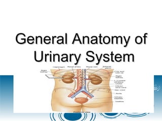
Urinary system .pdf

General Anatomy of
General Anatomy of
Urinary System
Urinary System
Introduction
Introduction
 The
The urinary system
urinary system, also known as
, also known as
the
the renal system
renal system
 The urinary system refers to the structures
The urinary system refers to the structures
that produce and conduct
that produce and conduct urine
urine to the
to the
point of excretion.
point of excretion.
URINARY SYSTEM ORGANS
 Kidneys (2)
Kidneys (2)
 Ureters (2)
Ureters (2)
 Urinary bladder
Urinary bladder
 Urethra
Urethra
Urinary system .pdf
Kidney
Kidney
 The human body normally has two paired
The human body normally has two paired
kidneys
kidneys, one on the left and one on the
, one on the left and one on the
right.
right.
 The functional unit of the kidney is
The functional unit of the kidney is
nephron.
nephron.
 Urine is formed by
Urine is formed by nephrons
nephrons
Location and External Anatomy of
Kidneys
Kidneys
 Located
Located
retroperitoneally
retroperitoneally
 Lateral to T
Lateral to T12
12–L
–L3
3
vertebrae
vertebrae
 Average kidney
Average kidney

12 cm tall, 6 cm wide,
3 cm thick
3 cm thick
Protected by three connective tissue
layers
layers
 Renal fascia
Renal fascia

Attaches to abdominal wall
 Adipose capsule
Adipose capsule

Fat cushioning kidney
 Renal capsule
Renal capsule

Fibrous sac
Fibrous sac

Protects from trauma and infection
Urinary system .pdf
KIDNEY
KIDNEY
Gross anatomy
Gross anatomy
 Renal parenchyma
Renal parenchyma
 Renal sinus
Renal sinus
KIDNEY ANATOMY
KIDNEY ANATOMY
Renal parenchyma
Renal parenchyma
 Two zones
Two zones

Outer cortex
Outer cortex

Inner medulla
Inner medulla
Urinary system .pdf
Anatomy of the kidneys
 Superficial outer cortex and inner medulla
Superficial outer cortex and inner medulla

The medulla consists of 6-18 renal
pyramids
pyramids

The cortex is composed of roughly 1.25
million nephrons
million nephrons
KIDNEY ANATOMY
KIDNEY ANATOMY
Renal parenchyma
Renal parenchyma
 Renal pyramids
Renal pyramids

Extensions of cortex (renal columns)
divide medulla into 6 – 10
divide medulla into 6 – 10 renal
renal
pyramids
pyramids

Pyramid + overlying cortex = Lobe

Point of pyramid = Papilla

Papilla nested in cup (minor calyx)

2 – 3 minor calices
2 – 3 minor calices 
 Major calyx
Major calyx

2 – 3 major calices
2 – 3 major calices 
 Renal pelvis
Renal pelvis

Renal pelvis
Renal pelvis 
 Ureter
Ureter
KIDNEY ANATOMY
KIDNEY ANATOMY
Renal sinus
Renal sinus
 Surrounded by renal parenchyma
Surrounded by renal parenchyma
 Contains blood & lymph vessels, nerves,
Contains blood & lymph vessels, nerves,
urine-collecting structures
 Hilus
Hilus

On concave surface
On concave surface

Vessels and nerves enter and exit
 Major and minor calyces along with the
Major and minor calyces along with the
pelvis drain urine to the ureters
Human Anatomy, 3rd edition
Prentice Hall, © 2001
Structure of the Kidney
Urinary system .pdf
NEPHRONS
NEPHRONS
Nephrons
Nephrons
 Functional units of kidney
Functional units of kidney
 ~1.2 million per kidney
~1.2 million per kidney
 Three main parts
Three main parts

Blood vessels
Blood vessels

Renal corpuscle
Renal corpuscle

Renal tubule
Renal tubule
Blood vessels servicing kidney
 Glomerulus
Glomerulus

Fenestrated capillaries

Capillary filtration in glomerulus initiates urine
production
production

Filtrate lacks cells & proteins

Drained by efferent arteriole


Peritubular capillaries
Peritubular capillaries


 
 
 
 Renal vein
Renal vein
Urinary system .pdf
Renal corpuscle
Renal corpuscle
 Composed of a
Composed of a glomerulus
glomerulus and the
and the
Bowman's capsule
Bowman's capsule,
,
 The
The renal corpuscle
renal corpuscle is the beginning of
is the beginning of
the nephron.
the nephron.
 It is the nephron's initial filtering
It is the nephron's initial filtering
component.
component.
Glomerulus
Glomerulus
 The
The glomerulus
glomerulus is a
is a capillary
capillary tuft that
tuft that
receives its blood supply from an afferent
arteriole
arteriole of the
of the renal circulation
renal circulation.
.
 The glomerular blood pressure provides
The glomerular blood pressure provides
the driving force for water and solutes to
be filtered out of the blood and into the
space made by
space made byBowman's capsule
Bowman's capsule
 The remainder of the blood passes into
The remainder of the blood passes into
the efferent arteriole.
 The diameter of efferent arterioles is
The diameter of efferent arterioles is
smaller than that of afferent arterioles,
increasing the hydrostatic pressure in the
glomerulus.
glomerulus.
Bowman's capsule
Bowman's capsule
 The Bowman's capsule, also called the
The Bowman's capsule, also called the
glomerular capsule.
glomerular capsule.
 surrounds the glomerulus.
surrounds the glomerulus.
 It is composed of a visceral inner layer
It is composed of a visceral inner layer
formed by specialized cells
called podocytes.
called podocytes.
 Parietal outer layer composed of simple
Parietal outer layer composed of simple
squamous epithelium.

 Fluids from blood in the glomerulus are
Fluids from blood in the glomerulus are
filtered through the visceral layer of
podocytes, resulting in the glomerular
filtrate.
filtrate.
NOTE
NOTE
Renal corpuscle
Renal corpuscle
 Glomerulus plus capsule
Glomerulus plus capsule
 Glomerulus enclosed in two-layered glomerular
Glomerulus enclosed in two-layered glomerular
capsule
capsule

“
“Bowman’s capsule”
Bowman’s capsule”
 Fluid filters from glomerular capillaries
Fluid filters from glomerular capillaries

“
“Glomerular filtrate”
Glomerular filtrate”
 Fluid collects in capsular space
Fluid collects in capsular space
 Fluid flows into renal tubule
Fluid flows into renal tubule
Urinary system .pdf
Renal tubule
Renal tubule
 Leads from glomerular capsule
Leads from glomerular capsule
 Ends at tip of medullary pyramid
Ends at tip of medullary pyramid
 ~3 cm long
~3 cm long
 Four major regions
Four major regions

Proximal convoluted tubule

Nephron loop
Nephron loop

Distal convoluted tubule

Collecting duct
Collecting duct
Urinary system .pdf
Proximal convoluted tubule
(PCT)
(PCT)
 Arises from glomerular capsule
Arises from glomerular capsule
 Longest, most coiled region
Longest, most coiled region
 lies in cortex
lies in cortex
 lined by simple cuboidal epithelium with brush
lined by simple cuboidal epithelium with brush
borders which help to increase the area of
absorption greatly.)
 Prominent microvilli
Prominent microvilli

Function in absorption
Urinary system .pdf
Nephron loop (“Loop of Henle”)
 “
“U” – shaped, distal to PCT
U” – shaped, distal to PCT
 lies in medulla
lies in medulla
2 parts
2 parts
 Descending limb of loop of Henle
Descending limb of loop of Henle
 Ascending limb of loop of Henle
Ascending limb of loop of Henle

The ascending limb of loop of Henle is divided
into 2 segments:
into 2 segments:

Lower end of ascending limb
Lower end of ascending limb is very thin and
is very thin and
is lined by simple squamous epithelium.

The distal portion of ascending limb
The distal portion of ascending limb is thick
is thick
and is lined by simple cuboidal epithelium.

Thin ascending limb of loop of Henle

Thick ascending limb of loop of Henle (enters
cortex and becomes DCT-distal convoluted
tubule.)
tubule.)
 Thick segments
Thick segments

Active transport of salts

High metabolism, many mitochondria
 Thin segments
Thin segments

Permeable to water
Permeable to water

Low metabolism
Low metabolism
Distal convoluted tubule (DCT)
 Coiled, distal to nephron loop
Coiled, distal to nephron loop
 Shorter than PCT
Shorter than PCT
 Less coiled than PCT
Less coiled than PCT
 Very few microvilli
Very few microvilli
 Contacts afferent and efferent arterioles
Contacts afferent and efferent arterioles
 Contact with peritubular capillaries
Contact with peritubular capillaries
Urinary system .pdf
Collecting duct
Collecting duct
 DCTs of several nephrons empty into a
DCTs of several nephrons empty into a
collecting duct
collecting duct
 Passes into medulla
Passes into medulla
 Several merge into papillary duct (~30 per
Several merge into papillary duct (~30 per
papilla)
papilla)
 Drain into minor calyx
Drain into minor calyx
Urinary system .pdf
CLASSES
CLASSES
The two general classes of nephrons are
 Cortical nephrons
Cortical nephrons
 Juxtamedullary nephrons
Juxtamedullary nephrons
which are classified according to the
 length of their Loop of Henle
length of their Loop of Henle
 location of their
location of their renal corpuscle
renal corpuscle.
.
 All nephrons have their renal corpuscles
All nephrons have their renal corpuscles
in the cortex.
in the cortex.
 Cortical
Cortical nephrons have their Loop of
nephrons have their Loop of
Henle in the renal medulla near its junction
with the renal cortex,
 Loop of Henle of juxtamedullary nephrons
Loop of Henle of juxtamedullary nephrons
is located deep in the renal medulla;
Urinary system .pdf
URINE FORMATION
URINE FORMATION
Overview
Overview
 Blood plasma
Blood plasma 
 Urine
Urine
 Four steps
Four steps

Glomerular filtration

Tubular reabsorption

Tubular secretion
Tubular secretion

Water conservation
Water conservation
Urinary system .pdf
Urinary system .pdf
Urinary system .pdf
KIDNEY FUNCTIONS
KIDNEY FUNCTIONS
 Regulate blood volume, pressure
Regulate blood volume, pressure
 Regulate fluid osmolarity
Regulate fluid osmolarity
 Secrete renin
Secrete renin
 Secrete erythropoietin (EPO)
Secrete erythropoietin (EPO)
 Regulate P
Regulate PCO
CO2
2, Acid-Base balance
, Acid-Base balance
 Synthesize calcitriol (Vitamin D)
Synthesize calcitriol (Vitamin D)
 Detoxify free radicals, drugs
Detoxify free radicals, drugs
 Gluconeogenesis
Gluconeogenesis
The Ureters
The Ureters
 Pair of muscular tubes
Pair of muscular tubes
 Extend from renal pelvis to the bladder
Extend from renal pelvis to the bladder
 Oblique entry into bladder prevents backflow of urine
Oblique entry into bladder prevents backflow of urine
Histology of Ureter
Histology of Ureter
 Mucosa –
Mucosa – transitional
transitional
epithelium
epithelium
 Muscularis
Muscularis – two layers
– two layers

Inner longitudinal layer

Outer circular layer
 Adventitia
Adventitia – typical
– typical
connective tissue
connective tissue
 Carry urine from kidneys to urinary
Carry urine from kidneys to urinary
bladder via peristalsis

Rhythmic contraction of smooth muscle
 Enter bladder from below
Enter bladder from below
 Pressure from full bladder compresses
Pressure from full bladder compresses
ureters and prevents backflow
 Small diameter
Small diameter
 Easily obstructed or injured by kidney
Easily obstructed or injured by kidney
stones (renal calculi)
Urinary Bladder
Urinary Bladder
 A collapsible muscular
A collapsible muscular
sac
sac
 Stores and expels urine
Stores and expels urine

Full bladder –
Full bladder –
spherical
spherical
• Expands into the
Expands into the
abdominal cavity
abdominal cavity

Empty bladder – lies
entirely within the
entirely within the
pelvis
pelvis
Figure 23.13
Urinary Bladder
Urinary Bladder
 Wall of bladder
Wall of bladder

Mucosa - transitional epithelium

Muscular layer - detrusor muscle

Adventitia
Adventitia
 Wrinkles termed rugae
Wrinkles termed rugae
 Openings of ureters common site for
Openings of ureters common site for
bladder infection
bladder infection
Urinary bladder
Urinary bladder
Urethra
Urethra
 Conveys urine from body
Conveys urine from body
 Internal urethral sphincter
Internal urethral sphincter

Retains urine in bladder

Smooth muscle, involuntary
 External urethral sphincter
External urethral sphincter

Provides voluntary control over voiding of urine
Urethra in female
Urethra in female
 3 – 4 cm long in females
3 – 4 cm long in females

Bound by connective tissue to anterior wall of
vagina
vagina

Urethral orifice exits body between vaginal
orifice and clitoris
Urinary system .pdf
Urethra in male
Urethra in male
 ~
~18 cm long in males
18 cm long in males

Prostatic urethra
Prostatic urethra
• ~2.5 cm long, urinary bladder
~2.5 cm long, urinary bladder 

prostate
prostate

Membranous urethra
Membranous urethra
• ~0.5 cm, passes through floor of
~0.5 cm, passes through floor of
pelvic cavity
pelvic cavity

Penile urethra
Penile urethra
• ~15 cm long, passes through penis
~15 cm long, passes through penis
Urinary system .pdf
URINE ELIMINATION
URINE ELIMINATION
Urination (micturition)
 ~200 ml of urine held
~200 ml of urine held
 Distension initiates desire to void
Distension initiates desire to void
 Internal sphincter relaxes involuntarily
Internal sphincter relaxes involuntarily

Smooth muscle
Smooth muscle
 External sphincter voluntarily relaxes
External sphincter voluntarily relaxes

Skeletal muscle
Skeletal muscle

Poor control in infants
 Bladder muscle contracts
Bladder muscle contracts
 Urine forces through urethra
Urine forces through urethra
Figure 26.1
Urinary System
Urinary System
 Kidneys – produce
Kidneys – produce
urine
urine
 Ureters –transport
Ureters –transport
urine to bladder
urine to bladder
 Urinary bladder -
Urinary bladder -
stores urine
stores urine
 Urethra transports
Urethra transports
urine to exterior
urine to exterior
Functions of the urinary system
 Homeostatic regulation of blood plasma
Homeostatic regulation of blood plasma

Regulating blood volume and pressure

Regulating plasma ion concentrations

Stabilizing blood pH

Conserving nutrients
 Filter many liters of fluid from blood
Filter many liters of fluid from blood
 Excretion - The removal of organic waste
Excretion - The removal of organic waste
products from body fluids

Urea
Urea

Uric acid
Uric acid

Creatinine
Creatinine
 Elimination - The discharge of waste products
Elimination - The discharge of waste products
into the environment
1 sur 62

Recommandé

General anatomy of urinary system ppt par
General anatomy of urinary system pptGeneral anatomy of urinary system ppt
General anatomy of urinary system pptjagan _jaggi
165.4K vues62 diapositives
URINARY SYSTEM par
URINARY  SYSTEMURINARY  SYSTEM
URINARY SYSTEMGrandhi sandeep ganesh
184 vues31 diapositives
URINARY SYSTEM.pptx par
URINARY SYSTEM.pptxURINARY SYSTEM.pptx
URINARY SYSTEM.pptxRameeThj
42 vues93 diapositives
Urinary System Presentationyorkville College 1203376567476185 2 par
Urinary System Presentationyorkville College 1203376567476185 2Urinary System Presentationyorkville College 1203376567476185 2
Urinary System Presentationyorkville College 1203376567476185 2guest8b2ebd
2.2K vues64 diapositives
Urinary system par
Urinary systemUrinary system
Urinary systemTRUMAN COLLEGE
3.6K vues35 diapositives
Uri A par
Uri AUri A
Uri Axtrm nurse
1.2K vues125 diapositives

Contenu connexe

Similaire à Urinary system .pdf

Genitourinary System Anatomy and Physiology..pptx par
Genitourinary System Anatomy and Physiology..pptxGenitourinary System Anatomy and Physiology..pptx
Genitourinary System Anatomy and Physiology..pptxDR .PALLAVI PATHANIA
1.4K vues186 diapositives
Urinary System Presentation-Yorkville College par
Urinary System Presentation-Yorkville CollegeUrinary System Presentation-Yorkville College
Urinary System Presentation-Yorkville Collegeguestae94d9
5K vues57 diapositives
Excretory Products and their Elimination par
Excretory Products and their Elimination Excretory Products and their Elimination
Excretory Products and their Elimination BhoomikaDhiman2
3.3K vues106 diapositives
Urinary system-presentationyorkville-college-1203376567476185-2 par
Urinary system-presentationyorkville-college-1203376567476185-2Urinary system-presentationyorkville-college-1203376567476185-2
Urinary system-presentationyorkville-college-1203376567476185-2Ksenja Ljashenko
1.3K vues57 diapositives
Anatomy of urinary system par
Anatomy of urinary systemAnatomy of urinary system
Anatomy of urinary systemjafarqamar
1.5K vues51 diapositives
Excretory system par
Excretory systemExcretory system
Excretory systemHafiz M Waseem
1K vues38 diapositives

Similaire à Urinary system .pdf(20)

Urinary System Presentation-Yorkville College par guestae94d9
Urinary System Presentation-Yorkville CollegeUrinary System Presentation-Yorkville College
Urinary System Presentation-Yorkville College
guestae94d95K vues
Excretory Products and their Elimination par BhoomikaDhiman2
Excretory Products and their Elimination Excretory Products and their Elimination
Excretory Products and their Elimination
BhoomikaDhiman23.3K vues
Urinary system-presentationyorkville-college-1203376567476185-2 par Ksenja Ljashenko
Urinary system-presentationyorkville-college-1203376567476185-2Urinary system-presentationyorkville-college-1203376567476185-2
Urinary system-presentationyorkville-college-1203376567476185-2
Ksenja Ljashenko1.3K vues
Anatomy of urinary system par jafarqamar
Anatomy of urinary systemAnatomy of urinary system
Anatomy of urinary system
jafarqamar1.5K vues
7 excretory system par Home
7 excretory system7 excretory system
7 excretory system
Home12 vues
Excretory system:Kidneys,ureter,urethra and urinary bladder par Jasleen Kaur
Excretory system:Kidneys,ureter,urethra and urinary bladderExcretory system:Kidneys,ureter,urethra and urinary bladder
Excretory system:Kidneys,ureter,urethra and urinary bladder
Jasleen Kaur524 vues
Urinary System.pptx par krimscott
Urinary System.pptxUrinary System.pptx
Urinary System.pptx
krimscott15 vues
16. The Urinary System par SUNY Ulster
16. The Urinary System16. The Urinary System
16. The Urinary System
SUNY Ulster3.1K vues
the urinary system par Deb Smu
the urinary systemthe urinary system
the urinary system
Deb Smu6.1K vues
The urinary system par Sohel Daria
The urinary systemThe urinary system
The urinary system
Sohel Daria4.2K vues
Anatomy of Urinary system par drsukriti1
Anatomy of Urinary systemAnatomy of Urinary system
Anatomy of Urinary system
drsukriti1403 vues

Plus de ssuserbf4af22

Эндокрин.pdf par
Эндокрин.pdfЭндокрин.pdf
Эндокрин.pdfssuserbf4af22
4 vues38 diapositives
Эндо.pdf par
Эндо.pdfЭндо.pdf
Эндо.pdfssuserbf4af22
7 vues34 diapositives
444.ppt par
444.ppt444.ppt
444.pptssuserbf4af22
1 vue27 diapositives
1-190128092525.pdf par
1-190128092525.pdf1-190128092525.pdf
1-190128092525.pdfssuserbf4af22
5 vues21 diapositives
malereproductivesystem-140511011110-phpapp01.pdf par
malereproductivesystem-140511011110-phpapp01.pdfmalereproductivesystem-140511011110-phpapp01.pdf
malereproductivesystem-140511011110-phpapp01.pdfssuserbf4af22
5 vues54 diapositives
Математика .ppt par
Математика .pptМатематика .ppt
Математика .pptssuserbf4af22
1 vue17 diapositives

Plus de ssuserbf4af22(20)

malereproductivesystem-140511011110-phpapp01.pdf par ssuserbf4af22
malereproductivesystem-140511011110-phpapp01.pdfmalereproductivesystem-140511011110-phpapp01.pdf
malereproductivesystem-140511011110-phpapp01.pdf
ssuserbf4af225 vues
брюшина лекция 22.pptx par ssuserbf4af22
брюшина лекция 22.pptxбрюшина лекция 22.pptx
брюшина лекция 22.pptx
ssuserbf4af223 vues

Dernier

BUSINESS ETHICS MODULE 1 UNIT I_A.pdf par
BUSINESS ETHICS MODULE 1 UNIT I_A.pdfBUSINESS ETHICS MODULE 1 UNIT I_A.pdf
BUSINESS ETHICS MODULE 1 UNIT I_A.pdfDr Vijay Vishwakarma
106 vues25 diapositives
UNIT NO 13 ORGANISMS AND POPULATION.pptx par
UNIT NO 13 ORGANISMS AND POPULATION.pptxUNIT NO 13 ORGANISMS AND POPULATION.pptx
UNIT NO 13 ORGANISMS AND POPULATION.pptxMadhuri Bhande
59 vues33 diapositives
ANGULARJS.pdf par
ANGULARJS.pdfANGULARJS.pdf
ANGULARJS.pdfArthyR3
54 vues10 diapositives
Boston In The American Revolution par
Boston In The American RevolutionBoston In The American Revolution
Boston In The American RevolutionMary Brown
38 vues78 diapositives
Interaction of microorganisms with vascular plants.pptx par
Interaction of microorganisms with vascular plants.pptxInteraction of microorganisms with vascular plants.pptx
Interaction of microorganisms with vascular plants.pptxMicrobiologyMicro
77 vues33 diapositives
PRELIMS ANSWER.pptx par
PRELIMS ANSWER.pptxPRELIMS ANSWER.pptx
PRELIMS ANSWER.pptxsouravkrpodder
58 vues60 diapositives

Dernier(20)

UNIT NO 13 ORGANISMS AND POPULATION.pptx par Madhuri Bhande
UNIT NO 13 ORGANISMS AND POPULATION.pptxUNIT NO 13 ORGANISMS AND POPULATION.pptx
UNIT NO 13 ORGANISMS AND POPULATION.pptx
Madhuri Bhande59 vues
ANGULARJS.pdf par ArthyR3
ANGULARJS.pdfANGULARJS.pdf
ANGULARJS.pdf
ArthyR354 vues
Boston In The American Revolution par Mary Brown
Boston In The American RevolutionBoston In The American Revolution
Boston In The American Revolution
Mary Brown38 vues
Interaction of microorganisms with vascular plants.pptx par MicrobiologyMicro
Interaction of microorganisms with vascular plants.pptxInteraction of microorganisms with vascular plants.pptx
Interaction of microorganisms with vascular plants.pptx
Artificial Intelligence and The Sustainable Development Goals (SDGs) Adoption... par BC Chew
Artificial Intelligence and The Sustainable Development Goals (SDGs) Adoption...Artificial Intelligence and The Sustainable Development Goals (SDGs) Adoption...
Artificial Intelligence and The Sustainable Development Goals (SDGs) Adoption...
BC Chew55 vues
From social media to generative media (3).pptx par Mark Carrigan
From social media to generative media (3).pptxFrom social media to generative media (3).pptx
From social media to generative media (3).pptx
Mark Carrigan88 vues
Introduction to Physiotherapy and Electrotherapy par Sreeraj S R
Introduction to Physiotherapy and ElectrotherapyIntroduction to Physiotherapy and Electrotherapy
Introduction to Physiotherapy and Electrotherapy
Sreeraj S R82 vues
NodeJS and ExpressJS.pdf par ArthyR3
NodeJS and ExpressJS.pdfNodeJS and ExpressJS.pdf
NodeJS and ExpressJS.pdf
ArthyR360 vues
The Future of Micro-credentials: Is Small Really Beautiful? par Mark Brown
The Future of Micro-credentials:  Is Small Really Beautiful?The Future of Micro-credentials:  Is Small Really Beautiful?
The Future of Micro-credentials: Is Small Really Beautiful?
Mark Brown131 vues
JRN 362 - Lecture Twenty-Two par Rich Hanley
JRN 362 - Lecture Twenty-TwoJRN 362 - Lecture Twenty-Two
JRN 362 - Lecture Twenty-Two
Rich Hanley41 vues
Introduction to AERO Supply Chain - #BEAERO Trainning program par Guennoun Wajih
Introduction to AERO Supply Chain  - #BEAERO Trainning programIntroduction to AERO Supply Chain  - #BEAERO Trainning program
Introduction to AERO Supply Chain - #BEAERO Trainning program
Guennoun Wajih142 vues
Guidelines & Identification of Early Sepsis DR. NN CHAVAN 02122023.pptx par Niranjan Chavan
Guidelines & Identification of Early Sepsis DR. NN CHAVAN 02122023.pptxGuidelines & Identification of Early Sepsis DR. NN CHAVAN 02122023.pptx
Guidelines & Identification of Early Sepsis DR. NN CHAVAN 02122023.pptx
Niranjan Chavan48 vues
Geology of Ethiopia and the Horn Chapter 2 GeES 1011.pptx par behailugetu
Geology of Ethiopia and the Horn Chapter 2 GeES 1011.pptxGeology of Ethiopia and the Horn Chapter 2 GeES 1011.pptx
Geology of Ethiopia and the Horn Chapter 2 GeES 1011.pptx
behailugetu104 vues

Urinary system .pdf

  • 1. General Anatomy of General Anatomy of Urinary System Urinary System
  • 2. Introduction Introduction  The The urinary system urinary system, also known as , also known as the the renal system renal system  The urinary system refers to the structures The urinary system refers to the structures that produce and conduct that produce and conduct urine urine to the to the point of excretion. point of excretion.
  • 3. URINARY SYSTEM ORGANS URINARY SYSTEM ORGANS  Kidneys (2) Kidneys (2)  Ureters (2) Ureters (2)  Urinary bladder Urinary bladder  Urethra Urethra
  • 5. Kidney Kidney  The human body normally has two paired The human body normally has two paired kidneys kidneys, one on the left and one on the , one on the left and one on the right. right.  The functional unit of the kidney is The functional unit of the kidney is nephron. nephron.  Urine is formed by Urine is formed by nephrons nephrons
  • 6. Location and External Anatomy of Location and External Anatomy of Kidneys Kidneys  Located Located retroperitoneally retroperitoneally  Lateral to T Lateral to T12 12–L –L3 3 vertebrae vertebrae  Average kidney Average kidney  12 cm tall, 6 cm wide, 12 cm tall, 6 cm wide, 3 cm thick 3 cm thick
  • 7. Protected by three connective tissue Protected by three connective tissue layers layers  Renal fascia Renal fascia  Attaches to abdominal wall Attaches to abdominal wall  Adipose capsule Adipose capsule  Fat cushioning kidney Fat cushioning kidney  Renal capsule Renal capsule  Fibrous sac Fibrous sac  Protects from trauma and infection Protects from trauma and infection
  • 9. KIDNEY KIDNEY Gross anatomy Gross anatomy  Renal parenchyma Renal parenchyma  Renal sinus Renal sinus
  • 10. KIDNEY ANATOMY KIDNEY ANATOMY Renal parenchyma Renal parenchyma  Two zones Two zones  Outer cortex Outer cortex  Inner medulla Inner medulla
  • 12. Anatomy of the kidneys Anatomy of the kidneys  Superficial outer cortex and inner medulla Superficial outer cortex and inner medulla  The medulla consists of 6-18 renal The medulla consists of 6-18 renal pyramids pyramids  The cortex is composed of roughly 1.25 The cortex is composed of roughly 1.25 million nephrons million nephrons
  • 13. KIDNEY ANATOMY KIDNEY ANATOMY Renal parenchyma Renal parenchyma  Renal pyramids Renal pyramids  Extensions of cortex (renal columns) Extensions of cortex (renal columns) divide medulla into 6 – 10 divide medulla into 6 – 10 renal renal pyramids pyramids  Pyramid + overlying cortex = Lobe Pyramid + overlying cortex = Lobe  Point of pyramid = Papilla Point of pyramid = Papilla  Papilla nested in cup (minor calyx) Papilla nested in cup (minor calyx)  2 – 3 minor calices 2 – 3 minor calices   Major calyx Major calyx  2 – 3 major calices 2 – 3 major calices   Renal pelvis Renal pelvis  Renal pelvis Renal pelvis   Ureter Ureter
  • 14. KIDNEY ANATOMY KIDNEY ANATOMY Renal sinus Renal sinus  Surrounded by renal parenchyma Surrounded by renal parenchyma  Contains blood & lymph vessels, nerves, Contains blood & lymph vessels, nerves, urine-collecting structures urine-collecting structures  Hilus Hilus  On concave surface On concave surface  Vessels and nerves enter and exit Vessels and nerves enter and exit
  • 15.  Major and minor calyces along with the Major and minor calyces along with the pelvis drain urine to the ureters pelvis drain urine to the ureters
  • 16. Human Anatomy, 3rd edition Human Anatomy, 3rd edition Prentice Hall, © 2001 Prentice Hall, © 2001 Structure of the Kidney Structure of the Kidney
  • 18. NEPHRONS NEPHRONS Nephrons Nephrons  Functional units of kidney Functional units of kidney  ~1.2 million per kidney ~1.2 million per kidney  Three main parts Three main parts  Blood vessels Blood vessels  Renal corpuscle Renal corpuscle  Renal tubule Renal tubule
  • 19. Blood vessels servicing kidney Blood vessels servicing kidney  Glomerulus Glomerulus  Fenestrated capillaries Fenestrated capillaries  Capillary filtration in glomerulus initiates urine Capillary filtration in glomerulus initiates urine production production  Filtrate lacks cells & proteins Filtrate lacks cells & proteins  Drained by efferent arteriole Drained by efferent arteriole   Peritubular capillaries Peritubular capillaries          Renal vein Renal vein
  • 21. Renal corpuscle Renal corpuscle  Composed of a Composed of a glomerulus glomerulus and the and the Bowman's capsule Bowman's capsule, ,  The The renal corpuscle renal corpuscle is the beginning of is the beginning of the nephron. the nephron.  It is the nephron's initial filtering It is the nephron's initial filtering component. component.
  • 22. Glomerulus Glomerulus  The The glomerulus glomerulus is a is a capillary capillary tuft that tuft that receives its blood supply from an afferent receives its blood supply from an afferent arteriole arteriole of the of the renal circulation renal circulation. .  The glomerular blood pressure provides The glomerular blood pressure provides the driving force for water and solutes to the driving force for water and solutes to be filtered out of the blood and into the be filtered out of the blood and into the space made by space made byBowman's capsule Bowman's capsule
  • 23.  The remainder of the blood passes into The remainder of the blood passes into the efferent arteriole. the efferent arteriole.  The diameter of efferent arterioles is The diameter of efferent arterioles is smaller than that of afferent arterioles, smaller than that of afferent arterioles, increasing the hydrostatic pressure in the increasing the hydrostatic pressure in the glomerulus. glomerulus.
  • 24. Bowman's capsule Bowman's capsule  The Bowman's capsule, also called the The Bowman's capsule, also called the glomerular capsule. glomerular capsule.  surrounds the glomerulus. surrounds the glomerulus.  It is composed of a visceral inner layer It is composed of a visceral inner layer formed by specialized cells formed by specialized cells called podocytes. called podocytes.  Parietal outer layer composed of simple Parietal outer layer composed of simple squamous epithelium. squamous epithelium. 
  • 25.  Fluids from blood in the glomerulus are Fluids from blood in the glomerulus are filtered through the visceral layer of filtered through the visceral layer of podocytes, resulting in the glomerular podocytes, resulting in the glomerular filtrate. filtrate.
  • 26. NOTE NOTE Renal corpuscle Renal corpuscle  Glomerulus plus capsule Glomerulus plus capsule  Glomerulus enclosed in two-layered glomerular Glomerulus enclosed in two-layered glomerular capsule capsule  “ “Bowman’s capsule” Bowman’s capsule”  Fluid filters from glomerular capillaries Fluid filters from glomerular capillaries  “ “Glomerular filtrate” Glomerular filtrate”  Fluid collects in capsular space Fluid collects in capsular space  Fluid flows into renal tubule Fluid flows into renal tubule
  • 28. Renal tubule Renal tubule  Leads from glomerular capsule Leads from glomerular capsule  Ends at tip of medullary pyramid Ends at tip of medullary pyramid  ~3 cm long ~3 cm long  Four major regions Four major regions  Proximal convoluted tubule Proximal convoluted tubule  Nephron loop Nephron loop  Distal convoluted tubule Distal convoluted tubule  Collecting duct Collecting duct
  • 30. Proximal convoluted tubule Proximal convoluted tubule (PCT) (PCT)  Arises from glomerular capsule Arises from glomerular capsule  Longest, most coiled region Longest, most coiled region  lies in cortex lies in cortex  lined by simple cuboidal epithelium with brush lined by simple cuboidal epithelium with brush borders which help to increase the area of borders which help to increase the area of absorption greatly.) absorption greatly.)  Prominent microvilli Prominent microvilli  Function in absorption Function in absorption
  • 32. Nephron loop (“Loop of Henle”) Nephron loop (“Loop of Henle”)  “ “U” – shaped, distal to PCT U” – shaped, distal to PCT  lies in medulla lies in medulla 2 parts 2 parts  Descending limb of loop of Henle Descending limb of loop of Henle  Ascending limb of loop of Henle Ascending limb of loop of Henle
  • 33. Ascending limb of loop of Henle Ascending limb of loop of Henle  The ascending limb of loop of Henle is divided The ascending limb of loop of Henle is divided into 2 segments: into 2 segments:  Lower end of ascending limb Lower end of ascending limb is very thin and is very thin and is lined by simple squamous epithelium. is lined by simple squamous epithelium.  The distal portion of ascending limb The distal portion of ascending limb is thick is thick and is lined by simple cuboidal epithelium. and is lined by simple cuboidal epithelium.  Thin ascending limb of loop of Henle Thin ascending limb of loop of Henle  Thick ascending limb of loop of Henle (enters Thick ascending limb of loop of Henle (enters cortex and becomes DCT-distal convoluted cortex and becomes DCT-distal convoluted tubule.) tubule.)
  • 34.  Thick segments Thick segments  Active transport of salts Active transport of salts  High metabolism, many mitochondria High metabolism, many mitochondria  Thin segments Thin segments  Permeable to water Permeable to water  Low metabolism Low metabolism
  • 35. Distal convoluted tubule (DCT) Distal convoluted tubule (DCT)  Coiled, distal to nephron loop Coiled, distal to nephron loop  Shorter than PCT Shorter than PCT  Less coiled than PCT Less coiled than PCT  Very few microvilli Very few microvilli  Contacts afferent and efferent arterioles Contacts afferent and efferent arterioles  Contact with peritubular capillaries Contact with peritubular capillaries
  • 37. Collecting duct Collecting duct  DCTs of several nephrons empty into a DCTs of several nephrons empty into a collecting duct collecting duct  Passes into medulla Passes into medulla  Several merge into papillary duct (~30 per Several merge into papillary duct (~30 per papilla) papilla)  Drain into minor calyx Drain into minor calyx
  • 39. CLASSES CLASSES The two general classes of nephrons are The two general classes of nephrons are  Cortical nephrons Cortical nephrons  Juxtamedullary nephrons Juxtamedullary nephrons which are classified according to the which are classified according to the  length of their Loop of Henle length of their Loop of Henle  location of their location of their renal corpuscle renal corpuscle. .
  • 40.  All nephrons have their renal corpuscles All nephrons have their renal corpuscles in the cortex. in the cortex.  Cortical Cortical nephrons have their Loop of nephrons have their Loop of Henle in the renal medulla near its junction Henle in the renal medulla near its junction with the renal cortex, with the renal cortex,  Loop of Henle of juxtamedullary nephrons Loop of Henle of juxtamedullary nephrons is located deep in the renal medulla; is located deep in the renal medulla;
  • 42. URINE FORMATION URINE FORMATION Overview Overview  Blood plasma Blood plasma   Urine Urine  Four steps Four steps  Glomerular filtration Glomerular filtration  Tubular reabsorption Tubular reabsorption  Tubular secretion Tubular secretion  Water conservation Water conservation
  • 46. KIDNEY FUNCTIONS KIDNEY FUNCTIONS  Regulate blood volume, pressure Regulate blood volume, pressure  Regulate fluid osmolarity Regulate fluid osmolarity  Secrete renin Secrete renin  Secrete erythropoietin (EPO) Secrete erythropoietin (EPO)  Regulate P Regulate PCO CO2 2, Acid-Base balance , Acid-Base balance  Synthesize calcitriol (Vitamin D) Synthesize calcitriol (Vitamin D)  Detoxify free radicals, drugs Detoxify free radicals, drugs  Gluconeogenesis Gluconeogenesis
  • 47. The Ureters The Ureters  Pair of muscular tubes Pair of muscular tubes  Extend from renal pelvis to the bladder Extend from renal pelvis to the bladder  Oblique entry into bladder prevents backflow of urine Oblique entry into bladder prevents backflow of urine
  • 48. Histology of Ureter Histology of Ureter  Mucosa – Mucosa – transitional transitional epithelium epithelium  Muscularis Muscularis – two layers – two layers  Inner longitudinal layer Inner longitudinal layer  Outer circular layer Outer circular layer  Adventitia Adventitia – typical – typical connective tissue connective tissue
  • 49.  Carry urine from kidneys to urinary Carry urine from kidneys to urinary bladder via peristalsis bladder via peristalsis  Rhythmic contraction of smooth muscle Rhythmic contraction of smooth muscle  Enter bladder from below Enter bladder from below  Pressure from full bladder compresses Pressure from full bladder compresses ureters and prevents backflow ureters and prevents backflow
  • 50.  Small diameter Small diameter  Easily obstructed or injured by kidney Easily obstructed or injured by kidney stones (renal calculi) stones (renal calculi)
  • 51. Urinary Bladder Urinary Bladder  A collapsible muscular A collapsible muscular sac sac  Stores and expels urine Stores and expels urine  Full bladder – Full bladder – spherical spherical • Expands into the Expands into the abdominal cavity abdominal cavity  Empty bladder – lies Empty bladder – lies entirely within the entirely within the pelvis pelvis Figure 23.13
  • 52. Urinary Bladder Urinary Bladder  Wall of bladder Wall of bladder  Mucosa - transitional epithelium Mucosa - transitional epithelium  Muscular layer - detrusor muscle Muscular layer - detrusor muscle  Adventitia Adventitia
  • 53.  Wrinkles termed rugae Wrinkles termed rugae  Openings of ureters common site for Openings of ureters common site for bladder infection bladder infection Urinary bladder Urinary bladder
  • 54. Urethra Urethra  Conveys urine from body Conveys urine from body  Internal urethral sphincter Internal urethral sphincter  Retains urine in bladder Retains urine in bladder  Smooth muscle, involuntary Smooth muscle, involuntary  External urethral sphincter External urethral sphincter  Provides voluntary control over voiding of urine Provides voluntary control over voiding of urine
  • 55. Urethra in female Urethra in female  3 – 4 cm long in females 3 – 4 cm long in females  Bound by connective tissue to anterior wall of Bound by connective tissue to anterior wall of vagina vagina  Urethral orifice exits body between vaginal Urethral orifice exits body between vaginal orifice and clitoris orifice and clitoris
  • 57. Urethra in male Urethra in male  ~ ~18 cm long in males 18 cm long in males  Prostatic urethra Prostatic urethra • ~2.5 cm long, urinary bladder ~2.5 cm long, urinary bladder   prostate prostate  Membranous urethra Membranous urethra • ~0.5 cm, passes through floor of ~0.5 cm, passes through floor of pelvic cavity pelvic cavity  Penile urethra Penile urethra • ~15 cm long, passes through penis ~15 cm long, passes through penis
  • 59. URINE ELIMINATION URINE ELIMINATION Urination (micturition) Urination (micturition)  ~200 ml of urine held ~200 ml of urine held  Distension initiates desire to void Distension initiates desire to void  Internal sphincter relaxes involuntarily Internal sphincter relaxes involuntarily  Smooth muscle Smooth muscle  External sphincter voluntarily relaxes External sphincter voluntarily relaxes  Skeletal muscle Skeletal muscle  Poor control in infants Poor control in infants  Bladder muscle contracts Bladder muscle contracts  Urine forces through urethra Urine forces through urethra
  • 60. Figure 26.1 Urinary System Urinary System  Kidneys – produce Kidneys – produce urine urine  Ureters –transport Ureters –transport urine to bladder urine to bladder  Urinary bladder - Urinary bladder - stores urine stores urine  Urethra transports Urethra transports urine to exterior urine to exterior
  • 61. Functions of the urinary system Functions of the urinary system  Homeostatic regulation of blood plasma Homeostatic regulation of blood plasma  Regulating blood volume and pressure Regulating blood volume and pressure  Regulating plasma ion concentrations Regulating plasma ion concentrations  Stabilizing blood pH Stabilizing blood pH  Conserving nutrients Conserving nutrients
  • 62.  Filter many liters of fluid from blood Filter many liters of fluid from blood  Excretion - The removal of organic waste Excretion - The removal of organic waste products from body fluids products from body fluids  Urea Urea  Uric acid Uric acid  Creatinine Creatinine  Elimination - The discharge of waste products Elimination - The discharge of waste products into the environment into the environment