SlideShare une entreprise Scribd logo
1  sur  103
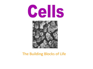
The Building Blocks of Life
Definition 
= Three part theory about cells 
1. All living things are made of cells.
Part 2 of the Theory 
2. The cell is the basic structural and functional 
unit of life.
Part 3 of the Theory 
3. All cells come from pre-existing cells. 
yeast cells dividing
Cells are Us 
A person contains about 100 trillion cells. That’s 
100,000,000,000,000 or 1 x 10¹⁴ cells. 
There are about 200 different cell types in 
mammals (one of us). 
Cells are teeny, tiny, measuring on average 
about 0.002 cm (20 um) across. That’s about 
1250 cells, “shoulder-to-shoulder” per inch. 
nerve cell
Why Study Cell 
Biology? 
The key to every biological 
problem must finally be 
sought in the cell, for every 
living organism is, or at some 
time has been, a cell.
The Cell Theory (review) 
The Cell Theory (proposed independently in 1838 and 
1839) is a cornerstone of biology. 
 Cells are the basic unit of life 
 All Cells arise from previously existing cells
Two Types of Cells 
Two Fundamentally Different Cell Architectures: 
1) A prokaryotic cell 
2) A eukaryotic cell
Prokaryotic cells 
• have no nucleus or organelles 
enclosed within membranes. 
• All species in the domains Archaea and 
Eubacteria have prokaryotic cells.
Eukaryotic cells 
• have a nucleus and organelles that are 
surrounded by membranes. 
• Each organelle does a specific cell function. 
• All species in the Eukaryota domain (protists, 
fungi, plants, and animals) have eukaryotic cells. 
Individual protists have only one cell, while plants 
and animals can have trillions of cells.
Prokaryotes vs. Eukaryotes
Size comparison??
• 1. A few types of cells 
are large enough to be 
seen by the unaided 
eye. 
• 2. Most cells are 
visible only with a 
microscope. 
•The Female Egg is the largest cell in the body, and can be seen without the aid of a 
microscope.
PROKARYOTES vs. EUKARYOTES: Differences 
(Location)
Similarities: Common Features of 
ALL CELLS!
The common features of prokaryotic 
and eukaryotic cells are: 
• 1. DNA 
2. Plasma membrane (a.k.a. “cell membrane”) 
• 3. Cytoplasm 
• 4. Ribosomes
An idealized animal cell.
A rat liver cell (with color enhancement to show 
organelles).
What Do Those Parts 
Do?
Structure of cells
HeLa Cells
Structure of Eukaryotic Cell
Pathways of Differentiation
Relative sizes of cells and cell components
Cell replacement therapy
Prokaryotic and Eukaryotic Cellular Structure
Prokaryotic and Eukaryotic Cells 
• Prokaryote comes from 
the Greek words for 
prenucleus. 
• Eukaryote comes from 
the Greek words for 
true nucleus.
Prokaryotic Cells: Shapes 
• Average size: 0.2 –1.0 μm ´ 2 – 8 μm 
• Most bacteria are monomorphic 
• A few are pleomorphic
Basic Shapes 
• Bacillus (rod-shaped) 
• Coccus (spherical) 
• Spiral 
– Spirillum 
– Vibrio 
– Spirochete
Prokaryotic & Eukaryotic Cells: An Overview 
 Prokaryotes 
 Do not have membrane surrounding their 
DNA 
 lack a nucleus 
 Lack various internal structures bound 
with phospholipid membranes 
 Are small, ~1.0 μm in diameter 
 Have a simple structure 
 Composed of bacteria and archaea
Prokaryotic & Eukaryotic Cells: An Overview 
 Eukaryotes 
 Have membrane surrounding their 
DNA 
 Have a nucleus 
 Have internal membrane-bound 
organelles 
 Are larger, 10-100 μm in diameter 
 Have more complex structure 
 Composed of algae, protozoa, fungi, 
animals, and plants
Prokaryotic & Eukaryotic Cells: An Overview 
[INSERT FIGURE 3.1]
Prokaryotic Cell Membrane 
• Structure 
– Referred to as phospholipid bilayer; 
composed of lipids and associated 
proteins 
– Approximately half composed of 
proteins that act as recognition 
proteins, enzymes, receptors, 
carriers, or channels 
• Integral proteins 
• Peripheral proteins 
• Glycoproteins 
– Fluid mosaic model describes current 
understanding of membrane 
structure
Cell Membrane 
Membranes contain a 
hydrophilic and 
hydrophobic side 
Composed of many 
different types of 
proteins 
Proteins in the lipid 
bilayer move freely 
within the membrane
Cell Membrane 
Thin pliable lipid and protein envelope 
that defines a cell. 
Phospholipid bilayer 
Functions: 
• Regulates nutrient and water intake 
• Regulates waste removal 
• Site of prokaryotic respiration 
• Site of prokaryotic flagella 
attachment 
• Involved in the distribution of genetic 
material during binary fission
Prokaryotic Cytoplasmic Membranes 
• Function 
– Energy storage 
– Harvest light energy in 
photosynthetic prokaryotes 
– Selectively permeable 
– Naturally impermeable to most 
substances 
– Proteins allow substances to cross 
membrane 
• Occurs by passive or active 
processes 
– Maintain concentration and electrical 
gradient 
• Chemicals concentrated on one 
side of the membrane or the 
other 
• Voltage exists across the 
membrane
Cell Membrane
Trilaminar appearance of membranes
Structure of the Plasma membrane
• Plasma Membrane 
• A lipid/protein/carbohydrate complex, providing a 
barrier and containing transport and signaling 
systems.
Cell membrane - Function 
• The cell membrane's function, in general, revolves 
around is membrane proteins. General functions 
include: 
• Receptor proteins which allow cells to 
communicate, 
• transport proteins regulate what enters or leaves 
the cell, 
• and marker proteins which identify the cell
Cell membrane - Function - Regulation of 
transport 
• Transport Proteins come in two forms: 
Carrier proteins are peripheral proteins which 
do not extend all the way through the 
membrane. They move specific molecules 
through the membrane one at a time.
Channel proteins extend through the 
bilipid layer. They form a pore through 
the membrane that can move 
molecules in several ways.
• The cell membrane can also engulf structures 
that are much too large to fit through the 
pores in the membrane proteins. 
• This process is known as endocytosis. 
• In this process the membrane itself wraps 
around the particle and pinches off a vesicle 
inside the cell.
Structure of Cholestrol molecule
STEALTH LIPOSOMES
External Structures of Prokaryotic Cells 
• Types of Glycocalyces 
– Capsule 
• Composed of organized 
repeating units of organic 
chemicals 
• Firmly attached to cell surface 
• Protects cells from drying out 
• May prevent bacteria from 
being recognized and destroyed 
by host
External Structures of Prokaryotic Cells 
• Glycocalyces 
– Gelatinous, sticky substance 
surrounding the outside of 
the cell 
– Composed of 
polysaccharides, 
polypeptides, or both
Capsule 
Polysaccharides or 
polypeptides in composition. 
Surround the cell wall in some 
bacteria. 
Function: 
•Protection from phagocytosis 
•Osmotic barrier 
•Reservoir for nutrients 
•Virulence factor
Capsule Stain
Slime Layer 
Consist of polysaccharide 
fibers that extend form the 
bacterial surface 
Functions: 
•Protection 
•Attachment 
•Associated with biofilms
External Structures of Prokaryotic Cells 
• Types of Glycocalyces 
– Slime layer 
• Loosely attached to cell surface 
• Water soluble 
• Protects cells from drying out 
• Sticky layer that allows 
prokaryotes to attach to 
surfaces
Bacterial 
Appendages 
Flagella 
Axial Filaments 
Pili (Fimbriae)
Bacterial Appendages Flagella 
Structures of locomotion 
Originate in the plasma 
membrane 
In bacteria rotate like a 
propellar 
Many different 
arrangements
External Structures of Prokaryotic Cells 
• Flagella 
– Are responsible for 
movement 
– Have long structures that 
extend beyond cell surface 
– Are not present on all 
prokaryotes
External Structures of Prokaryotic Cells 
 Flagella 
 Structure 
 Composed of filament, hook, and 
basal body 
 Flagellin protein (filament) 
deposited in a helix at the 
lengthening tip 
 Base of filament inserts into hook 
 Basal body anchors filament and 
hook to cell wall by a rod and a 
series of either two or four rings 
of integral proteins 
 Filament capable of rotating 360º
Bacterial Appendages Arrangements of Flagella 
A.Monotrichous 
B.Lophotrichous 
C.Amphitrichous 
D.Peritrichous
Bacterial Appendages 
Axial filament (endoflagella) 
Originates in the cell membrane and 
transverses the length of the cell in the 
periplasmic space. 
As the endoflagella rotate to move the cell the 
characteristic shape is formed . 
Endoflagella are associated with spirochetes.
External Structures of Prokaryotic Cells 
Endoflagellum is also know as an 
axial filament. 
Attached to the plasma embrane 
and transverses the entire cell. 
Responsible for the spirochete 
morphology.
External Structures of Prokaryotic Cells 
• Flagella 
– Function 
• Rotation propels bacterium 
through environment 
• Rotation reversible, can be 
clockwise or counterclockwise 
• Bacteria move in response to 
stimuli (taxis) 
– Runs 
– Tumbles
Bacterial Appendages 
• Fimbriae and Pili 
– Rod-like proteinaceous 
extensions
Bacterial 
Appendages Fimbriae 
Hollow tubes that 
protrude from some 
bacteria 
Compose of protein
External Structures of Prokaryotic Cells 
• Fimbriae 
• Sticky, bristlelike projections 
• Used by bacteria to adhere 
to one another, to hosts, and 
to substances in 
environment 
• Shorter than flagella 
• May be hundreds per cell 
• Serve an important function 
in biofilms 
• Virulence factor
External Structures of Prokaryotic Cells 
• Pili 
– Tubules composed of pilin 
– Also known as conjugation pili 
– Longer than fimbriae but shorter 
than flagella 
– Bacteria typically only have one or 
two per cell 
– Mediate the transfer of DNA from 
one cell to another (conjugation)
Bacterial Conjugation 
Transfer of plasmid DNA 
from a donor to a 
recipient. 
Process strengthens the 
bacterial cell and alows for 
survival in a competitive 
environment.
Bacterial Inclusion Bodies 
1. poly-Beta-hydroxybutyric acid - stores lipids for use in plasma membrane 
2. glycogen - stores starch like polymer of sugar for energy production 
3. Polyphosphate granules (metachromatic granules) - storage for 
phosphates for plasma membrane and the formation of ATP from ADP. 
4. Sulfur granules - stores sulfur which is necessary for the metabolic 
reactions in biosynthesis.
5. Mesosome 
Mesosomes - invagination of the 
plasma membrane that increases the 
surfaces area of the plasma membrane 
during binary fission. 
The mesosome also serves as a site 
for the attachment and distribution of 
genetic material during binary fission.
Mesosome 
In prokaryotic cell division, called 
binary fission. 
A diagram of the attachment of 
bacterial chromosomes, indicating the 
possible role of the mesosome (an 
inward fold of the cell membrane) in 
ensuring the distribution of the 
"chromosomes" in a dividing cell. 
Upon attachment to the plasma 
membrane, the DNA replicates and 
reattaches at separate points. 
Continued growth of the cell gradually 
separates the chromosomes and 
allocates chromosome copies to the 
two daughter cells.
Inclusion Bodies 
6. gas vacuoles - storage of metabolic gases such as methane or hydrogen gas. The 
gas vacuoles help in the buoyancy of the cell and aids in it motility. 
7. ribosomes - responsible for the synthesis of proteins. 
8. nucleoid material - the genetic material of bacteria, which usually is balled up in 
the cell. During binary fission the nucleoid material unravels within the cell in 
order to be copied and distributed to the daughter cells. 
9. Plasmid - small fragments of self-replicating extrachromosomal DNA that codes for 
the resistance to antibiotics or for the productions of a specific metabolite, i.e. 
toxins, pigments. These plasmids may be transferred from one bacterial cell to 
another by the F-pili.
Inclusion Bodies 
9. Plasmid - small fragments of self-replicating extrachromosomal DNA that codes 
for the resistance to antibiotics or for the productions of a specific metabolite, i.e. 
toxins, pigments. These plasmids may be transferred from one bacterial cell to 
another by the F-pili.
Inclusion Bodies 
These plasmids may be transferred from one bacterial cell to another by the 
F-pili.
Inclusion Bodies 
10. Endospores - a survival mechanism of certain genera of bacteria such as 
Clostridium and Bacillus. 
The endospores are composed of a complex of dipicolinc acid and 
calcium and the function of the endospore is to protect the bacterial 
chromosome. 
The endospores are very resistant to heat, desiccation, freezing, and 
other physical properties such as pesticides, antibiotics, dyes, and acids.
Inclusion Bodies 
The endospores may remain dormant for many years until the 
environment becomes suitable to sustain the life of the bacteria. 
The endospore will then germinate to form an exact copy of the parent 
cell that produced it.
Eukaryotic Cell Walls & Cytoplasmic Membranes 
• Fungi, algae, plants, and some protozoa 
have cell walls but no glycocalyx 
• Composed of various polysaccharides 
– Cellulose found in plant cell walls 
– Fungal cell walls composed of 
cellulose, chitin, and/or 
glucomannan 
– Algal cell walls composed of 
cellulose, proteins, agar, 
carrageenan, silicates, algin, 
calcium carbonate, or a 
combination of these
Cell Walls 
Three different types of cell walls and 
their compositions: 
Fungal cell walls are composed of 
cellulose and/or chitin. 
Plant cell walls are composed of 
cellulose. 
Algal cell walls are composed of 
cellulose, silicon, and calcium 
carbonate.
Plasma Membrane 
Consist of a lipid bilayer and 
associated proteins. The Plasma 
Membrane of Eukaryotic cells 
resembles and functions in the same 
manner as the prokaryotic plasma 
membrane with the following 
exceptions; 
Contains high levels of sterols such as 
cholesterol. 
No respiratory enzymes are located in 
the eukaryotic plasma membrane. 
Respiration occurs in the 
mitochondria.
External Structure of Eukaryotic Cells 
• Glycocalyces 
– Never as organized as prokaryotic 
capsules 
– Help anchor animal cells to each 
other 
– Strengthen cell surface 
– Provide protection against 
dehydration 
– Function in cell-to-cell recognition 
and communication
Eukaryotic Appendages 
Flagella 
There are several different 
arrangements of flagella in eucaryotes. 
This diagram represents a 
biflagellated eukaryotic cell. 
One of the flagella aids in movement 
laterally and the other aids in up and 
down movement. 
The eukaryotic flagella move like a 
whip. 
See Flagellar handout.
Eukaryotic Appendages 
• Flagella 
– Function 
• Do not rotate, but undulate 
rhythmically
Eukaryotic Appendages 
Cilia 
Similar to flagella both structurally and 
functionally but are much shorter and 
more numerous. 
Cilia are found peritrichously to the cell. 
Move in an undulating manner and 
motility by those organisms with cilia is 
much more rapid than those with flagella.
Intracellular Structures of Eukaryotic Organisms (organelles) 
 Membranous Organelles 
 Nucleus 
 Often largest organelle in cell 
 Contains most of the cell’s DNA 
 Semi-liquid portion called 
nucleoplasm 
 One or more nucleoli present in 
nucleoplasm; RNA synthesized in 
nucleoli 
 Nucleoplasm contains chromatin – 
masses of DNA associated with 
histones 
 Surrounded by nuclear envelope – 
double membrane composed of two 
phospholipid bilayers 
 Nuclear envelope contains nuclear 
pores
Intracellular Structures of Eukaryotic Organisms (organelles) 
Nucleus - double membraned 
organelle that houses the genetic 
material of cell. 
Nuclear membrane contains numerous 
pores through which proteins and RNA 
can move.
Intracellular Structures of Eukaryotic Organisms (organelles) 
 Membranous Organelles 
 Endoplasmic reticulum 
 Netlike arrangement of flattened, hollow 
tubules continuous with nuclear 
envelope 
 Functions as transport system 
 Two forms 
 Smooth endoplasmic reticulum 
(SER) – plays role in lipid 
synthesis 
 Rough endoplasmic reticulum 
(RER) – ribosomes attached to 
its outer surface; transports 
proteins produced by 
ribosomes
Intracellular Structures of Eukaryotic Organisms (organelles) 
Endoplasmic reticulum - network of 
cytoplasmic membranes where lipids 
and proteins are produced. 
Smooth ER - synthesis of lipids 
Rough ER - associated with ribosomes 
and is responsible for the synthesis of 
proteins. 
.
Intracellular Structures of Eukaryotic Organisms (organelles) 
 Membranous Organelles 
 Golgi body 
 Receives, processes, and 
packages large molecules for 
export from cell 
 Packages molecules in secretory 
vesicles that fuse with 
cytoplasmic membrane 
 Composed of flattened hollow 
sacs surrounded by 
phospholipid bilayer 
 Not in all eukaryotic cells
Intracellular Structures of Eukaryotic Organisms (organelles) 
Golgi apparatus (dictyosome) is 
associated with the ER. 
It modifies and packages the lipids and 
proteins manufactured by the ER and 
places them in vesicles for cellular use.
Intracellular Structures of Eukaryotic Organisms (organelles) 
• Membranous Organelles 
– Lysosomes, peroxisomes,vacuoles, and 
vesicles 
• Store and transfer chemicals within 
cells 
• May store nutrients in cell 
• Lysosomes contain catabolic 
enzymes 
• Peroxisomes contain enzymes that 
degrade poisonous wastes
Intracellular Structures of Eukaryotic Organisms (organelles) 
• Membranous Organelles 
– Mitochondria 
• Have two membranes 
composed of phospholipid 
bilayer 
• Produce most of cell’s ATP 
• Interior matrix contains 70S 
ribosomes and circular 
molecule of DNA
Intracellular Structures of Eukaryotic Organisms (organelles) 
mitochondria - involved in the 
production of chemical energy in the 
form of ATP. 
Consist of convoluted inner membrane 
and outer membrane. Invaginations 
are called cristae and contain enzymes 
used to synthesis ATP. 
All respiratory enzymes are located in 
the inner membrane of the 
mitochondria.
Cytoplasm of Eukaryotes 
• Membranous Organelles 
– Chloroplasts 
• Light-harvesting structures 
found in photosynthetic 
eukaryotes 
• Have two phospholipid 
bilayer membranes and DNA 
• Have 70S ribosomes
Cytoplasm of Eukaryotes 
• Endosymbiotic Theory 
– Eukaryotes formed from union of small aerobic 
prokaryotes with larger anaerobic prokaryotes 
– smaller prokaryotes became internal parasites 
• Parasites lost ability to exist independently; retained 
portion of DNA, ribosomes, and cytoplasmic 
membranes 
• Larger cell became dependent on parasites for aerobic 
ATP production 
• Aerobic prokaryotes evolved into mitochondria 
• Similar scenario for origin of chloroplasts 
– Not universally accepted
Cytoplasm of Eukaryotes 
[INSERT TABLE 3.5]
Cytoplasm of Eukaryotes 
[INSERT TABLE 3.4]
Eukaryotic Cell Walls & Cytoplasmic 
Membranes 
[INSERT TABLE 3.3]

Contenu connexe

Tendances

Prokaryotes & eukaryotes
Prokaryotes & eukaryotesProkaryotes & eukaryotes
Prokaryotes & eukaryotesAftab Badshah
 
Difference between prokaryotic and eukaryotic cell.pptx
Difference between prokaryotic and eukaryotic cell.pptxDifference between prokaryotic and eukaryotic cell.pptx
Difference between prokaryotic and eukaryotic cell.pptxAmjad Afridi
 
Structure & function of cell membrane
Structure & function of cell membraneStructure & function of cell membrane
Structure & function of cell membranePriyanka Gohil
 
Cell structure and functions
Cell structure and functionsCell structure and functions
Cell structure and functionsJoy Batang Ü
 
Cell theory & types of cells
Cell theory & types of cellsCell theory & types of cells
Cell theory & types of cellsOhMiss
 
Cell membrane (Plasma Membrane) & Cellular Junctions
Cell membrane (Plasma Membrane) & Cellular JunctionsCell membrane (Plasma Membrane) & Cellular Junctions
Cell membrane (Plasma Membrane) & Cellular JunctionsPradeep Singh Narwat
 
Structure and function of plasma membrane 2
Structure and function of plasma membrane 2Structure and function of plasma membrane 2
Structure and function of plasma membrane 2ICHHA PURAK
 
Cell Cycle and Cell Division
Cell Cycle and Cell DivisionCell Cycle and Cell Division
Cell Cycle and Cell DivisionShivang Patel
 
Chapter 4 ppt
Chapter 4 pptChapter 4 ppt
Chapter 4 pptmjnepa
 
Prokaryotes vs Eukaryotes
Prokaryotes vs EukaryotesProkaryotes vs Eukaryotes
Prokaryotes vs Eukaryotesmgsonline
 
Plasma/Cell Membrane
Plasma/Cell MembranePlasma/Cell Membrane
Plasma/Cell MembraneGul Muneer
 

Tendances (20)

Plasma membrane
Plasma membranePlasma membrane
Plasma membrane
 
Prokaryotes & eukaryotes
Prokaryotes & eukaryotesProkaryotes & eukaryotes
Prokaryotes & eukaryotes
 
Difference between prokaryotic and eukaryotic cell.pptx
Difference between prokaryotic and eukaryotic cell.pptxDifference between prokaryotic and eukaryotic cell.pptx
Difference between prokaryotic and eukaryotic cell.pptx
 
Intro to cells
Intro to cellsIntro to cells
Intro to cells
 
The cell membrane function
The cell membrane functionThe cell membrane function
The cell membrane function
 
Structure & function of cell membrane
Structure & function of cell membraneStructure & function of cell membrane
Structure & function of cell membrane
 
Cell structure and functions
Cell structure and functionsCell structure and functions
Cell structure and functions
 
Cell theory & types of cells
Cell theory & types of cellsCell theory & types of cells
Cell theory & types of cells
 
Plasma membrane
Plasma membranePlasma membrane
Plasma membrane
 
Lysosome
LysosomeLysosome
Lysosome
 
Animal cell
Animal cellAnimal cell
Animal cell
 
Cell membrane (Plasma Membrane) & Cellular Junctions
Cell membrane (Plasma Membrane) & Cellular JunctionsCell membrane (Plasma Membrane) & Cellular Junctions
Cell membrane (Plasma Membrane) & Cellular Junctions
 
Structure and function of plasma membrane 2
Structure and function of plasma membrane 2Structure and function of plasma membrane 2
Structure and function of plasma membrane 2
 
Cell
CellCell
Cell
 
Cell Cycle and Cell Division
Cell Cycle and Cell DivisionCell Cycle and Cell Division
Cell Cycle and Cell Division
 
Chapter 4 ppt
Chapter 4 pptChapter 4 ppt
Chapter 4 ppt
 
The cell
The cellThe cell
The cell
 
Cell: Structure & Function
Cell: Structure & FunctionCell: Structure & Function
Cell: Structure & Function
 
Prokaryotes vs Eukaryotes
Prokaryotes vs EukaryotesProkaryotes vs Eukaryotes
Prokaryotes vs Eukaryotes
 
Plasma/Cell Membrane
Plasma/Cell MembranePlasma/Cell Membrane
Plasma/Cell Membrane
 

Similaire à Cell introduction

Structure of Bacteria
Structure of BacteriaStructure of Bacteria
Structure of BacteriaAnand365588
 
Cell: The Unit of Life
Cell: The Unit of LifeCell: The Unit of Life
Cell: The Unit of LifeDrHeenaDevnani
 
La celula: la teoría celular, estructura y función. La división celular
La celula: la teoría celular, estructura y función. La división celularLa celula: la teoría celular, estructura y función. La división celular
La celula: la teoría celular, estructura y función. La división celularJosué Moreno Marquina
 
Parts and Functions of the Cell
Parts and Functions of the CellParts and Functions of the Cell
Parts and Functions of the CellJayson Gula
 
CELL BIOLOGY LECTURE. Cells described in the purest of forms.
CELL BIOLOGY LECTURE. Cells described in the purest of forms.CELL BIOLOGY LECTURE. Cells described in the purest of forms.
CELL BIOLOGY LECTURE. Cells described in the purest of forms.ndugbu34
 
cellthefundamentalunitoflife-180820182355 (1)_1.pptx
cellthefundamentalunitoflife-180820182355 (1)_1.pptxcellthefundamentalunitoflife-180820182355 (1)_1.pptx
cellthefundamentalunitoflife-180820182355 (1)_1.pptxEktaBhatt10
 
Lecture 3 Cell_structure_function.ppt
Lecture 3 Cell_structure_function.pptLecture 3 Cell_structure_function.ppt
Lecture 3 Cell_structure_function.pptMuhammadAbbasWali
 
Cell-biology-1.pptx (1).pdf
Cell-biology-1.pptx (1).pdfCell-biology-1.pptx (1).pdf
Cell-biology-1.pptx (1).pdfSachinPokhrel2
 
Cell: it’s structure and functions
Cell: it’s structure and functionsCell: it’s structure and functions
Cell: it’s structure and functionsdigvijay3344
 
Topic 2 –cells
Topic 2 –cellsTopic 2 –cells
Topic 2 –cellsnjj0hnson
 
bcterialultrastructureall-201118081937.pdf
bcterialultrastructureall-201118081937.pdfbcterialultrastructureall-201118081937.pdf
bcterialultrastructureall-201118081937.pdfdawitg2
 

Similaire à Cell introduction (20)

Structure of Bacteria
Structure of BacteriaStructure of Bacteria
Structure of Bacteria
 
Cell biology
Cell biologyCell biology
Cell biology
 
Cell: The Unit of Life
Cell: The Unit of LifeCell: The Unit of Life
Cell: The Unit of Life
 
La celula: la teoría celular, estructura y función. La división celular
La celula: la teoría celular, estructura y función. La división celularLa celula: la teoría celular, estructura y función. La división celular
La celula: la teoría celular, estructura y función. La división celular
 
Cell biology
Cell biology Cell biology
Cell biology
 
Biology Theory Lec. 3
Biology Theory Lec. 3Biology Theory Lec. 3
Biology Theory Lec. 3
 
Cells
CellsCells
Cells
 
Parts and Functions of the Cell
Parts and Functions of the CellParts and Functions of the Cell
Parts and Functions of the Cell
 
CELL BIOLOGY LECTURE. Cells described in the purest of forms.
CELL BIOLOGY LECTURE. Cells described in the purest of forms.CELL BIOLOGY LECTURE. Cells described in the purest of forms.
CELL BIOLOGY LECTURE. Cells described in the purest of forms.
 
cellthefundamentalunitoflife-180820182355 (1)_1.pptx
cellthefundamentalunitoflife-180820182355 (1)_1.pptxcellthefundamentalunitoflife-180820182355 (1)_1.pptx
cellthefundamentalunitoflife-180820182355 (1)_1.pptx
 
Lecture 3 Cell_structure_function.ppt
Lecture 3 Cell_structure_function.pptLecture 3 Cell_structure_function.ppt
Lecture 3 Cell_structure_function.ppt
 
Antibiotics
AntibioticsAntibiotics
Antibiotics
 
Cell-biology-1.pptx (1).pdf
Cell-biology-1.pptx (1).pdfCell-biology-1.pptx (1).pdf
Cell-biology-1.pptx (1).pdf
 
Cell: it’s structure and functions
Cell: it’s structure and functionsCell: it’s structure and functions
Cell: it’s structure and functions
 
Seminar on cell
Seminar on cellSeminar on cell
Seminar on cell
 
Topic 2 –cells
Topic 2 –cellsTopic 2 –cells
Topic 2 –cells
 
Atv what is biology
Atv what is biologyAtv what is biology
Atv what is biology
 
bcterialultrastructureall-201118081937.pdf
bcterialultrastructureall-201118081937.pdfbcterialultrastructureall-201118081937.pdf
bcterialultrastructureall-201118081937.pdf
 
Bcterial ultrastructure all
Bcterial ultrastructure allBcterial ultrastructure all
Bcterial ultrastructure all
 
2.1 Cells
2.1 Cells2.1 Cells
2.1 Cells
 

Plus de Subramaniya Sharma (6)

Cytoskeleton
CytoskeletonCytoskeleton
Cytoskeleton
 
Ecm
EcmEcm
Ecm
 
Copy of cell introduction
Copy of cell introductionCopy of cell introduction
Copy of cell introduction
 
Copy of cytoskeleton
Copy of cytoskeletonCopy of cytoskeleton
Copy of cytoskeleton
 
Copy of plasma membrane
Copy of plasma membraneCopy of plasma membrane
Copy of plasma membrane
 
Copy of ecm
Copy of ecmCopy of ecm
Copy of ecm
 

Dernier

Separation of Lanthanides/ Lanthanides and Actinides
Separation of Lanthanides/ Lanthanides and ActinidesSeparation of Lanthanides/ Lanthanides and Actinides
Separation of Lanthanides/ Lanthanides and ActinidesFatimaKhan178732
 
APM Welcome, APM North West Network Conference, Synergies Across Sectors
APM Welcome, APM North West Network Conference, Synergies Across SectorsAPM Welcome, APM North West Network Conference, Synergies Across Sectors
APM Welcome, APM North West Network Conference, Synergies Across SectorsAssociation for Project Management
 
How to Make a Pirate ship Primary Education.pptx
How to Make a Pirate ship Primary Education.pptxHow to Make a Pirate ship Primary Education.pptx
How to Make a Pirate ship Primary Education.pptxmanuelaromero2013
 
A Critique of the Proposed National Education Policy Reform
A Critique of the Proposed National Education Policy ReformA Critique of the Proposed National Education Policy Reform
A Critique of the Proposed National Education Policy ReformChameera Dedduwage
 
18-04-UA_REPORT_MEDIALITERAСY_INDEX-DM_23-1-final-eng.pdf
18-04-UA_REPORT_MEDIALITERAСY_INDEX-DM_23-1-final-eng.pdf18-04-UA_REPORT_MEDIALITERAСY_INDEX-DM_23-1-final-eng.pdf
18-04-UA_REPORT_MEDIALITERAСY_INDEX-DM_23-1-final-eng.pdfssuser54595a
 
Concept of Vouching. B.Com(Hons) /B.Compdf
Concept of Vouching. B.Com(Hons) /B.CompdfConcept of Vouching. B.Com(Hons) /B.Compdf
Concept of Vouching. B.Com(Hons) /B.CompdfUmakantAnnand
 
Introduction to AI in Higher Education_draft.pptx
Introduction to AI in Higher Education_draft.pptxIntroduction to AI in Higher Education_draft.pptx
Introduction to AI in Higher Education_draft.pptxpboyjonauth
 
Q4-W6-Restating Informational Text Grade 3
Q4-W6-Restating Informational Text Grade 3Q4-W6-Restating Informational Text Grade 3
Q4-W6-Restating Informational Text Grade 3JemimahLaneBuaron
 
Arihant handbook biology for class 11 .pdf
Arihant handbook biology for class 11 .pdfArihant handbook biology for class 11 .pdf
Arihant handbook biology for class 11 .pdfchloefrazer622
 
Measures of Central Tendency: Mean, Median and Mode
Measures of Central Tendency: Mean, Median and ModeMeasures of Central Tendency: Mean, Median and Mode
Measures of Central Tendency: Mean, Median and ModeThiyagu K
 
Contemporary philippine arts from the regions_PPT_Module_12 [Autosaved] (1).pptx
Contemporary philippine arts from the regions_PPT_Module_12 [Autosaved] (1).pptxContemporary philippine arts from the regions_PPT_Module_12 [Autosaved] (1).pptx
Contemporary philippine arts from the regions_PPT_Module_12 [Autosaved] (1).pptxRoyAbrique
 
URLs and Routing in the Odoo 17 Website App
URLs and Routing in the Odoo 17 Website AppURLs and Routing in the Odoo 17 Website App
URLs and Routing in the Odoo 17 Website AppCeline George
 
Call Girls in Dwarka Mor Delhi Contact Us 9654467111
Call Girls in Dwarka Mor Delhi Contact Us 9654467111Call Girls in Dwarka Mor Delhi Contact Us 9654467111
Call Girls in Dwarka Mor Delhi Contact Us 9654467111Sapana Sha
 
Alper Gobel In Media Res Media Component
Alper Gobel In Media Res Media ComponentAlper Gobel In Media Res Media Component
Alper Gobel In Media Res Media ComponentInMediaRes1
 
Software Engineering Methodologies (overview)
Software Engineering Methodologies (overview)Software Engineering Methodologies (overview)
Software Engineering Methodologies (overview)eniolaolutunde
 
The Most Excellent Way | 1 Corinthians 13
The Most Excellent Way | 1 Corinthians 13The Most Excellent Way | 1 Corinthians 13
The Most Excellent Way | 1 Corinthians 13Steve Thomason
 
Sanyam Choudhary Chemistry practical.pdf
Sanyam Choudhary Chemistry practical.pdfSanyam Choudhary Chemistry practical.pdf
Sanyam Choudhary Chemistry practical.pdfsanyamsingh5019
 
“Oh GOSH! Reflecting on Hackteria's Collaborative Practices in a Global Do-It...
“Oh GOSH! Reflecting on Hackteria's Collaborative Practices in a Global Do-It...“Oh GOSH! Reflecting on Hackteria's Collaborative Practices in a Global Do-It...
“Oh GOSH! Reflecting on Hackteria's Collaborative Practices in a Global Do-It...Marc Dusseiller Dusjagr
 

Dernier (20)

Separation of Lanthanides/ Lanthanides and Actinides
Separation of Lanthanides/ Lanthanides and ActinidesSeparation of Lanthanides/ Lanthanides and Actinides
Separation of Lanthanides/ Lanthanides and Actinides
 
APM Welcome, APM North West Network Conference, Synergies Across Sectors
APM Welcome, APM North West Network Conference, Synergies Across SectorsAPM Welcome, APM North West Network Conference, Synergies Across Sectors
APM Welcome, APM North West Network Conference, Synergies Across Sectors
 
How to Make a Pirate ship Primary Education.pptx
How to Make a Pirate ship Primary Education.pptxHow to Make a Pirate ship Primary Education.pptx
How to Make a Pirate ship Primary Education.pptx
 
A Critique of the Proposed National Education Policy Reform
A Critique of the Proposed National Education Policy ReformA Critique of the Proposed National Education Policy Reform
A Critique of the Proposed National Education Policy Reform
 
18-04-UA_REPORT_MEDIALITERAСY_INDEX-DM_23-1-final-eng.pdf
18-04-UA_REPORT_MEDIALITERAСY_INDEX-DM_23-1-final-eng.pdf18-04-UA_REPORT_MEDIALITERAСY_INDEX-DM_23-1-final-eng.pdf
18-04-UA_REPORT_MEDIALITERAСY_INDEX-DM_23-1-final-eng.pdf
 
Concept of Vouching. B.Com(Hons) /B.Compdf
Concept of Vouching. B.Com(Hons) /B.CompdfConcept of Vouching. B.Com(Hons) /B.Compdf
Concept of Vouching. B.Com(Hons) /B.Compdf
 
Introduction to AI in Higher Education_draft.pptx
Introduction to AI in Higher Education_draft.pptxIntroduction to AI in Higher Education_draft.pptx
Introduction to AI in Higher Education_draft.pptx
 
Código Creativo y Arte de Software | Unidad 1
Código Creativo y Arte de Software | Unidad 1Código Creativo y Arte de Software | Unidad 1
Código Creativo y Arte de Software | Unidad 1
 
Q4-W6-Restating Informational Text Grade 3
Q4-W6-Restating Informational Text Grade 3Q4-W6-Restating Informational Text Grade 3
Q4-W6-Restating Informational Text Grade 3
 
Arihant handbook biology for class 11 .pdf
Arihant handbook biology for class 11 .pdfArihant handbook biology for class 11 .pdf
Arihant handbook biology for class 11 .pdf
 
Measures of Central Tendency: Mean, Median and Mode
Measures of Central Tendency: Mean, Median and ModeMeasures of Central Tendency: Mean, Median and Mode
Measures of Central Tendency: Mean, Median and Mode
 
Contemporary philippine arts from the regions_PPT_Module_12 [Autosaved] (1).pptx
Contemporary philippine arts from the regions_PPT_Module_12 [Autosaved] (1).pptxContemporary philippine arts from the regions_PPT_Module_12 [Autosaved] (1).pptx
Contemporary philippine arts from the regions_PPT_Module_12 [Autosaved] (1).pptx
 
URLs and Routing in the Odoo 17 Website App
URLs and Routing in the Odoo 17 Website AppURLs and Routing in the Odoo 17 Website App
URLs and Routing in the Odoo 17 Website App
 
Call Girls in Dwarka Mor Delhi Contact Us 9654467111
Call Girls in Dwarka Mor Delhi Contact Us 9654467111Call Girls in Dwarka Mor Delhi Contact Us 9654467111
Call Girls in Dwarka Mor Delhi Contact Us 9654467111
 
Alper Gobel In Media Res Media Component
Alper Gobel In Media Res Media ComponentAlper Gobel In Media Res Media Component
Alper Gobel In Media Res Media Component
 
Software Engineering Methodologies (overview)
Software Engineering Methodologies (overview)Software Engineering Methodologies (overview)
Software Engineering Methodologies (overview)
 
The Most Excellent Way | 1 Corinthians 13
The Most Excellent Way | 1 Corinthians 13The Most Excellent Way | 1 Corinthians 13
The Most Excellent Way | 1 Corinthians 13
 
Sanyam Choudhary Chemistry practical.pdf
Sanyam Choudhary Chemistry practical.pdfSanyam Choudhary Chemistry practical.pdf
Sanyam Choudhary Chemistry practical.pdf
 
Model Call Girl in Tilak Nagar Delhi reach out to us at 🔝9953056974🔝
Model Call Girl in Tilak Nagar Delhi reach out to us at 🔝9953056974🔝Model Call Girl in Tilak Nagar Delhi reach out to us at 🔝9953056974🔝
Model Call Girl in Tilak Nagar Delhi reach out to us at 🔝9953056974🔝
 
“Oh GOSH! Reflecting on Hackteria's Collaborative Practices in a Global Do-It...
“Oh GOSH! Reflecting on Hackteria's Collaborative Practices in a Global Do-It...“Oh GOSH! Reflecting on Hackteria's Collaborative Practices in a Global Do-It...
“Oh GOSH! Reflecting on Hackteria's Collaborative Practices in a Global Do-It...
 

Cell introduction

  • 2. Definition = Three part theory about cells 1. All living things are made of cells.
  • 3. Part 2 of the Theory 2. The cell is the basic structural and functional unit of life.
  • 4. Part 3 of the Theory 3. All cells come from pre-existing cells. yeast cells dividing
  • 5. Cells are Us A person contains about 100 trillion cells. That’s 100,000,000,000,000 or 1 x 10¹⁴ cells. There are about 200 different cell types in mammals (one of us). Cells are teeny, tiny, measuring on average about 0.002 cm (20 um) across. That’s about 1250 cells, “shoulder-to-shoulder” per inch. nerve cell
  • 6. Why Study Cell Biology? The key to every biological problem must finally be sought in the cell, for every living organism is, or at some time has been, a cell.
  • 7. The Cell Theory (review) The Cell Theory (proposed independently in 1838 and 1839) is a cornerstone of biology.  Cells are the basic unit of life  All Cells arise from previously existing cells
  • 8. Two Types of Cells Two Fundamentally Different Cell Architectures: 1) A prokaryotic cell 2) A eukaryotic cell
  • 9. Prokaryotic cells • have no nucleus or organelles enclosed within membranes. • All species in the domains Archaea and Eubacteria have prokaryotic cells.
  • 10. Eukaryotic cells • have a nucleus and organelles that are surrounded by membranes. • Each organelle does a specific cell function. • All species in the Eukaryota domain (protists, fungi, plants, and animals) have eukaryotic cells. Individual protists have only one cell, while plants and animals can have trillions of cells.
  • 13. • 1. A few types of cells are large enough to be seen by the unaided eye. • 2. Most cells are visible only with a microscope. •The Female Egg is the largest cell in the body, and can be seen without the aid of a microscope.
  • 14. PROKARYOTES vs. EUKARYOTES: Differences (Location)
  • 16. The common features of prokaryotic and eukaryotic cells are: • 1. DNA 2. Plasma membrane (a.k.a. “cell membrane”) • 3. Cytoplasm • 4. Ribosomes
  • 18. A rat liver cell (with color enhancement to show organelles).
  • 19. What Do Those Parts Do?
  • 21.
  • 25. Relative sizes of cells and cell components
  • 27.
  • 28. Prokaryotic and Eukaryotic Cellular Structure
  • 29. Prokaryotic and Eukaryotic Cells • Prokaryote comes from the Greek words for prenucleus. • Eukaryote comes from the Greek words for true nucleus.
  • 30.
  • 31. Prokaryotic Cells: Shapes • Average size: 0.2 –1.0 μm ´ 2 – 8 μm • Most bacteria are monomorphic • A few are pleomorphic
  • 32. Basic Shapes • Bacillus (rod-shaped) • Coccus (spherical) • Spiral – Spirillum – Vibrio – Spirochete
  • 33. Prokaryotic & Eukaryotic Cells: An Overview  Prokaryotes  Do not have membrane surrounding their DNA  lack a nucleus  Lack various internal structures bound with phospholipid membranes  Are small, ~1.0 μm in diameter  Have a simple structure  Composed of bacteria and archaea
  • 34. Prokaryotic & Eukaryotic Cells: An Overview  Eukaryotes  Have membrane surrounding their DNA  Have a nucleus  Have internal membrane-bound organelles  Are larger, 10-100 μm in diameter  Have more complex structure  Composed of algae, protozoa, fungi, animals, and plants
  • 35. Prokaryotic & Eukaryotic Cells: An Overview [INSERT FIGURE 3.1]
  • 36.
  • 37. Prokaryotic Cell Membrane • Structure – Referred to as phospholipid bilayer; composed of lipids and associated proteins – Approximately half composed of proteins that act as recognition proteins, enzymes, receptors, carriers, or channels • Integral proteins • Peripheral proteins • Glycoproteins – Fluid mosaic model describes current understanding of membrane structure
  • 38. Cell Membrane Membranes contain a hydrophilic and hydrophobic side Composed of many different types of proteins Proteins in the lipid bilayer move freely within the membrane
  • 39. Cell Membrane Thin pliable lipid and protein envelope that defines a cell. Phospholipid bilayer Functions: • Regulates nutrient and water intake • Regulates waste removal • Site of prokaryotic respiration • Site of prokaryotic flagella attachment • Involved in the distribution of genetic material during binary fission
  • 40. Prokaryotic Cytoplasmic Membranes • Function – Energy storage – Harvest light energy in photosynthetic prokaryotes – Selectively permeable – Naturally impermeable to most substances – Proteins allow substances to cross membrane • Occurs by passive or active processes – Maintain concentration and electrical gradient • Chemicals concentrated on one side of the membrane or the other • Voltage exists across the membrane
  • 42.
  • 44. Structure of the Plasma membrane
  • 45. • Plasma Membrane • A lipid/protein/carbohydrate complex, providing a barrier and containing transport and signaling systems.
  • 46. Cell membrane - Function • The cell membrane's function, in general, revolves around is membrane proteins. General functions include: • Receptor proteins which allow cells to communicate, • transport proteins regulate what enters or leaves the cell, • and marker proteins which identify the cell
  • 47. Cell membrane - Function - Regulation of transport • Transport Proteins come in two forms: Carrier proteins are peripheral proteins which do not extend all the way through the membrane. They move specific molecules through the membrane one at a time.
  • 48. Channel proteins extend through the bilipid layer. They form a pore through the membrane that can move molecules in several ways.
  • 49. • The cell membrane can also engulf structures that are much too large to fit through the pores in the membrane proteins. • This process is known as endocytosis. • In this process the membrane itself wraps around the particle and pinches off a vesicle inside the cell.
  • 52. External Structures of Prokaryotic Cells • Types of Glycocalyces – Capsule • Composed of organized repeating units of organic chemicals • Firmly attached to cell surface • Protects cells from drying out • May prevent bacteria from being recognized and destroyed by host
  • 53. External Structures of Prokaryotic Cells • Glycocalyces – Gelatinous, sticky substance surrounding the outside of the cell – Composed of polysaccharides, polypeptides, or both
  • 54. Capsule Polysaccharides or polypeptides in composition. Surround the cell wall in some bacteria. Function: •Protection from phagocytosis •Osmotic barrier •Reservoir for nutrients •Virulence factor
  • 56. Slime Layer Consist of polysaccharide fibers that extend form the bacterial surface Functions: •Protection •Attachment •Associated with biofilms
  • 57. External Structures of Prokaryotic Cells • Types of Glycocalyces – Slime layer • Loosely attached to cell surface • Water soluble • Protects cells from drying out • Sticky layer that allows prokaryotes to attach to surfaces
  • 58. Bacterial Appendages Flagella Axial Filaments Pili (Fimbriae)
  • 59. Bacterial Appendages Flagella Structures of locomotion Originate in the plasma membrane In bacteria rotate like a propellar Many different arrangements
  • 60. External Structures of Prokaryotic Cells • Flagella – Are responsible for movement – Have long structures that extend beyond cell surface – Are not present on all prokaryotes
  • 61. External Structures of Prokaryotic Cells  Flagella  Structure  Composed of filament, hook, and basal body  Flagellin protein (filament) deposited in a helix at the lengthening tip  Base of filament inserts into hook  Basal body anchors filament and hook to cell wall by a rod and a series of either two or four rings of integral proteins  Filament capable of rotating 360º
  • 62. Bacterial Appendages Arrangements of Flagella A.Monotrichous B.Lophotrichous C.Amphitrichous D.Peritrichous
  • 63. Bacterial Appendages Axial filament (endoflagella) Originates in the cell membrane and transverses the length of the cell in the periplasmic space. As the endoflagella rotate to move the cell the characteristic shape is formed . Endoflagella are associated with spirochetes.
  • 64. External Structures of Prokaryotic Cells Endoflagellum is also know as an axial filament. Attached to the plasma embrane and transverses the entire cell. Responsible for the spirochete morphology.
  • 65. External Structures of Prokaryotic Cells • Flagella – Function • Rotation propels bacterium through environment • Rotation reversible, can be clockwise or counterclockwise • Bacteria move in response to stimuli (taxis) – Runs – Tumbles
  • 66. Bacterial Appendages • Fimbriae and Pili – Rod-like proteinaceous extensions
  • 67. Bacterial Appendages Fimbriae Hollow tubes that protrude from some bacteria Compose of protein
  • 68. External Structures of Prokaryotic Cells • Fimbriae • Sticky, bristlelike projections • Used by bacteria to adhere to one another, to hosts, and to substances in environment • Shorter than flagella • May be hundreds per cell • Serve an important function in biofilms • Virulence factor
  • 69. External Structures of Prokaryotic Cells • Pili – Tubules composed of pilin – Also known as conjugation pili – Longer than fimbriae but shorter than flagella – Bacteria typically only have one or two per cell – Mediate the transfer of DNA from one cell to another (conjugation)
  • 70. Bacterial Conjugation Transfer of plasmid DNA from a donor to a recipient. Process strengthens the bacterial cell and alows for survival in a competitive environment.
  • 71. Bacterial Inclusion Bodies 1. poly-Beta-hydroxybutyric acid - stores lipids for use in plasma membrane 2. glycogen - stores starch like polymer of sugar for energy production 3. Polyphosphate granules (metachromatic granules) - storage for phosphates for plasma membrane and the formation of ATP from ADP. 4. Sulfur granules - stores sulfur which is necessary for the metabolic reactions in biosynthesis.
  • 72. 5. Mesosome Mesosomes - invagination of the plasma membrane that increases the surfaces area of the plasma membrane during binary fission. The mesosome also serves as a site for the attachment and distribution of genetic material during binary fission.
  • 73. Mesosome In prokaryotic cell division, called binary fission. A diagram of the attachment of bacterial chromosomes, indicating the possible role of the mesosome (an inward fold of the cell membrane) in ensuring the distribution of the "chromosomes" in a dividing cell. Upon attachment to the plasma membrane, the DNA replicates and reattaches at separate points. Continued growth of the cell gradually separates the chromosomes and allocates chromosome copies to the two daughter cells.
  • 74. Inclusion Bodies 6. gas vacuoles - storage of metabolic gases such as methane or hydrogen gas. The gas vacuoles help in the buoyancy of the cell and aids in it motility. 7. ribosomes - responsible for the synthesis of proteins. 8. nucleoid material - the genetic material of bacteria, which usually is balled up in the cell. During binary fission the nucleoid material unravels within the cell in order to be copied and distributed to the daughter cells. 9. Plasmid - small fragments of self-replicating extrachromosomal DNA that codes for the resistance to antibiotics or for the productions of a specific metabolite, i.e. toxins, pigments. These plasmids may be transferred from one bacterial cell to another by the F-pili.
  • 75. Inclusion Bodies 9. Plasmid - small fragments of self-replicating extrachromosomal DNA that codes for the resistance to antibiotics or for the productions of a specific metabolite, i.e. toxins, pigments. These plasmids may be transferred from one bacterial cell to another by the F-pili.
  • 76. Inclusion Bodies These plasmids may be transferred from one bacterial cell to another by the F-pili.
  • 77.
  • 78. Inclusion Bodies 10. Endospores - a survival mechanism of certain genera of bacteria such as Clostridium and Bacillus. The endospores are composed of a complex of dipicolinc acid and calcium and the function of the endospore is to protect the bacterial chromosome. The endospores are very resistant to heat, desiccation, freezing, and other physical properties such as pesticides, antibiotics, dyes, and acids.
  • 79. Inclusion Bodies The endospores may remain dormant for many years until the environment becomes suitable to sustain the life of the bacteria. The endospore will then germinate to form an exact copy of the parent cell that produced it.
  • 80.
  • 81.
  • 82. Eukaryotic Cell Walls & Cytoplasmic Membranes • Fungi, algae, plants, and some protozoa have cell walls but no glycocalyx • Composed of various polysaccharides – Cellulose found in plant cell walls – Fungal cell walls composed of cellulose, chitin, and/or glucomannan – Algal cell walls composed of cellulose, proteins, agar, carrageenan, silicates, algin, calcium carbonate, or a combination of these
  • 83. Cell Walls Three different types of cell walls and their compositions: Fungal cell walls are composed of cellulose and/or chitin. Plant cell walls are composed of cellulose. Algal cell walls are composed of cellulose, silicon, and calcium carbonate.
  • 84. Plasma Membrane Consist of a lipid bilayer and associated proteins. The Plasma Membrane of Eukaryotic cells resembles and functions in the same manner as the prokaryotic plasma membrane with the following exceptions; Contains high levels of sterols such as cholesterol. No respiratory enzymes are located in the eukaryotic plasma membrane. Respiration occurs in the mitochondria.
  • 85. External Structure of Eukaryotic Cells • Glycocalyces – Never as organized as prokaryotic capsules – Help anchor animal cells to each other – Strengthen cell surface – Provide protection against dehydration – Function in cell-to-cell recognition and communication
  • 86. Eukaryotic Appendages Flagella There are several different arrangements of flagella in eucaryotes. This diagram represents a biflagellated eukaryotic cell. One of the flagella aids in movement laterally and the other aids in up and down movement. The eukaryotic flagella move like a whip. See Flagellar handout.
  • 87. Eukaryotic Appendages • Flagella – Function • Do not rotate, but undulate rhythmically
  • 88. Eukaryotic Appendages Cilia Similar to flagella both structurally and functionally but are much shorter and more numerous. Cilia are found peritrichously to the cell. Move in an undulating manner and motility by those organisms with cilia is much more rapid than those with flagella.
  • 89.
  • 90. Intracellular Structures of Eukaryotic Organisms (organelles)  Membranous Organelles  Nucleus  Often largest organelle in cell  Contains most of the cell’s DNA  Semi-liquid portion called nucleoplasm  One or more nucleoli present in nucleoplasm; RNA synthesized in nucleoli  Nucleoplasm contains chromatin – masses of DNA associated with histones  Surrounded by nuclear envelope – double membrane composed of two phospholipid bilayers  Nuclear envelope contains nuclear pores
  • 91. Intracellular Structures of Eukaryotic Organisms (organelles) Nucleus - double membraned organelle that houses the genetic material of cell. Nuclear membrane contains numerous pores through which proteins and RNA can move.
  • 92. Intracellular Structures of Eukaryotic Organisms (organelles)  Membranous Organelles  Endoplasmic reticulum  Netlike arrangement of flattened, hollow tubules continuous with nuclear envelope  Functions as transport system  Two forms  Smooth endoplasmic reticulum (SER) – plays role in lipid synthesis  Rough endoplasmic reticulum (RER) – ribosomes attached to its outer surface; transports proteins produced by ribosomes
  • 93. Intracellular Structures of Eukaryotic Organisms (organelles) Endoplasmic reticulum - network of cytoplasmic membranes where lipids and proteins are produced. Smooth ER - synthesis of lipids Rough ER - associated with ribosomes and is responsible for the synthesis of proteins. .
  • 94. Intracellular Structures of Eukaryotic Organisms (organelles)  Membranous Organelles  Golgi body  Receives, processes, and packages large molecules for export from cell  Packages molecules in secretory vesicles that fuse with cytoplasmic membrane  Composed of flattened hollow sacs surrounded by phospholipid bilayer  Not in all eukaryotic cells
  • 95. Intracellular Structures of Eukaryotic Organisms (organelles) Golgi apparatus (dictyosome) is associated with the ER. It modifies and packages the lipids and proteins manufactured by the ER and places them in vesicles for cellular use.
  • 96. Intracellular Structures of Eukaryotic Organisms (organelles) • Membranous Organelles – Lysosomes, peroxisomes,vacuoles, and vesicles • Store and transfer chemicals within cells • May store nutrients in cell • Lysosomes contain catabolic enzymes • Peroxisomes contain enzymes that degrade poisonous wastes
  • 97. Intracellular Structures of Eukaryotic Organisms (organelles) • Membranous Organelles – Mitochondria • Have two membranes composed of phospholipid bilayer • Produce most of cell’s ATP • Interior matrix contains 70S ribosomes and circular molecule of DNA
  • 98. Intracellular Structures of Eukaryotic Organisms (organelles) mitochondria - involved in the production of chemical energy in the form of ATP. Consist of convoluted inner membrane and outer membrane. Invaginations are called cristae and contain enzymes used to synthesis ATP. All respiratory enzymes are located in the inner membrane of the mitochondria.
  • 99. Cytoplasm of Eukaryotes • Membranous Organelles – Chloroplasts • Light-harvesting structures found in photosynthetic eukaryotes • Have two phospholipid bilayer membranes and DNA • Have 70S ribosomes
  • 100. Cytoplasm of Eukaryotes • Endosymbiotic Theory – Eukaryotes formed from union of small aerobic prokaryotes with larger anaerobic prokaryotes – smaller prokaryotes became internal parasites • Parasites lost ability to exist independently; retained portion of DNA, ribosomes, and cytoplasmic membranes • Larger cell became dependent on parasites for aerobic ATP production • Aerobic prokaryotes evolved into mitochondria • Similar scenario for origin of chloroplasts – Not universally accepted
  • 101. Cytoplasm of Eukaryotes [INSERT TABLE 3.5]
  • 102. Cytoplasm of Eukaryotes [INSERT TABLE 3.4]
  • 103. Eukaryotic Cell Walls & Cytoplasmic Membranes [INSERT TABLE 3.3]