Lilliquist Membrane
•Télécharger en tant que PPTX, PDF•
14 j'aime•5,097 vues

Liliequist membrane may be understood as a projection formed by an arachnoid membrane extending from the dorsum sellae to the mammillary bodies coined after Liliequist (1956). It has surgical importance in Endoscopic third ventriculostomy and cisternostomy.

Recommandé
Contenu connexe
Tendances
Tendances (20)
Similaire à Lilliquist Membrane
Similaire à Lilliquist Membrane (20)
Plus de suresh Bishokarma
Plus de suresh Bishokarma (20)
Dernier
Dernier (20)
Lilliquist Membrane
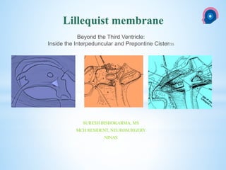
- 1. cka SURESH BISHOKARMA, MS MCH RESIDENT, NEUROSURGERY NINAS Lillequist membrane Beyond the Third Ventricle: Inside the Interpeduncular and Prepontine Cisterns
- 2. Initially described by Key and Retzius in 1875, it was further investigated by Liliequist in 1956 in his studies with pneumoencephalography in cadavers. It may be considered a remnant of the primary tentorium History
- 3. Liliequist membrane may be understood as a projection formed by an arachnoid membrane extending from the dorsum sellae to the mammillary bodies. The LM can be identified as a thin structure (≤ 1 mm) with a thickness that is ever inferior to that of the tuber cinereum, located under the floor of the third ventricle, anteriorly extending from the dorsum sellae to the mammillary bodies. The membrane presents lateral insertions into the oculomotor nerves or adjacent to them, generally into the circumjacent arachnoid sheaths Lillequist membrane
- 4. Schematic illustration of the LM anatomy, demonstrating the three segments of the Liliequist membrane in the sagittal plane THREE SEGMENTS OF THE LILIEQUIST MEMBRANE It is formed by either a single or double arachnoid layer and divided into three segments. Sellar, Diencephalic and Mesencephalic segments
- 5. The subarachnoid space below the third ventricle is reached after opening the ependymal layer just ahead of the mammillary bodies (tuber cinereum) when an endoscopic third ventriculostomy (ETV) is performed. The structure in this region that divides the space into individual cisterns is the membrane of Liliequist. the LM isolates the interpedun- cular cistern from the chiasmatic cistern, with complete blockage in about 10-30% of cases.
- 6. Visualisation of basilar artery after opening the membrane of liliequist.
- 7. This membrane presents a sellar portion, which is inserted in the dorsum sellae, and this sellar portion is subdivided into a posterior projection, a diencephalic portion, and a mesencephalic portion. The diencephalic portion is in close contact with the ependymal layer and extends to the mammillary bodies, and the mesencephalic portion has a posterior inferior projection, surrounding the mesencephalon
- 8. Liliequist just below the third ventricle Pars profunda of the interpeduncular cistern (1), pars superficialis of the interpeduncular cistern (2), prepontine cistern (3), and pia mater (4)
- 9. The membrane of Liliequist limits the interpeduncular cistern. This cistern has a pars profunda, adjacent to the ependymal layer, and a pars superficialis, just below the pars profunda. The diencephalic portion of the membrane of Liliequist divides the two segments
- 10. The pars profunda contains the anterior group of thalamoperforating arteries. Located in the pars superficialis is the bifurcation of the basilar artery with its two main branches, the posterior cerebral arteries (P1) and the superior cerebellar arteries (located immediately before the bifurcation of the basilar artery), and the oculomotor nerves (CN III). The lower limit of the pars superficialis is the mesencephalic portion of the membrane of Liliequist. The recess below the latter is the prepontine cistern
- 11. the key to the success of an ETV is the opening of the membrane of Liliequist, at least of its diencephalic portion. In certain cases progression of the endoscope inside the prepontine cistern is possible, enabling visualization of the trajectory of the basilar artery, and the nerves that emerge from the anterior side of the brainstem, such as the abducens nerve (CN VI), in the transition between the pons and the medulla oblongata, and the hypoglossal nerve (CN XII), at the medulla oblongata
- 12. The endoscopic viewing angle for the interpeduncular and prepontine cisterns The endoscopic viewing angle for the interpeduncular and prepontine cisterns
- 13. Trajectory of the neuroendoscope through the pars profunda of the interpeduncular cistern. This step is mandatory for ETV success. TRAJECTORY OF THE NEUROENDOSCOPE Insertion of the mesencephalic portion of the membrane of Liliequist at the pontomesencephalic rim (a), insertion of the diencephalic portion of the membrane of Liliequist at mammillary body (b), bulging of this segment against the chiasmatic cistern (c), insertion of the membrane of Liliequist at the dorsum sellae (d), gap of the mesencephalic segment (e). Bifurcation of the basilar artery (1), posterior bundle of the thalamoperforating arteries, penetrating the posterior perforated substance (2), anterior bundle of the thalamoperforating arteries, crossing pars superficialis of the interpeduncular cistern (3), prepontine cistern (4), pars superficialis of the interpeduncular cistern (5), pars profunda of the interpeduncular cistern (6), chiasmatic cistern (7)
- 14. In such a procedure, the neurosurgeon performs the puncture of the floor of the third ventricle, by direct visualization, communicating the third ventricle with the basal cisterns. Intraoperatively, the CSF flow can already be seen through the orifice. Also, in order to guarantee the success of the treatment, it is necessary to create a patent pathway from the interpeduncular and pre- pontine cisterns to the chiasmatic/suprasellar cistern.
- 15. Occlusion caused by the LM or even by other pre-pon- tine arachnoid trabeculae is already a well established cause of failure of endoscopic third- ventriculostomy, a surgical approach classically utilized for obstructive hydrocephalus resulting from aqueductal stenosis. Surgical implication
- 16. 1. Dias DA, castro FLO, yared JH, júnior AL, filho LAF, nelson fortes ferreira PD. Liliequist membrane: radiological evaluation, clinical and therapeutic implications. Radiol bras. 2014;47(3):182–5. 2. Fushimi y. Et al. Liliequist membrane: three-dimensional constructive interference in steady state MR imaging. Neuroradiology;22:360-5. REFERENCES
- 17. Thank you NATIONAL INSTITUTE OF NEUROLOGICAL AND ALLIED SCIENCES, BANSBARI, KATHMANDU TOPIC NATIONAL INSTITUTE OF NEUROLOGICAL AND ALLIED SCIENCES, BANSBARI, KATHMANDU